ACHD first look
Name the anatomySitus → AV connection → VA connection → associated lesions
Name every repairOperation + age + conduit/valve/baffle + reinterventions
Name the physiologySystemic ventricle · shunt · obstruction · cyanosis · Fontan
Call ACHD earlyNew arrhythmia, HF, cyanosis, pregnancy, procedure, infection
A
The one-line ACHD summary
anatomy + repair + residue + physiologySay it this way“Situs solitus, d-loop ventricles, d-TGA after Senning atrial switch; unobstructed systemic and pulmonary venous baffles, moderately dysfunctional systemic morphologic RV, moderate systemic TR, prior IART ablation.” This prevents the common error of treating the label while missing the circulation.
1 · Segmental anatomy
Atria → ventricles → great arteries; venous returns; pulmonary veins
Atria → ventricles → great arteries; venous returns; pulmonary veins
›
2 · Surgical map
What was connected, baffled, divided, patched, valved, or left as a shunt?
What was connected, baffled, divided, patched, valved, or left as a shunt?
›
3 · Current lesions
Residual shunt/obstruction/regurgitation; ventricular function; aorta; PA pressure
Residual shunt/obstruction/regurgitation; ventricular function; aorta; PA pressure
›
4 · Physiology
NYHA/CPET, rhythm, oxygenation, end-organ burden, pregnancy goals
NYHA/CPET, rhythm, oxygenation, end-organ burden, pregnancy goals
B
Complexity is anatomy × physiology
do not use anatomy alone| AP anatomy | Examples | Physiologic stage | What moves a patient up |
|---|---|---|---|
| Simple | Small ASD/VSD/PDA; repaired isolated lesions | A no sequelae → D severe | Symptoms, cyanosis, PH, ventricular dysfunction, valve disease, arrhythmia, end-organ disease |
| Moderate | Repaired TOF, AVSD, coarctation, Ebstein, PAPVR, congenital valve disease | Follow-up intensity is set by the combined anatomic–physiologic class, not by childhood “repair.” | |
| Complex | TGA, ccTGA, Fontan/single ventricle, truncus, PA/VSD, heterotaxy, cyanotic CHD | At least one ACHD specialist evaluation for every adult with CHD; ongoing ACHD-center care for moderate/complex disease. | |
C
Records that change management
- Original diagnosis: catheterization, echo, operative drawings.
- All operative notes: patch/baffle material, conduit size, coronary transfer.
- Device history: lead route, abandoned leads, epicardial system, venous access.
- Imaging trend: CMR volumes/flows; aortic dimensions; baffle/conduit patency.
- Rhythm history: IART/AF/VT, ablation map, surgical scars, prior EP study.
- Noncardiac burden: liver, kidney, lung, neurologic, hematologic, mental health.
D
Choose the test that answers the anatomy
| Tool | Best ACHD questions | Common limitation |
|---|---|---|
| TTE/TEE | Valves, gradients, shunts, pressure estimates, baffles; TEE for rims/thrombus/intraoperative detail | Unusual windows and geometric RV assumptions |
| CMR | Ventricular volumes/EF, flow/Qp:Qs, PR fraction, branch-PA split, baffles/Fontan, fibrosis | Devices, arrhythmia, claustrophobia; local congenital expertise |
| Cardiac CT | Coronaries, pulmonary veins, aorta, conduits/stents, calcification, airway and surgical relationships | Radiation/contrast; Fontan needs a tailored contrast protocol |
| CPET | Objective trajectory, chronotropic response, ventilatory efficiency, desaturation, transplant/therapy timing | Compare serial tests with the same protocol and lesion context |
| Holter / rhythm monitor | 24–48 h Holter for daily events; patch/event/telemetry or ILR when events are less frequent | Match duration to event frequency; a normal short monitor does not exclude episodic high-risk rhythm |
| Catheterization | PVR/filling pressure, gradients, collateral/shunt flows, test occlusion, intervention planning | One resting snapshot; interpret with anatomy and oxygen-consumption assumptions |
E
Baseline adult inventory
Clinical
Trajectory
NYHA symptoms, exercise, syncope/palpitations, saturation, all-limb BP, volume status, scars, pulses, murmurs, contraception/pregnancy goals.Data
Trend, do not snapshot
ECG/QRS, BNP, CBC/iron, kidney/liver tests; lesion-specific CMR/CT, CPET, rhythm and end-organ surveillance.Context
Life course
Genetics/family, neurocognitive and mental health, dental care, vaccination, employment/insurance, exercise and transition gaps.Safety ruleUnexplained deterioration is an anatomic problem until proven otherwise: look for baffle/conduit obstruction, residual shunt, valve failure, collateral flow, new PH, thrombosis, endocarditis, and arrhythmia before escalating generic HF therapy.
Why history mattersMany adults carry a circulation created by the best operation available in their era. The date and eponym often reveal the route of blood flow, scar substrate, prosthetic material, and expected failure mode.
A
Milestones you will see in adult charts
1938
Gross ligates a PDA — first successful congenital cardiac operation.
1944
Blalock–Taussig systemic-to-PA shunt — pulmonary-flow palliation for cyanotic CHD.
1952
Lewis closes an ASD under hypothermia; direct-vision intracardiac surgery begins.
1953–55
Gibbon pump / Lillehei cross-circulation enables open repair of ASD, VSD, and TOF.
1958–64
Senning then Mustard atrial switch creates physiologic correction of d-TGA; systemic RV remains.
1968
Rastelli tunnels LV through VSD to aorta and places an RV–PA conduit for complex TGA/DORV.
1971
Fontan–Baudet routes systemic venous blood to pulmonary arteries for tricuspid atresia.
1975
Jatene arterial switch restores LV-to-aorta and RV-to-PA continuity with coronary transfer.
1981
Lecompte maneuver brings branch PAs anterior to the aorta; now integral to most arterial switches.
1980s
Norwood / bidirectional Glenn / total cavopulmonary connection establish staged single-ventricle palliation.
1990s
Double-switch and cone reconstruction favor anatomic ventricles and valve preservation in selected patients.
2000s+
Transcatheter era: ASD/PDA devices, stents, transcatheter pulmonary valves, hybrid palliation, covered-stent sinus venosus repair.
B
Era clues at the bedside
Thoracotomy scar
Closed-heart palliation
Think classic BTT, Potts, Waterston, coarctation repair, PDA ligation. Check unequal arm pulses/BP and distortion or aneurysm at the old connection.Atrial switch era
Born before mid-1980s
Mustard/Senning is likely in d-TGA. Expect systemic RV failure, systemic TR, sinus-node disease, IART, and baffle leak/stenosis.Conduit-dependent
Multiple sternotomies
Rastelli, truncus, PA/VSD, Ross, or Yasui physiology. Every conduit degenerates: define type, size, age, calcification, regurgitation, and coronary proximity.C
Historical operations that may still shape flow
| Era operation | Original goal | Adult consequence if present or previously taken down |
|---|---|---|
| PA band | Restrict excess pulmonary flow/protect PVR | Residual supravalvar PS, PA distortion, pulmonary-valve injury, subaortic obstruction in some SV hearts |
| Systemic–PA shunt | Increase pulmonary blood flow | Unequal pulses, PA distortion, collateral/aneurysm/endarteritis, pulmonary vascular injury |
| Atrial septectomy | Mix or decompress atria | Large residual communication, paradoxical embolic route, atrial scar |
| Atriopulmonary Fontan | Route systemic venous blood to PA | Giant RA, IART, thrombus, energy loss; selected conversion to TCPC |
| Valveless conduit | Create RV–PA continuity when options were limited | Severe PR and RV load in addition to stenosis/calcification |
| Patch aortoplasty | Enlarge coarcted arch | Late repair-site aneurysm/pseudoaneurysm; cross-sectional imaging is mandatory |
D
Three surgical revolutions
Palliation
Survive with altered physiology
Shunts, bands, septectomy and cavopulmonary connections changed flow without normalizing anatomy.Physiologic repair
Correct the streams
Mustard/Senning and classic ccTGA repairs produced serial flow but left a morphologic RV systemic.Anatomic repair
Restore ventricular roles
Arterial/double switch, root translocation and modern valve repair improve anatomy but create coronary/root/baffle/conduit surveillance.E
Translate the operative note into current anatomy
Verb
patched · baffled · divided · translocated · unifocalized
patched · baffled · divided · translocated · unifocalized
›
Structure
septum · vein · root · PA · arch · AV valve
septum · vein · root · PA · arch · AV valve
›
Material
pericardium · PTFE · Dacron · homograft · prosthesis
pericardium · PTFE · Dacron · homograft · prosthesis
›
Failure
leak · stenosis · degeneration · dilation · scar/arrhythmia
leak · stenosis · degeneration · dilation · scar/arrhythmia
READ
Never infer anatomy from a sternotomy scar or an eponym alone. Obtain the operative report and reconstruct current blood flow; named operations have variants and may have been revised.
A
Pulmonary blood-flow operations
systemic-to-pulmonary shunts| Operation | Connection / purpose | Adult fingerprint |
|---|---|---|
| Classic BTT | Subclavian artery divided and connected to ipsilateral PA | Weak/absent pulse and falsely low BP in that arm; PA distortion; shunt occlusion |
| Modified BTT | Gore-Tex graft from innominate/subclavian to PA; preserves arm flow | Residual/occluded graft, PA stenosis, aneurysm, endarteritis |
| Central shunt | Ascending aorta-to-main PA graft | Prior pulmonary overcirculation; proximal PA scarring |
| Waterston / Potts | Ascending aorta–RPA / descending aorta–LPA window | Preferential PA hypertension, branch distortion; rarely left patent in adults |
| Sano | RV-to-PA conduit in Norwood stage I | Ventriculotomy scar, conduit stenosis/regurgitation; usually taken down at stage II |
B
Mixing and decompression
Rashkind balloon atrial septostomy
Tears the atrial septum to improve mixing in neonatal TGA or decompress a restrictive atrium. Adult clue: residual ASD or later atrial-switch history.
Tears the atrial septum to improve mixing in neonatal TGA or decompress a restrictive atrium. Adult clue: residual ASD or later atrial-switch history.
Blalock–Hanlon septectomy
Surgical atrial septectomy, historically used for mixing. Leaves broad interatrial communication and atrial scar.
Surgical atrial septectomy, historically used for mixing. Leaves broad interatrial communication and atrial scar.
Potts shunt for suprasystemic PAH
Modern selected use: descending aorta-to-LPA connection as a pressure-relief “pop-off.” Lower-body cyanosis is expected.
Modern selected use: descending aorta-to-LPA connection as a pressure-relief “pop-off.” Lower-body cyanosis is expected.
Atrial septostomy in PAH
Selected bridge/palliation only; trades systemic saturation for cardiac output. Never a routine bedside intervention.
Selected bridge/palliation only; trades systemic saturation for cardiac output. Never a routine bedside intervention.
C
Cavopulmonary connections
no subpulmonary pump| Operation | Flow created | Key late issues |
|---|---|---|
| Classic Glenn | SVC end-to-side to RPA; often excludes contralateral lung | Pulmonary AVMs, venovenous collaterals, unilateral PA distortion |
| Bidirectional Glenn | SVC to undivided PAs; flow to both lungs | Glenn obstruction, collaterals, pulmonary AVMs if hepatic flow excluded |
| Hemi-Fontan | Stage-II cavopulmonary connection prepared for lateral-tunnel Fontan | Similar Glenn physiology; atrial/pulmonary-artery patch material |
| Kawashima | For interrupted IVC with azygos continuation: SVC/azygos flow to PAs | Hepatic veins remain excluded → pulmonary AVMs until hepatic flow is added |
| 1.5-ventricle repair | Bidirectional Glenn unloads a borderline RV while preserving RV-to-PA flow | SVC hypertension, pulsatile PA flow, residual TR/RV dysfunction |
D
What a palliative connection changes today
| Connection | Pressure/flow consequence | Procedure implication |
|---|---|---|
| Patent systemic–PA shunt | Continuous runoff; pulmonary overcirculation or protected flow if stenotic | Do not occlude without defining native pulmonary supply and oxygen response |
| Occluded classic BTT | Arm supplied by collaterals; ipsilateral pressure misleading | Use contralateral arm for systemic BP; avoid assuming radial line reflects central pressure |
| Glenn | SVC pressure becomes pulmonary-artery pressure; flow depends on low PVR | IJ line can obstruct circuit; high PEEP/hypoxia/acidosis reduce flow |
| Kawashima | Most venous return reaches PAs except hepatic veins | Late cyanosis suggests PAVMs from absent/maldistributed hepatic factor |
| Fenestration/ASD pop-off | Right-to-left decompression maintains output at cost of saturation | Air embolism risk; closure needs test occlusion and full hemodynamics |
| PA band remnant | Protective obstruction can become excessive or distort branches | Gradient is flow-dependent; CT/CMR anatomy before dilation/removal |
E
Old shunt bedside clues
Look
Scar and cyanosis
Posterolateral thoracotomy, clavicular scar, differential cyanosis, venous collaterals, clubbing.Feel
Four pulses/BPs
Absent arm pulse after classic BTT/subclavian flap; radiofemoral delay with arch disease.Listen
Murmur can disappear
A silent old shunt may be occluded—or pulmonary pressures may have equalized.F
When an old communication may be intentional
Decompression
Pressure pop-off
Residual VSD/ASD/fenestration can protect a ventricle or venous circuit; test before closure.Pulmonary supply
Only source of flow
Patent shunt/duct/collateral may supply a lung segment with absent or stenotic native PA.Mixing
Required for output
Restrictive atrial communication in parallel or single-ventricle physiology can be life-threatening.Before occlusion: perform a complete saturation/pressure map, define alternate blood supply, and predict the effect on preload, ventricular pressure, cardiac output and oxygenation.
Line and device warningA Glenn/Fontan/old shunt changes venous pressure, access, and embolic risk. Define the circuit before central access, transvenous pacing, or air-containing infusion.
A
Switch, redirect, or route
| Operation | Core maneuver | Dominant adult surveillance |
|---|---|---|
| Mustard / Senning | Atrial baffle redirects caval blood to LV→PA and pulmonary venous blood to RV→aorta | Baffles, systemic RV/TR, sinus node/IART, device access |
| Jatene arterial switch | Great arteries transected/reconnected; coronaries transferred; usually Lecompte | Coronaries, neo-aortic root/valve, branch PA stenosis |
| Rastelli | LV-to-aorta intraventricular baffle + RV–PA conduit | Conduit, baffle leak, LVOTO, ventricular function |
| REV | LV-to-aorta tunnel; anterior PA connection without prosthetic conduit when feasible | RVOT/PA obstruction or PR; LV tunnel |
| Nikaidoh | Aortic root translocation over LV + RVOT reconstruction | Coronaries, neo-aortic valve/root, RVOT |
| Double switch | Atrial switch + arterial switch or Rastelli so morphologic LV becomes systemic | Baffles, LVOTO/RV–PA conduit, neo-aorta/coronaries, rhythm |
B
Outflow, valve, and root operations
Transannular patch
RVOT incision crosses pulmonary annulus: relieves obstruction but creates chronic PR; substrate for RV dilation and scar-related VT.
RVOT incision crosses pulmonary annulus: relieves obstruction but creates chronic PR; substrate for RV dilation and scar-related VT.
Valved RV–PA conduit
Homograft or bioprosthetic tube from RV to PA. Expect stenosis, regurgitation, calcification, endocarditis, and serial replacement.
Homograft or bioprosthetic tube from RV to PA. Expect stenosis, regurgitation, calcification, endocarditis, and serial replacement.
Ross / Ross–Konno
Pulmonary autograft replaces aortic valve; RV–PA conduit inserted. Konno enlarges LVOT. Two valves now require surveillance.
Pulmonary autograft replaces aortic valve; RV–PA conduit inserted. Konno enlarges LVOT. Two valves now require surveillance.
Konno–Rastan
Ventricular septal incision/patch enlarges LVOT and annulus for an aortic prosthesis. Watch AV block, VSD leak, valve/root.
Ventricular septal incision/patch enlarges LVOT and annulus for an aortic prosthesis. Watch AV block, VSD leak, valve/root.
Yasui
Damus–Kaye–Stansel connects pulmonary root to aorta; LV routed through VSD to pulmonary root; RV–PA conduit supplies lungs.
Damus–Kaye–Stansel connects pulmonary root to aorta; LV routed through VSD to pulmonary root; RV–PA conduit supplies lungs.
David / Bentall
Valve-sparing root replacement or composite valve-graft root. Know coronary button anatomy and anticoagulation needs.
Valve-sparing root replacement or composite valve-graft root. Know coronary button anatomy and anticoagulation needs.
C
Valve and venous reconstruction names
| Cone reconstruction | Mobilizes and rotates Ebstein tricuspid leaflets into a 360° cone at the true annulus; often with plication/ring ± Glenn. |
| Warden procedure | Divides SVC above anomalous pulmonary veins; baffled pulmonary veins → LA; cephalad SVC → right atrial appendage. |
| Unifocalization | Collects MAPCAs/native PAs into one pulmonary arterial tree, then connects RV–PA ± closes VSD. |
| Takeuchi repair | ALCAPA: intrapulmonary tunnel directs aortic blood through an aortopulmonary window to left coronary ostium. |
| Sutureless PV repair | Pericardium marsupialized around pulmonary venous confluence; avoids suturing directly on small veins. |
D
Prosthetic material is a future lesion
| Material | Where encountered | What to anticipate |
|---|---|---|
| Dacron/PTFE patch | Baffle, septum, RVOT, arch, PA | Calcification, shrinkage, leak, pseudoaneurysm, adjacent obstruction, IE substrate |
| PTFE tube/graft | Modified BTT, extracardiac Fontan, vascular graft | Fixed caliber, thrombosis/stenosis, lack of growth, infection |
| Homograft | RV–PA conduit, Ross pulmonary position | Calcific stenosis, regurgitation, endocarditis; TPVR landing zone in selected anatomy |
| Bioprosthetic valve | Pulmonary, tricuspid, aortic, conduit | Structural degeneration, thrombosis/IE; possible valve-in-valve strategy |
| Mechanical valve | Aortic/AV-valve replacement | Lifelong anticoagulation, pregnancy complexity, thrombosis/hemolysis |
| Device/stent/coil | ASD/PDA, PA/CoA, collaterals, conduit | Fracture/migration/erosion, restenosis, adjacent compression, access/CMR artifact |
E
Redo intervention planning
Chest entry
Map what lies behind sternum
CT for RV–PA conduit, aorta/neo-aorta, atrium, collaterals, calcification and prior grafts.Cannulation
Map arteries and veins
Baffles, bilateral SVC, interrupted IVC, femoral vessels, prior cutdown/occlusion, Fontan/Glenn.One operation
Bundle the lesions
Valve/conduit, PA, residual shunt, aorta, arrhythmia substrate and device needs should be planned together.F
Common combination operations
| Rastelli + conduit revisions | Recurrent RV–PA work may coexist with LV-tunnel obstruction and residual VSD; interrogate both outflows every time. |
| Ross–Konno | Autograft/root/coronaries + enlarged LVOT + RV–PA conduit; a single “aortic valve” label omits half the repair. |
| Hemi-Mustard–Glenn–Rastelli | Systemic/pulmonary venous baffle + cavopulmonary flow + LV tunnel + conduit; EP/access planning spans all components. |
| Fontan conversion + maze | Atriopulmonary circuit converted to TCPC with atrial reduction/arrhythmia surgery ± pacemaker; recurrent rhythm and pathway disease remain possible. |
A
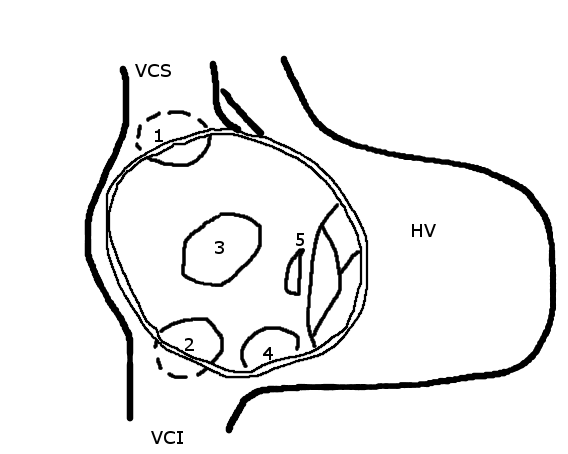
Type determines the repair
| Type | Anatomic clue | Usual closure |
|---|---|---|
| Secundum | Fossa ovalis; assess size and SVC/IVC/aortic/posterior rims | Device preferred when anatomy is suitable; surgery otherwise |
| Primum | Inferior atrial septum; AVSD spectrum; cleft left AV valve | Surgical patch + AV-valve repair |
| Sinus venosus | Usually SVC–RA junction; frequently right PAPVR | Warden/patch repair; selected superior defects by covered stent |
| Coronary sinus | Unroofing ± persistent left SVC | Surgery tailored to systemic venous anatomy |
| PFO | Flap valve, not missing septal tissue | Close for evidence-based secondary stroke prevention—not for RV volume load |
B
Secundum ASD closure pathway
2025 ACC/AHAConfirm anatomyTTE plus CMR, cardiac CT, or TEE to define defect, rims, pulmonary venous return, RV size/function, and Qp:Qs. Do not miss PAPVR.
Is the shunt important?Hemodynamically significant = chamber enlargement distal to the shunt and/or Qp:Qs ≥1.5. With RV dilation and PVR ≤2 WU, closure is recommended; isolated secundum anatomy usually favors device closure.
Borderline pulmonary vascular diseasePVR >2 to <5 WU: closure is reasonable in appropriate anatomy. PVR 5–8 WU: treat PAH first; closure can be beneficial only if PVR falls below 5 WU, generally in an expert center with consideration of fenestration. Continue PAH therapy peri-/post-closure—often long term—with repeat hemodynamics and ACHD–PH follow-up.
Do not closeEisenmenger physiology or net right-to-left shunt. A small ASD with normal/mild RV and Qp:Qs <1.5 has no volume-load indication.
C
Two physiology traps
Stiff left ventricle
ASD as an LA “pop-off”
Older age, hypertension, LVH, diastolic disease, or mitral disease can produce pulmonary edema after closure. If LA pressure is high or LV disease is significant, invasive hemodynamics and balloon test occlusion may be reasonable; consider fenestrated closure.Pulmonary hypertension
Qp:Qs alone is not enough
Measure PVR when PH is suspected. The 2025 definition of precapillary PAH is mPAP >20 mmHg, PCWP ≤15 mmHg, PVR >2 WU. Treat-to-repair decisions belong in ACHD–PH conference.D
How closure is performed
| Route | Requirements | Important complications |
|---|---|---|
| Secundum device | Adequate rims, size within device range, no unaddressed PAPVR; TEE/ICE guidance | Residual shunt, embolization, erosion, thrombus, atrial arrhythmia, venous access complication |
| Surgical patch | Large/deficient-rim secundum, primum, inferior SV/coronary sinus, associated valve/PV surgery | Residual shunt, sinus-node/atrial arrhythmia, venous or pulmonary-vein obstruction |
| Covered-stent SVASD | Superior defect with suitable SVC landing zone; balloon/TEE confirms anomalous PV remains open to LA | PV compression, SVC obstruction, stent migration, residual shunt |
| Fenestrated closure | Selected PAH or high-LA-pressure physiology needing a controlled pop-off | Persistent cyanosis/paradoxical embolic route; fenestration may close or require later revision |
E
PFO is not an ASD
PFO · see P6
Flap tunnel, no chronic RV load
Usually intermittent right-to-left passage, not a sustained volume shunt. Closure decisions follow secondary-stroke evidence after a complete competing-cause evaluation—not ACHD ASD thresholds.ASD
Missing septal tissue
Usually chronic left-to-right flow with RA/RV enlargement. Paradoxical embolism can be a separate closure indication, but pulmonary and LV filling physiology still matter.F
After repair: what should normalize?
- RV volume should fall; late repair may leave persistent dilation/TR.
- PA pressure may not normalize if pulmonary vascular disease is established.
- AF/IART risk persists with older age/atrial remodeling.
- Confirm no device/patch-adjacent residual shunt.
- Reassess LV filling pressure if dyspnea/pulmonary edema appears.
- Antiplatelet/endocarditis instructions depend on device and time since repair.
Common ≠ causalPFO is a flap/tunnel remnant present in about 25% of adults; most require no treatment. In selected patients younger than 60 years, the AAN estimated a 3.4% absolute recurrent-stroke reduction at 5 years, balanced against 3.9% periprocedural complications and an added nonprocedural AF rate of 0.33%/year.
A
Flap tunnel—not missing septal tissue
do not apply ASD closure criteria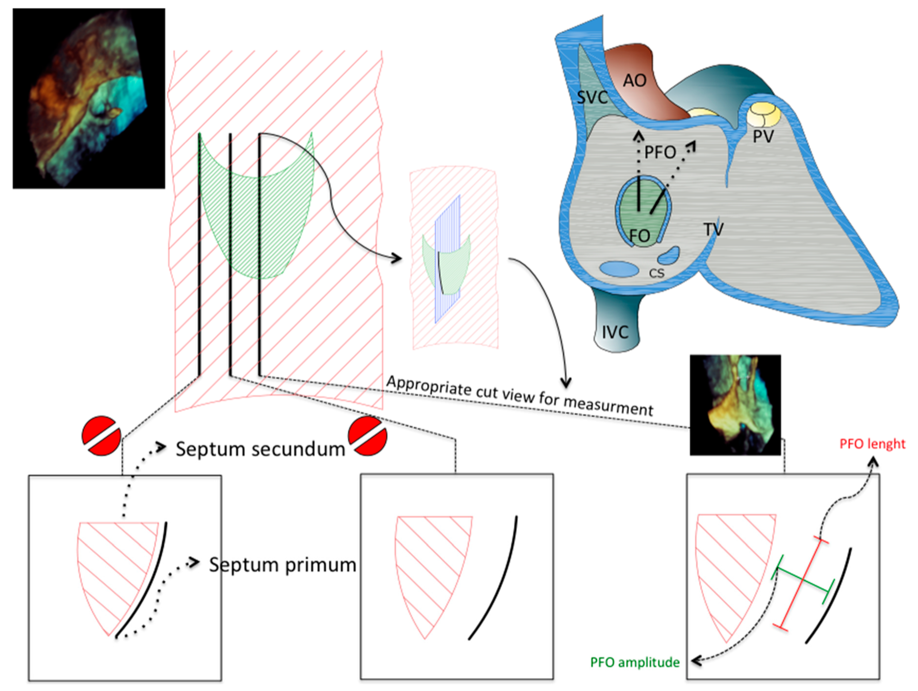
B
Six gates before stroke-prevention closure
heart–brain adjudication| Gate | Establish | Stop / redirect when |
|---|---|---|
| 1 · Real infarct | Brain imaging confirms an embolic/nonlacunar cerebral infarct; review territory and timing. SCAI includes retinal infarction in its PFO-associated stroke definition. Confirm retinal infarction and exclude competing mechanisms with ophthalmology/stroke specialists. The strongest randomized evidence concerns cerebral ischemic stroke. | Symptoms alone, migraine aura, or isolated TIA without infarction is not the trial-defined population. |
| 2 · No better cause | Brain + head/neck vascular imaging; vascular risks; TTE/TEE for cardiac/aortic sources; targeted DVT/PE or thrombophilia testing only when it changes therapy. | Lacunar disease, culprit atherosclerosis/dissection, LV/valve/aortic thromboembolism, or another convincing mechanism. |
| 3 · Exclude AF | ECG plus ≥24-hour telemetry/Holter; use ≥28 days in selected AF-risk patients and more intensive monitoring as age/risk rises. | Established AF usually explains the event and requires anticoagulation—not routine PFO closure. |
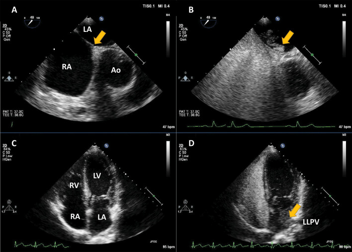
| 4 · Prove R→L shunt | Contrast TCD where available or contrast TTE at rest and with an effective Valsalva/cough. | Inadequate RA opacification or provocation makes a negative study nondiagnostic. |
| 5 · Define anatomy | TEE before closure: PFO versus ASD, tunnel, septal aneurysm, shunt, fenestrations, Eustachian valve/Chiari network and competing source. | TCD detects shunt but cannot localize cardiac versus pulmonary passage or size device anatomy. |
| 6 · Attribute causality | Use PASCAL (RoPE + large shunt/ASA) and a stroke–structural heart team; include patient values and bleeding/AF risk. | A coincidental PFO should not become the diagnosis merely because the workup is difficult. |
C
Interpret the study—and the patient
| Bubble finding | Meaning / pitfall |
|---|---|
| Early LA bubbles | Appearance across the septum within about 3 cycles favors intracardiac shunt; show the crossing if possible. |
| Delayed / from PV | Favors intrapulmonary shunt. Timing supports—but does not by itself prove—localization. |
| Large shunt | ESO convention: >20 LA bubbles within 3 cycles on TTE or >20 on TEE; laboratory/trial definitions vary. |
| Negative but suspicious | Repeat with effective provocation or an alternate modality. Sedation/poor Valsalva can make TEE falsely negative. |
| PASCAL | No large shunt / ASA | Large shunt and/or ASA |
|---|---|---|
| RoPE <7 | Unlikely | Possible |
| RoPE ≥7 | Possible | Probable |
Use scores correctly
RoPE is causal—not recurrence risk
Youth, cortical infarct, and absence of HTN, diabetes, smoking and prior stroke/TIA raise RoPE. Validated PASCAL high-risk anatomy is large shunt and/or ASA (septum-primum excursion generally ≥10 mm). Other tunnel features are contextual.D
Who should—and should not—be closed?
| Clinical setting | Position | Action |
|---|---|---|
| Age 18–60 + imaging-proven PFO-associated stroke | Offer | After a thorough negative competing-cause evaluation, closure plus antiplatelet therapy has the strongest evidence for PASCAL possible/probable patients. |
| Age >60 | Individualize | Evidence is insufficient/very low certainty. Intensify AF and atherosclerotic-source exclusion; use multidisciplinary review, preferably a registry/trial. |
| Incidental PFO; TIA alone; migraine; DCI prevention; ASA, thrombophilia or DVT alone | No routine closure | Do not turn an association into an indication. Treat the actual syndrome and risk factors. |
| Platypnea–orthodeoxia or unexplained systemic embolism | Rare exception | SCAI conditionally supports closure only after other causes of hypoxemia or embolism are excluded; certainty is very low. |
| Severe pulmonary vascular disease / RA pressure-relief physiology | Do not reflexively close | A PFO may be a pop-off; closure can worsen RV failure. Obtain invasive ACHD/PH hemodynamics. |
E
After device closure
Antithrombotics
State the uncertainty
ESO expert consensus used DAPT 1–6 months, then SAPT; SCAI found insufficient evidence for DAPT beyond 1 month. Follow the device/center protocol and bleeding risk. Anticoagulate only for an independent indication such as AF, VTE, or APS; continue vascular-risk management.Surveillance
Device + rhythm + recurrent event
Confirm device position, residual shunt, and thrombus per center/device protocol. New palpitations → AF monitoring. Watch access bleeding, embolization/erosion and endocarditis; use IE prophylaxis for 6 months and longer for a device-adjacent residual defect. Recurrent embolism restarts the entire etiologic workup.Core physiologyPAPVR is an obligatory left-to-right shunt at the venous level. Burden depends on number of anomalous veins, destination, ASD, obstruction, PVR, and associated lung/arterial anatomy—not the label alone.
A
PAPVR and sinus venosus ASD
| Pattern | Typical anatomy | Repair / late issue |
|---|---|---|
| Right upper/middle veins → SVC | Superior sinus venosus ASD; SVC “overrides” septum | Two-patch or Warden; late SVC or pulmonary venous obstruction, sinus-node dysfunction |
| Right veins → RA/IVC | May occur without ASD | Intracardiac baffle or reimplantation; avoid IVC/pulmonary-vein obstruction |
| Left upper vein → vertical vein | To innominate vein; often incidental | Reimplant/ligate vertical vein only when hemodynamically important and anatomy suitable |
| Covered-stent repair | Selected superior sinus venosus defects with suitable SVC/PV geometry | Expert-center option; exclude PV compression and secure landing zones |
B
When to repair anomalous pulmonary venous connection
Repair recommendedSymptoms or reduced exercise capacity + RV volume overload + Qp:Qs ≥1.5.
Repair with ASDWhen an ASD is being surgically repaired, repair associated APVC(s) when technically feasible; a small isolated or technically high-risk vein may be left when pulmonary-vein stenosis risk outweighs shunt benefit.
Repair reasonableAsymptomatic Qp:Qs ≥1.5 plus severe/progressive RV overload, RV dysfunction, invasive PAH, or objective exercise intolerance.
No repairNormal/mild RV enlargement and Qp:Qs <1.5; risk of repair exceeds physiologic benefit.
If repair is not indicated, continue ACHD imaging—echo and/or CMR about every 3–5 years—to reassess RV size/function and pulmonary hypertension.
C
Warden and patch-repair mechanics
| Technique | Flow created | Adult failure |
|---|---|---|
| Single intracardiac patch | Enlarges/uses ASD to baffle anomalous veins behind patch into LA | Long or narrow PV pathway; SVC narrowing; residual shunt |
| Two-patch | PV-to-LA baffle plus anterior SVC/RA enlargement patch | SVC or PV stenosis; sinus-node injury/atrial arrhythmia |
| Warden | SVC divided above highest anomalous PV; caudal SVC/PVs baffled to LA; cephalad SVC sewn to RAA | SVC–RAA anastomotic stenosis, PV baffle stenosis, sinus-node dysfunction |
| Direct reimplantation | Anomalous vein detached and connected to LA/appendage | Tension, kinking, anastomotic PV stenosis |
D
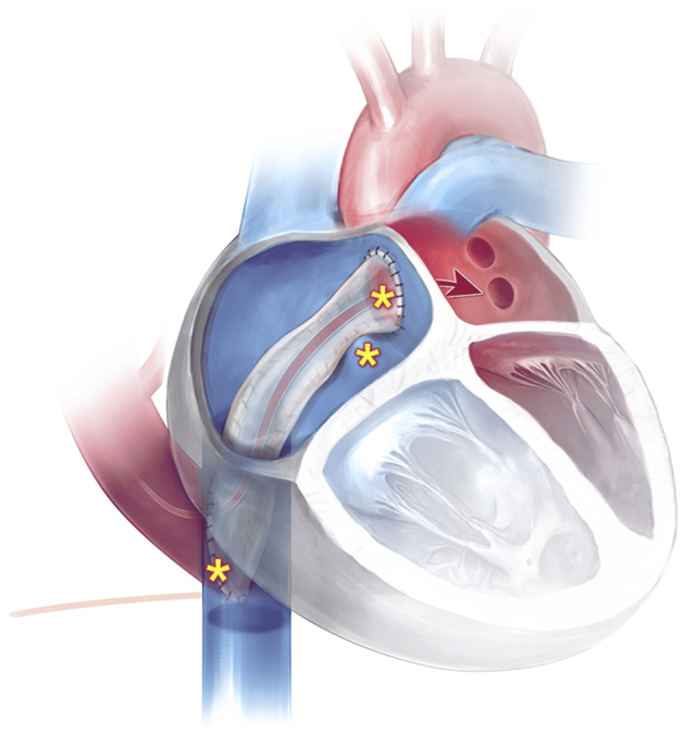
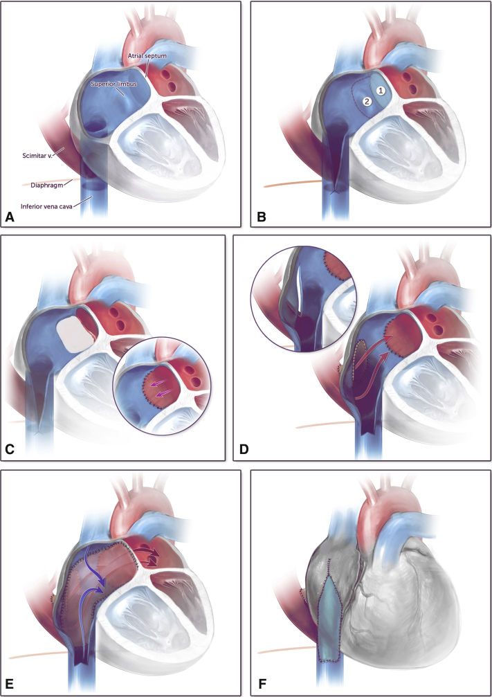
Scimitar syndrome
a syndrome, not just a vein- Anatomy: right pulmonary veins descend to IVC near diaphragm; right-lung hypoplasia/dextroposition, anomalous systemic arterial supply, ASD, sequestration, and PA hypoplasia may coexist.
- Workup: CMR/CTA for venous pathway, lung, systemic arterial supply, RV, Qp:Qs; catheterization if PH or collateral intervention is considered.
- Interventions: embolize important systemic collaterals selectively; repair venous return by reimplantation, intra-atrial baffle, or multipatch pathway. Recurrent pulmonary-vein obstruction is a central risk.
E
Repaired TAPVR / cor triatriatum
| Native route | Repair | Adult surveillance |
|---|---|---|
| Supracardiac | PV confluence-to-LA anastomosis; vertical vein usually ligated; ASD closed | Confluence/individual PV stenosis, vertical-vein persistence, PH, atrial rhythm |
| Cardiac / coronary sinus | Unroof coronary sinus or baffle venous return to LA; close ASD | Residual shunt, systemic/PV pathway obstruction, coronary-sinus anatomy |
| Infracardiac / mixed | Confluence connected to LA; descending channel ligated; mixed veins addressed separately | Highest concern for recurrent PV stenosis and segment-specific obstruction |
| Sutureless re-repair | Pericardium marsupialized around PVs rather than suturing directly on vein ostia | Recurrent stenosis still possible; CT/CMR/perfusion and PH follow-up |
| Cor triatriatum | Resect obstructing LA membrane and treat associated ASD/PV lesion | Residual membrane/gradient, pulmonary venous hypertension, AF |
After anomalous-vein repairNew dyspnea, hemoptysis, recurrent infection, desaturation, or PH warrants CMR/CTA for individual-vein/baffle stenosis, residual shunt, SVC obstruction, and differential lung perfusion.
A
VSD: location predicts the complication
| Type | What to inspect | Typical adult issue |
|---|---|---|
| Perimembranous | Aortic/right AV valve tissue; conduction axis; aneurysmal closure | Aortic regurgitation, subaortic ridge, residual shunt, AV block |
| Outlet / supracristal | Right/noncoronary cusp prolapse | Progressive AR even with a small restrictive shunt |
| Muscular | Single or multiple; apical accessibility | Often restrictive; device closure in selected residual defects |
| Inlet | AVSD spectrum and AV-valve tissue | AV-valve regurgitation; surgical repair |
| Post-repair | Patch margin, LVOT, RV pressure, IE | Residual jet may cause hemolysis or endocarditis |
B
VSD closure gates
Class IClose when Qp:Qs ≥1.5 with significant or progressive LV dilation and PVR ≤2 WU.
ReasonablePVR >2 to <5 WU; or perimembranous/outlet VSD with progressive moderate-or-greater AR. Closure may also be reasonable after VSD-associated infective endocarditis.
Do not closeQp:Qs <1.5 without another indication, or Eisenmenger physiology. PVR 5–8 WU after PAH therapy remains an expert, uncertain-benefit decision—not routine closure.
C
PDA and aortopulmonary window
Patent ductus arteriosus
Continuous systemic-to-PA runoff
Assess LV volume load, descending-aortic flow, PA pressure, PVR, and differential cyanosis. Device closure is usual when anatomy permits and there is LV enlargement/hemodynamic significance. Do not close with Eisenmenger/differential cyanosis. Calcified adult ducts may require covered stent or surgery.Aortopulmonary window
Separate valves, direct great-artery window
Large early shunt resembles truncus physiology but has two semilunar valves. Adults may have repaired patch leaks, branch-PA/aortic distortion, or pulmonary vascular disease. Residual intervention depends on Qp:Qs, chamber load, PVR, and anatomy.D
PDA operability pathway
CloseNet left-to-right PDA with attributable LA/LV enlargement and PVR ≤2 WU; closure is reasonable at PVR >2 to <5 WU.
Treat and reassessFor PVR 5–8 WU, the 2025 recommendation table and Figure 4 allow consideration only if PAH therapy lowers PVR below 5 WU. Evidence is limited. ACHD–PH experts should assess net left-to-right flow, left-heart enlargement and balloon-occlusion tolerance.
Do not closeEisenmenger physiology, PVR >10 WU, or ductal right-to-left flow/differential cyanosis. A tiny asymptomatic duct without LA/LV enlargement and no prior endarteritis has no routine closure benefit.
E
Choosing an intervention route
| Lesion | Catheter option | When surgery is favored |
|---|---|---|
| Muscular VSD | Selected device closure, including residual/postoperative defects | Large/multiple defects, associated valve/outflow operation, unsuitable rim/access |
| Perimembranous VSD | Selected expert use; conduction/valve risks; no U.S. FDA-approved dedicated device | Progressive AR, outlet anatomy, large defect, need for direct valve assessment |
| PDA | Coil/plug usually preferred | Very large, calcified, aneurysmal, infected, or unsuitable duct; selected covered stent |
| AP window | Selected small/residual anatomy in expert hands | Most native large windows; need PA/aortic reconstruction or associated repair |
F
Post-closure checklist
- Residual shunt and hemolysis; device/patch position.
- Aortic valve prolapse/regurgitation and subaortic obstruction.
- LV remodeling and filling pressure after unloading.
- PA pressure/PVR; confirm no late shunt reversal.
- Conduction disease after perimembranous repair.
- Endocarditis risk: residual defect adjacent to prosthetic material.
A
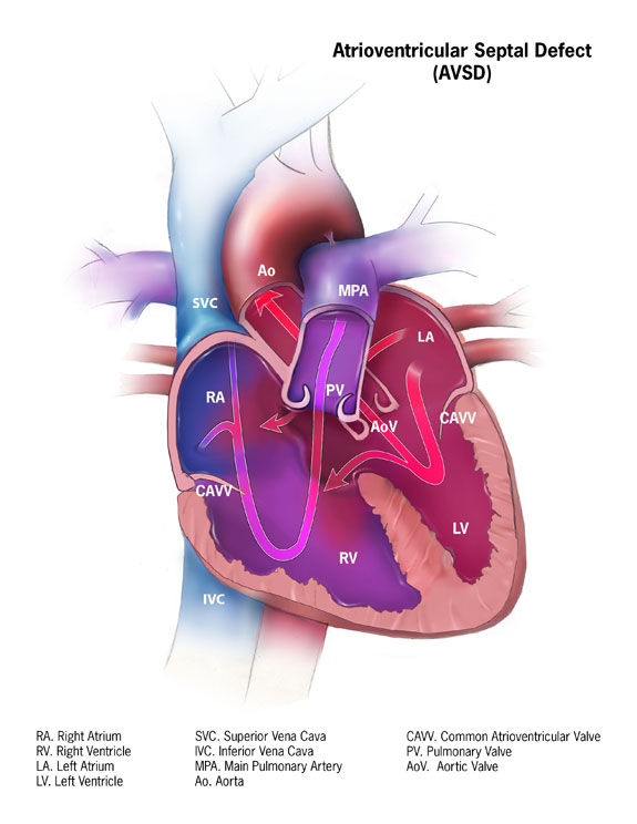
Anatomic spectrum
- Partial AVSD: primum ASD + cleft/zone of apposition in left AV valve; separate AV-valve orifices.
- Transitional/intermediate: primum ASD with restrictive inlet VSD and bridging leaflets.
- Complete AVSD: primum ASD + inlet VSD + common AV-valve orifice; balanced or ventricularly unbalanced.
- Associations: trisomy 21, heterotaxy, subaortic elongation (“gooseneck”), LVOTO, conduction disease.
B
Know the childhood repair
Single-patch / modified single-patch
Common patch or direct VSD closure plus ASD patch; AV-valve tissue attached to patch/crest.
Common patch or direct VSD closure plus ASD patch; AV-valve tissue attached to patch/crest.
Two-patch repair
Separate VSD and ASD patches reconstruct septa and divide common AV valve.
Separate VSD and ASD patches reconstruct septa and divide common AV valve.
Left AV-valve repair
Cleft/zone closure ± annuloplasty; residual stenosis and regurgitation must both be quantified.
Cleft/zone closure ± annuloplasty; residual stenosis and regurgitation must both be quantified.
Unbalanced pathway
May require single-ventricle palliation, biventricular conversion, or 1.5-ventricle strategy; do not assume standard AVSD physiology.
May require single-ventricle palliation, biventricular conversion, or 1.5-ventricle strategy; do not assume standard AVSD physiology.
C
Adult surveillance and intervention
| Target | What matters | Action trigger |
|---|---|---|
| Residual ASD/VSD | Qp:Qs, chamber dilation, PVR | Close/repair with Qp:Qs ≥1.5 and progressive load when PVR ≤2 WU; PVR >2 to <5 may still be reasonable. |
| Left AV valve | Regurgitation, stenosis, LV size/function | Operate for symptomatic severe regurgitation; in asymptomatic severe disease when LVESD ≥40 mm or LVEF <60% |
| LVOT | Subaortic narrowing, tunnel, membrane; Doppler + anatomy | Operate when symptomatic or with LVEF <50%; individualize asymptomatic severe obstruction |
| Conduction | PR prolongation, bundle branch disease, late AV block | Rhythm monitoring for syncope/palpitations; plan pacing around abnormal conduction anatomy |
Surgical nuanceThe “mitral” valve is a congenital left AV valve with abnormal leaflets, chordae, and papillary muscles. Repair/replacement should be planned by congenital valve surgeons; conventional degenerative-MR assumptions can mislead.
D
How adult AVSD fails
Valve
Regurgitation and stenosis
Residual cleft/zone leak, annular dilation, leaflet restriction, subvalvar abnormality, prior ring/patch distortion; quantify mean gradient as well as regurgitation.Outflow
Long narrow LVOT
Bridging leaflet/chordae, septal geometry, subaortic membrane or tunnel can produce progressive obstruction and AR.Electrical
Abnormal node/axis
First-degree block, bundle disease, surgical AV block, atrial scar/IART; late high-grade block can occur.E
Reoperation planning
| Repair versus replace | Define leaflet tissue, zone dehiscence, double-orifice anatomy, chordae/papillary muscles, annulus and prior patch; replacement may threaten LVOT or conduction. |
| Residual shunt | Map relation to AV-valve tissue and conduction system; close when hemodynamically important or when operating for another lesion. |
| LVOTO | Membrane resection/myectomy may be insufficient for long tunnel or valve/chordal obstruction; recurrence is common. |
| Pulmonary vascular disease | Invasive PVR and filling pressure guide operability; do not extrapolate simple-ASD device closure logic. |
| Pregnancy | Residual severe left AV-valve disease, PH, ventricular dysfunction or arrhythmia raises risk; plan before conception. |
A
What the operation did
Senning (1958)
Autologous atrial tissue flaps create systemic- and pulmonary-venous pathways. No prosthetic baffle, but complex atrial suture lines.
Autologous atrial tissue flaps create systemic- and pulmonary-venous pathways. No prosthetic baffle, but complex atrial suture lines.
Mustard (1964)
Pericardial or synthetic patch creates the atrial baffle. Easier geometry, more prosthetic material and potential late shrinkage/calcification.
Pericardial or synthetic patch creates the atrial baffle. Easier geometry, more prosthetic material and potential late shrinkage/calcification.
Both produce the same circulation
SVC/IVC → baffle → morphologic LV → PA; pulmonary veins → baffle → morphologic RV → aorta. The ventriculoarterial discordance remains.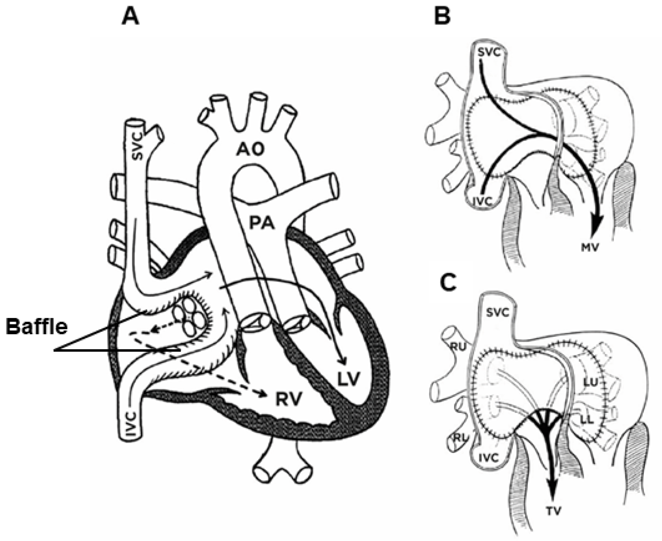
B
The five adult failure modes
| Problem | Clues | What to do |
|---|---|---|
| Systemic RV failure | Declining exercise, congestion, rising BNP, CMR RVEF/volumes | Exclude baffle/valve/rhythm lesions; ACHD-HF therapy and early advanced-care referral |
| Systemic TR | Tricuspid annular/RV dilation; leaflet tethering | Serial echo/CMR; intervene before advanced systemic RV failure when feasible |
| Baffle obstruction | SVC: upper-body venous collaterals; IVC: hepatic/lower-body congestion; PV: pulmonary edema/PH | CMR/CT + catheterization; stent or surgery at ACHD center |
| Baffle leak | Systemic venous→pulmonary venous: cyanosis/paradoxical embolus; reverse: left-to-right load | Quantify direction/size; close when clinically significant, especially before transvenous leads |
| Sinus node/IART | Bradycardia, flutter/IART, rapid deterioration | Rhythm control preferred; anticoagulate sustained IART/AF; congenital EP for ablation/access |
C
Imaging and device access
- Worsening exercise/HF: TTE + CMR for baffles, ventricles, valves, outflows; CPET; cath if hemodynamics remain unclear.
- Before pacemaker/ICD: cross-sectional baffle imaging + venography; treat stenosis/leak first.
- Transvenous leads traverse systemic venous baffle and subpulmonary mitral valve; obstruction and paradoxical embolic pathways matter.
- Systemic RV failure with high ventricular pacing burden/wide QRS may merit CRT or conduction-system strategy at expert center.
D
Baffle hemodynamics and interventions
| Finding | Physiology | Management concept |
|---|---|---|
| Superior systemic-venous stenosis | SVC hypertension, collaterals, facial/arm edema; may be silent | Stent when clinically significant; protect pulmonary-venous pathway and leads |
| Inferior systemic-venous stenosis | IVC/hepatic congestion, ascites, low preload | Catheter/surgical relief; define hepatic-vein relationships |
| Pulmonary-venous baffle stenosis | Pulmonary venous hypertension, edema, PH, reduced systemic-RV preload | Urgent expert intervention; may be focal or diffuse and technically difficult |
| Systemic→pulmonary venous leak | Desaturation, paradoxical embolus, brain abscess | Close when significant; use air precautions and evaluate before leads |
| Pulmonary→systemic venous leak | Left-to-right shunt and pulmonary overcirculation | Quantify Qp:Qs/chamber load; device or surgery as anatomy allows |
E
Systemic-RV care priorities
Unload causes
Fix what is fixable
Baffle obstruction/leak, sustained tachycardia, pacing dyssynchrony and systemic TR can drive failure.Medical therapy
Evidence is limited
Diuretics for congestion; selected HFrEF therapies may be reasonable, but standard LV trial benefits cannot be assumed.Advanced care
Refer early
Transplant/VAD planning is harder after severe RV failure, end-organ injury, collaterals and repeated sternotomy.IART is not benignRapid intra-atrial reentry can sharply reduce systemic-RV filling/output and precipitate shock. Restore rhythm promptly, anticoagulate as indicated, and search for baffle/hemodynamic triggers.
A
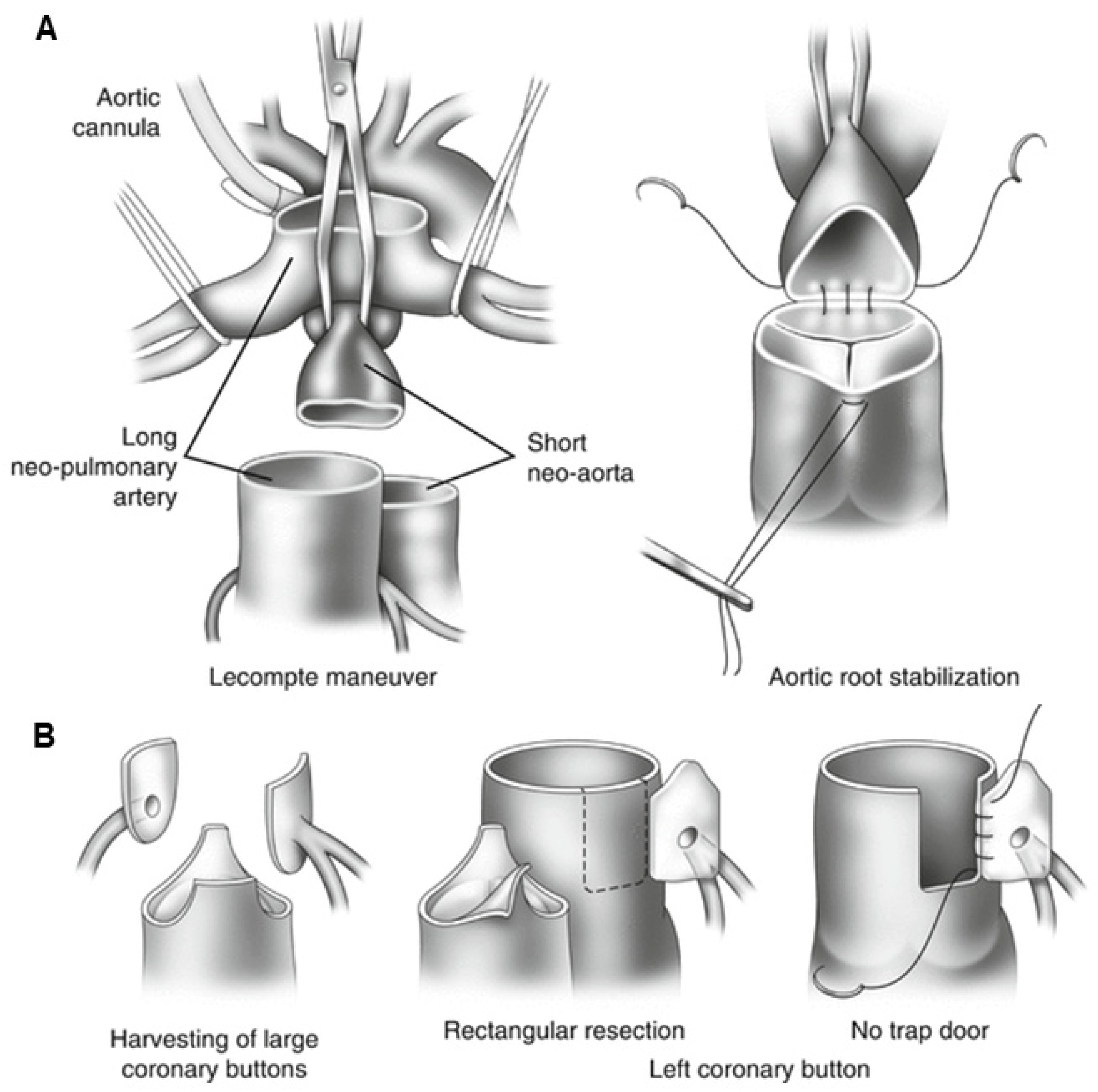
Arterial switch anatomy
- Great arteries are transected and connected to the correct ventricles.
- Native pulmonary root becomes the neo-aortic root; native aortic root becomes the neo-pulmonary root.
- Coronary “buttons” are excised and reimplanted into the neo-aorta.
- Lecompte: branch PAs are brought anterior to the ascending aorta.
Chest pain ruleTransferred coronaries can have ostial/proximal stenosis, kinking, or unusual courses. Ischemic symptoms, ventricular arrhythmia, or new dysfunction require coronary evaluation—young age does not reassure.
B
Arterial-switch adult surveillance
| Target | Modality | Late issue / action |
|---|---|---|
| Coronaries | Baseline gated coronary CT is reasonable even if asymptomatic; stress/anatomic testing for symptoms | Revascularize significant ischemia with congenital coronary team |
| Neo-aorta/valve | Serial TTE + CMR/CT root dimensions and AR | Root replacement may be considered for severe dilation with progressive AR, rapid growth, or valve indication |
| Branch PAs | CMR/CT flow split and anatomy | Stent or surgery for symptomatic or selected significant stenosis |
| Ventricles | TTE/CMR, CPET when functional change | Search coronary, valve, PA, rhythm, and residual-lesion causes before generic HF label |
C
When d-TGA has VSD + LVOTO
Rastelli
LV tunnel + conduit
LV→VSD→aorta baffle; RV–PA conduit. Watch conduit stenosis/PR, LV tunnel obstruction, patch leak, RV dysfunction, arrhythmia.REV
Réparation à l’étage ventriculaire
LV tunnel plus direct anterior RV–PA reconstruction, often avoiding a conduit. Watch RVOT/PA obstruction and PR.Nikaidoh
Aortic root translocation
Moves aortic root over LV for straighter LVOT; adds coronary-transfer, neo-aortic root/valve, and RVOT surveillance.D
Coronary-transfer anatomy
| Issue | Why it develops | Best question |
|---|---|---|
| Ostial stenosis | Scar, small button, intimal proliferation | Is there fixed proximal narrowing despite few conventional CAD risks? |
| Kink/stretch | Growth and root/PA geometry after transfer | Does 3-D CTA show acute angle or tension, especially with exercise symptoms? |
| Intramural/single ostium pattern | Complex native coronary origin makes transfer difficult | What exact pattern and technique were in the operative report? |
| Neo-aortic dilation | Pulmonary root tissue exposed to systemic pressure | Is root growth accompanied by AR or altered coronary geometry? |
| Branch-PA stenosis | Lecompte stretch/compression around enlarged neo-aorta | Is perfusion asymmetric and can PA intervention compromise a coronary? |
E
What the original operation report should contain
- Coronary pattern and button-transfer technique.
- Lecompte maneuver and PA reconstruction/patch material.
- Associated VSD/ASD/arch repair and residual lesions.
- Delayed arterial switch or LV retraining history.
- Neo-aortic root/valve and branch-PA reinterventions.
- Rastelli/REV/Nikaidoh tunnel and conduit type/size if used.
Noncoronary chest pain remains possibleEvaluate aorta, branch PAs, pulmonary hypertension, arrhythmia and conventional acquired CAD—but transferred-coronary disease must be addressed explicitly.
A
Double discordance
Systemic veins → morphologic RA → morphologic LV → PA
Pulmonary veins → morphologic LA → morphologic RV → aorta
- Systemic AV valve is tricuspid; systemic ventricle is morphologic RV.
- Associated VSD, pulmonary/subpulmonary stenosis, Ebsteinoid systemic TV, dextrocardia, and abnormal conduction are common.
- AV node/His anatomy is displaced; spontaneous complete AV block and pacing-related systemic-RV dyssynchrony rise over time.
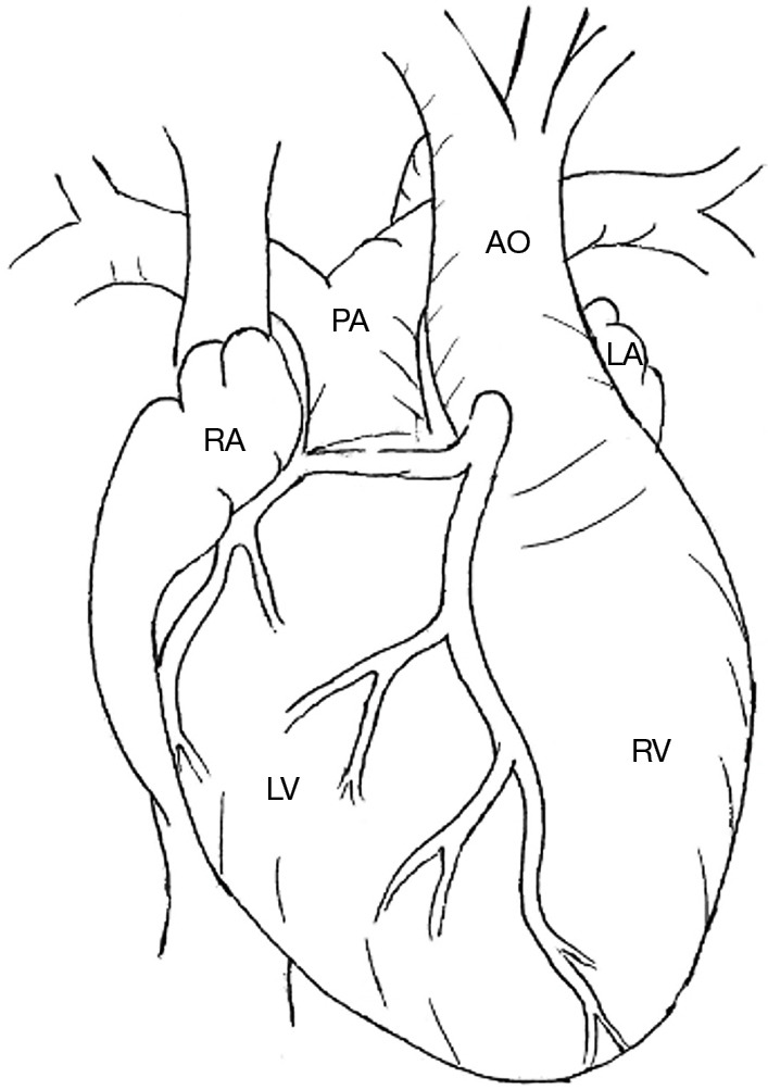
B
Which circulation was chosen?
| Strategy | What remains systemic | Late failure modes |
|---|---|---|
| No repair / physiologic repair | Morphologic RV + tricuspid valve | Systemic TR/RV failure; AV block; residual VSD/LVOTO; relief of LVOTO can worsen TR via septal shift |
| Double switch: atrial + arterial | Morphologic LV | Baffle leak/stenosis and IART plus coronary, neo-aortic, and branch-PA issues |
| Double switch: atrial + Rastelli | Morphologic LV through VSD tunnel to aorta | Baffles + LV tunnel/LVOTO + RV–PA conduit + arrhythmia |
| Hemi-Mustard + Glenn + Rastelli | Morphologic LV; SVC flows directly to the PAs via Glenn, bypassing the subpulmonary morphologic RV | Combined baffle, Glenn, tunnel, conduit, and rhythm complications |
C
Adult management
- Annual TTE and periodic CMR for systemic RV, systemic TR, outflow pathways; rhythm monitoring even if asymptomatic.
- Severe systemic TR: valve replacement is recommended when symptomatic with normal/mildly impaired systemic RV; reasonable in selected asymptomatic patients before advanced dysfunction.
- High-grade AV block: CRT or conduction-system pacing is reasonable to protect systemic RV synchrony.
- Late conversion to double switch is not routine; LV preparedness, age, TR/RV status, anatomy, and operative risk dominate.
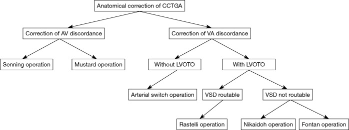
D
Why systemic TR accelerates
| Driver | Clinical implication |
|---|---|
| Ebsteinoid valve / abnormal leaflets | Primary valve disease can be severe before marked RV dilation |
| Systemic-RV pressure and geometry | Annular dilation/tethering create secondary TR and a failure spiral |
| Loss of subpulmonary pressure | Relieving LVOTO can shift septum away from systemic RV and worsen leaflet coaptation |
| Conventional RV pacing | Dyssynchrony can worsen systemic RV function/TR; physiologic/CRT strategy matters |
| Late valve operation | Replacement after advanced systemic-RV dysfunction has worse recovery; timing is preventive |
E
Anatomic-repair selection questions
LV preparedness
Can the LV be systemic?
Pressure/workload, mass, age and need for retraining; an unprepared low-pressure LV may fail after switch.Intracardiac anatomy
VSD and LVOTO
VSD size/location, coronary course across LVOT, AV-valve straddling and pulmonary annulus determine ASO versus Rastelli.Risk trade
Exchange one failure set
Double switch removes systemic RV/TV burden but adds baffle, coronary/root/PA or tunnel/conduit complications.Complete AV blockCan be spontaneous or procedural. Syncope, new bradycardia or exercise intolerance warrants prompt rhythm evaluation; pacing strategy should minimize systemic-RV dyssynchrony.
A
Four native components
- Anterior/malaligned VSD.
- Overriding aorta.
- RVOT/pulmonary obstruction.
- Secondary RV hypertrophy.
Variants matter
- TOF with pulmonary atresia/MAPCAs: conduit and unifocalization pathway.
- Absent pulmonary valve: massive branch-PAs and lifelong airway disease.
- AVSD–TOF: common AV valve and different patch/valve failure.
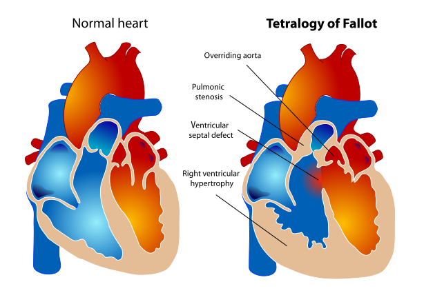
B
Repair phenotype → expected late lesion
| Repair | What was done | Adult fingerprint |
|---|---|---|
| Valve-sparing / transatrial-transpulmonary | VSD patch; muscle resection; pulmonary valvotomy/limited RVOT patch | Residual/recurrent RVOTO or mixed PS/PR; less free PR and less ventriculotomy scar |
| Transannular patch | Patch crosses pulmonary annulus into main PA | Chronic severe PR, RV dilation/TR; RVOT aneurysm; scar-related VT |
| RV–PA conduit | Valved tube bypasses native RVOT | Conduit stenosis/regurgitation/calcification/IE; repeated surgery or TPVR |
| Prior BTT shunt | Systemic-to-PA palliation before complete repair | Unequal arm BP/pulse, branch-PA distortion, old graft/aneurysm; late repair increases arrhythmic risk |
C
Every TOF study should answer
Right heart
RV and RVOT
CMR PR fraction; RVEDVi/RVESVi/RVEF; regional aneurysm/akinesia; RV pressure; TR; residual VSD; branch PA flow.Left heart/aorta
Not an isolated RV disease
LVEF, dyssynchrony, aortic-root dilation, AR, LV filling; coronary anatomy before RVOT intervention.Electrical substrate
Scar + hemodynamics
QRS duration/fragmentation, NSVT, syncope, atrial arrhythmia, prior ventriculotomy, CPET decline, LGE/scar.D
Variant-specific late problems
| Variant | Additional adult surveillance |
|---|---|
| Absent pulmonary valve | Aneurysmal branch PAs, airway compression/bronchomalacia, recurrent infection, RVOT/PR, prior PA reduction plasty |
| TOF + pulmonary atresia | Conduit and reconstructed PAs/MAPCAs, segmental PH, collaterals, hemoptysis, cyanosis |
| TOF + AVSD | Left/common AV-valve regurgitation, residual shunts, LVOT/RVOT, trisomy-21 and PH considerations |
| Double-chambered RV | Residual muscular obstruction below repaired RVOT/VSD; pressure load may coexist with PR |
| Major aortopathy | Root/ascending aortic growth, AR and conventional acquired aortic risk; surgery individualized |
E
Serial TOF dataset
Every visit
Clinical + ECG
Exercise/syncope/palpitations, saturation, murmur, congestion; QRS duration/morphology and rhythm.Periodic
Echo + CMR
PR/PS, RV/LV volumes and EF, TR, VSD, PA flows, aorta; keep volumetric method consistent.When trajectory changes
CPET + monitor + CT/cath
Objectify decline, quantify rhythm, map coronaries/RVOT and invasive hemodynamics.Before transcatheter pulmonary valve or conduit stentingFor RV–PA conduit stenting or balloon-expandable TPVR, test coronary compression at the intended implant diameter—typically balloon interrogation with simultaneous coronary angiography/aortography; CT/CMR alone cannot exclude compression. For native/patch RVOT TPVR, use ECG-gated CT for device-specific dynamic landing-zone geometry and coronary, aortic-root and branch-PA relationships; also assess conduit calcification/rupture risk, access and covered-stent rescue.
Moderate-or-greater pulmonary valve dysfunctionFor the 2025 ACHD algorithm: CMR pulmonary regurgitant fraction ≥25% and/or important RVOT obstruction (e.g., RV systolic pressure >2/3 systemic) defines the group needing structured reassessment.
A
Pulmonary valve replacement
surgical or transcatheterSymptomaticWith cardiovascular symptoms attributable to moderate-or-greater PR/PS, PVR is recommended. Symptoms include otherwise-unexplained dyspnea/chest pain, fatigue/exercise intolerance or below-expected objective exercise capacity, or saturation <90%. Confirm that arrhythmia, branch-PA disease, residual VSD, TR, or LV disease is not the dominant untreated driver.
Asymptomatic · 2025 ACC/AHAWith moderate-or-greater pulmonary valve dysfunction, PVR is reasonable when ≥2 are present: RVESVi >80 mL/m², RVEDV ≥2× LVEDV, RVEF ≤46%, LVEF ≤50%, or progressive decline in objective exercise capacity. Do not wait for self-reported symptoms; use serial CPET.
Asymptomatic · 2026 surgical guidelineWith PR fraction ≥25%, PVR is reasonable when ≥2 are present: RVEDVi ≥160 mL/m², RVESVi ≥80 mL/m², RVEF <47%, or LVEF <55%. RVESVi ≥80 mL/m² alone may support consideration in selected adults.
Choose routeTPVR: suitable conduit/bioprosthesis or selected native RVOT, no coronary compression, infection controlled. Surgery: need RVOT aneurysm remodeling, PA reconstruction, residual VSD closure, TV/aortic work, unsuitable landing zone, or active IE.
Progression and arrhythmia gatesRegardless of symptoms, PVR is reasonable with moderate-or-greater pulmonary valve dysfunction plus progressive RV or LV systolic dysfunction, and may be reasonable with progressive ≥moderate functional TR plus RV dilation. With ≥moderate PR and ventricular tachyarrhythmia requiring treatment, PVR may be considered only in addition to EP therapy; PVR does not remove scar. High SCD risk by clinical markers or a validated multivariable score supports ICD consideration.
B
PVR does not erase VT substrate
| Risk feature | Interpretation / response |
|---|---|
| Prior ventriculotomy or late repair | Anatomic isthmuses around VSD patch/RVOT scar support macroreentrant VT |
| QRS ≥180 ms or fragmentation | Marker of RV dilation/scar/dyssynchrony; trend, do not treat a number in isolation |
| NSVT, syncope, sustained palpitations | Urgent congenital EP evaluation; distinguish IART with aberrancy from VT |
| RV/LV dysfunction, atrial arrhythmia, scar | Raises SCD risk; integrate CMR, CPET, rhythm burden, and hemodynamics |
| Inducible sustained VA | Supports ICD/risk strategy in context; invasive EP/electroanatomic mapping before PVR is reasonable in selected at-risk adults |
C
At the time of PVR
Before
Map the substrate
Decide whether catheter ablation, surgical cryoablation, or substrate transection is needed before a prosthetic valve blocks access. Treat sustained atrial arrhythmia and optimize branch PA/RVOT hemodynamics.After
Continue surveillance
Track prosthetic valve gradients/regurgitation, RV remodeling, exercise, QRS/rhythm, IE, and recurrent VT. A normal new valve does not equal a normal myocardium.D
Prosthesis and procedure tradeoffs
| Choice | Strength | Limitation / future plan |
|---|---|---|
| Surgical bioprosthesis | Can remodel RVOT/PA and address multiple lesions | Redo sternotomy; structural degeneration; choose size for future valve-in-valve |
| Homograft conduit | Useful for complex RV–PA reconstruction | Calcific stenosis/PR and IE; variable future TPVR suitability |
| Balloon-expandable TPVR | Established for suitable conduit/bioprosthetic landing zone | Coronary compression, conduit rupture, stent fracture, IE; often prestenting |
| Self-expanding native-RVOT valve | Expands transcatheter option beyond fixed conduits | Large/variable RVOT geometry, PA branches/coronaries, long-term durability |
| Mechanical pulmonary valve | Rare selected durability strategy | Thrombosis/anticoagulation; generally not routine |
E
After PVR, ask more than “is the valve open?”
- Did RVESVi/RVEDVi and exercise capacity improve?
- Is residual RVOT/branch-PA obstruction present?
- Did TR or LV dysfunction persist?
- Is scar-related VT still inducible/clinical?
- Are gradients rising from thrombosis, degeneration or IE?
- Is antiplatelet/anticoagulation plan correct for prosthesis?
F
When replacing the valve is not enough
Hemodynamic
Residual load
Branch-PA stenosis, RVOT aneurysm, TR, VSD, aortic/left-heart disease can blunt remodeling.Electrical
Fixed substrate
Scar isthmuses and ventricular fibrosis persist; use ablation/ICD strategy when indicated.Timing
Late myocardium
Severe biventricular dysfunction or end-organ disease may require advanced-HF rather than isolated PVR.Endocarditis signalNew RVOT/conduit gradient, fever, septic pulmonary emboli, or valve dysfunction should trigger blood cultures and urgent imaging. Transcatheter and surgical pulmonary prostheses remain lifelong IE substrates.
A
Pulmonary atresia with VSD / MAPCAs
Reconstruct the supply
Native PAs versus MAPCAs
CTA/CMR/cath must map each lung segment: central PA continuity, stenoses, aortopulmonary collateral origins, prior coils/stents/shunts, and segmental pulmonary vascular disease. Saturation and RV pressure can hide unequal segmental physiology.Surgical pathway
Unifocalization ± complete repair
MAPCAs are gathered into a pulmonary tree; a valved RV–PA conduit is created; VSD is closed when the pulmonary vascular bed can accept full RV output. Some adults retain a fenestration or unrepaired VSD as a pressure pop-off.B
Pulmonary atresia with intact ventricular septum
Never decompress before defining coronariesRV sinusoids may supply coronary perfusion; in RV-dependent coronary circulation, lowering RV pressure can cause myocardial ischemia/infarction.
| Childhood route | Adult anatomy to expect | Key late problem |
|---|---|---|
| Biventricular | Valvotomy/RV decompression ± shunt; small/tripartite RV and abnormal TV | RV/TV dysfunction, PR/PS, ischemia from coronary abnormalities |
| 1.5 ventricle | Bidirectional Glenn + antegrade RV–PA flow | Combined SVC/Glenn and RV/TV physiology |
| Fontan | Single-ventricle circuit; RV may remain hypertensive | Fontan failure plus coronary/RV-pressure issues |
C
Truncus arteriosus after repair
VSD patch
routes LV to truncal root (systemic outflow)
routes LV to truncal root (systemic outflow)
+
PAs detached
from common trunk
from common trunk
+
RV–PA conduit
creates pulmonary outflow
creates pulmonary outflow
→
Adult surveillance
truncal valve/root + conduit/PAs + arch/VSD
truncal valve/root + conduit/PAs + arch/VSD
- Truncal valve regurgitation/stenosis and root dilation.
- Conduit degeneration and branch-PA stenosis.
- Residual VSD, arch lesion, ventricular dysfunction.
- 22q11-associated airway, immune, calcium, and aortic-arch issues.
D
The conduit rule
Rastelli, repaired truncus, PA/VSD, Ross, and some TOF repairs all create a biologic RV–PA conduit that will eventually stenose and/or regurgitate. New dysfunction requires exclusion of endocarditis, thrombus, calcification, stent fracture, branch-PA disease, and coronary compression risk before redo surgery or TPVR.
E
MAPCA and branch-PA questions
| Question | Why one global PA pressure is insufficient |
|---|---|
| Which segments connect to native PAs? | Some lung segments may have central PA supply while others depend on MAPCAs |
| Which collaterals are dual-supply? | Coil/ligation may be safe only when another unobstructed source supplies that segment |
| Where are stenoses? | Proximal stenosis can protect a segment; distal vascular disease may be irreversible |
| What was unifocalized? | Surgical neo-PA branches and anastomoses can narrow, disconnect or become aneurysmal |
| Is the VSD fully closed? | Fenestration/residual VSD may intentionally decompress a high-pressure RV |
| Why hemoptysis? | Hypertensive collateral, PA aneurysm, thrombosis, infection and bronchial collaterals require CTA/cath planning |
F
When a conduit needs action
Clinical/hemodynamic
Symptoms or important load
Declining exercise, HF, syncope, sustained arrhythmia, severe stenosis/PR, rising RV pressure, progressive RV dilation/dysfunction or TR support intervention.Choose the platform
TPVR versus surgery
TPVR for suitable landing zone without coronary compression/infection; surgery when conduit/PA reconstruction, aneurysm, residual shunt, valve work or anatomy requires open repair.G
Truncal valve and root
The truncal valve may have 2–4 leaflets and can be regurgitant or stenotic from birth or prior repair. Serial assessment includes valve severity, LV size/function, truncal-root growth, coronary origins, repaired arch, residual VSD and RV–PA conduit. Valve repair is specialized; replacement/root surgery may need to be combined with conduit/PA work.
1Urgent: hemoptysis, new cyanosis, fever/conduit gradient, syncope or RV-pressure crisis.
2Plan: CTA/cath must show segmental supply, collaterals, coronaries and all conduit/PA stenoses.
Infection is a unifying emergencyFever in a conduit-dependent patient can rapidly increase RV pressure or seed PAs/lungs. Obtain cultures promptly and image the entire RV–PA pathway—not only the valve leaflets.
DORV is a family, not one circulationBoth great arteries arise predominantly from the morphologic RV. Adult physiology depends on VSD position, outflow obstruction, great-artery relationship, ventricular balance, AV-valve straddling, and the surgical route created.
A
VSD relationship drives strategy
| Phenotype | Typical route | Adult surveillance |
|---|---|---|
| Subaortic VSD | Intraventricular LV-to-aorta tunnel ± RVOT work | Tunnel/subaortic obstruction, residual VSD, RVOT/PA, aortic valve |
| Subpulmonary VSD / Taussig–Bing | Arterial switch + VSD routing, sometimes arch repair | Coronaries, neo-aortic root/valve, branch PAs, arch, tunnel |
| DORV + PS (TOF-type) | LV-to-aorta tunnel + RVOT patch or RV–PA conduit | TOF/Rastelli phenotype: PR/PS, conduit, RV, arrhythmia |
| Noncommitted/distant VSD | Complex long tunnel, root translocation, or single-ventricle pathway | Tunnel narrowing, AV-valve interaction, Fontan physiology when used |
B
Read the tunnel
- Which ventricle does the patch commit to which great artery?
- Is the baffle narrow, calcified, aneurysmal, or leaking?
- Does it impinge on tricuspid/subpulmonary apparatus?
- Was the VSD enlarged? Where is the conduction axis?
- Is pulmonary flow via native RVOT, patch, conduit, or Fontan?
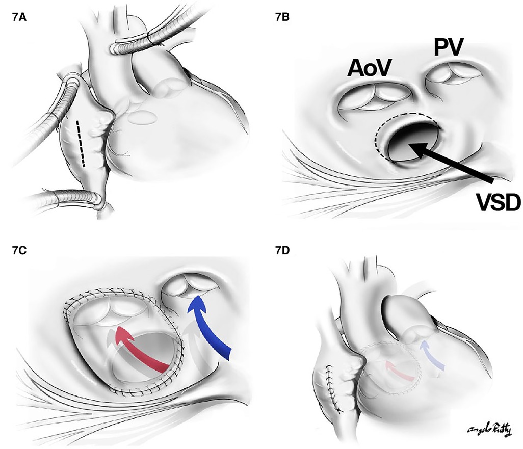
C
Clinical failure modes
Obstruction
Two outflows
LV tunnel/subaortic obstruction and RVOT/conduit/branch-PA obstruction can coexist. Echo gradients require cross-sectional anatomy.Valves/shunts
Patch neighbors
Residual VSD, aortic/neo-aortic regurgitation, and AV-valve regurgitation may reflect the tunnel or original anatomy.Rhythm/function
Scar burden
Ventriculotomy, VSD enlargement, conduits, multiple sternotomies, and ventricular pressure load create atrial/ventricular arrhythmia and HF risk.D
Routing decision anatomy
| Feature | Favors / prevents a pathway |
|---|---|
| VSD position and size | A short unobstructed tunnel is ideal; distant/restrictive VSD may require enlargement, long baffle or root translocation |
| Great-artery relation | Side-by-side/anterior-posterior geometry changes tunnel and arterial-switch feasibility |
| Pulmonary/LV outflow obstruction | Determines ASO versus Rastelli/REV/Nikaidoh and need for conduit |
| AV-valve straddling/chordae | Can cross proposed tunnel, preclude VSD enlargement or make biventricular repair unsafe |
| Ventricular adequacy | Hypoplasia, imbalance or dysfunction may lead to 1.5-ventricle or Fontan pathway |
| Coronary anatomy | Critical for arterial/root translocation and any RVOT/conduit intervention |
E
How to evaluate an adult DORV repair
Echo
Valves and gradients
VSD/tunnel Doppler, RVOT/conduit, AV valves, aortic/neo-aortic valve, ventricular function and pressure.CMR/CT
3-D route and flows
Ventricular volumes, Qp:Qs, tunnel/RVOT/PA geometry, coronaries, aorta and extracardiac relationships.Cath/EP
Resolve the uncertainty
Pressure pullbacks and PVR/collaterals; map scar/tunnel/conduit before ablation or reoperation.F
Intervention traps
- Relieving RVOT obstruction may expose severe PR or change VSD/tunnel flow.
- Tunnel enlargement can threaten AV-valve apparatus or conduction.
- Conduit stenting can compress a coronary.
- Residual VSD may be an intentional pressure relief.
- ASO-type DORV inherits neo-aortic/coronary/branch-PA issues.
- Fontan-type DORV must be managed as a Fontan circuit.
Do not use “DORV repaired” as the assessmentName the LV outlet, pulmonary outlet, systemic ventricle, residual VSD, AV-valve relation, coronary course, and any conduit/Fontan pathway.
A
Know the repair
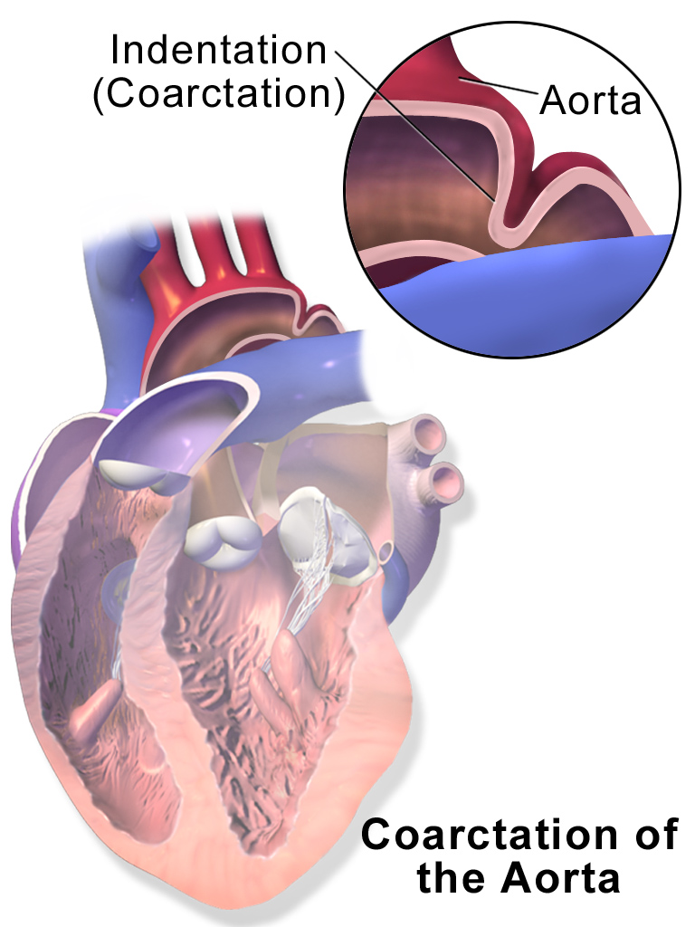
- End-to-end / extended end-to-end: resection and anastomosis; residual arch hypoplasia or recurrent narrowing.
- Subclavian flap: left subclavian incorporated into arch; left-arm pulse/BP can remain low.
- Patch aortoplasty: important late aneurysm/pseudoaneurysm risk.
- Interposition/extra-anatomic graft: fixed-caliber graft, anastomotic aneurysm, compression/infection.
- Balloon/stent/covered stent: restenosis, fracture, migration, aneurysm; CT/MR-compatible follow-up.
B
Lifelong assessment
| Domain | Do | Why |
|---|---|---|
| Arch anatomy | Periodic CMR/CTA of entire thoracic aorta; compare repair site and serial dimensions | Recoarctation, aneurysm, pseudoaneurysm, collateral vessels |
| Blood pressure | Both arms + a leg; ambulatory BP; exercise BP when useful | Masked/resting/exercise hypertension persists even after a good repair |
| Heart/aortic valve | TTE for LVH/function, BAV stenosis/regurgitation, root/ascending aorta | CoA is a diffuse arteriopathy; BAV is common |
| Brain | Intracranial aneurysm screening may be considered | Higher prevalence; individualize modality and repeat interval |
C
When residual/recurrent CoA is important
Integrate evidenceArm–leg systolic BP difference, Doppler mean gradient, cath peak-to-peak gradient, anatomic narrowing, collateral flow, LVH, and hypertension. A threshold around 20 mmHg is a common marker, but severe anatomy/collaterals or hypertension can justify intervention with a lower measured gradient.
Adult intervention
Stent often preferred
Covered stent when aneurysm, wall injury risk, or suitable anatomy; surgery for long-segment disease, complex arch, infected material, large aneurysm, or associated cardiac operation.Interrupted aortic arch survivor
More than repaired CoA
Inspect entire reconstructed arch, LVOT, aortic valve/root, VSD patch, conduit/graft, airway compression, and 22q11-associated conditions.D
Hypertension is part of the lesion
| Pattern | Interpretation / response |
|---|---|
| Resting upper-body HTN | Treat to guideline targets; confirm lower-limb pressure and repair anatomy; ACEi/ARB, beta-blocker or other agents individualized |
| Masked HTN | ABPM can reveal nocturnal/daytime load despite normal clinic BP |
| Exercise HTN | May reflect abnormal arch compliance/endothelial function even without severe recoarctation; informs counseling and treatment |
| Persistent gradient + HTN/LVH | Raises benefit of stent/surgical relief; treat BP before and after intervention |
| Low left-arm BP | Could reflect subclavian-flap or classic BTT anatomy—not systemic hypotension |
E
Aorta and life events
Exercise
Control pressure load
Avoid extreme static effort when uncontrolled HTN, significant obstruction or aneurysm; prescribe aerobic activity after anatomy/BP review.Pregnancy
Image before conception
Assess repair site, entire aorta, BAV, BP and cerebral history; manage hypertension and delivery with pregnancy-heart team.Acquired risk
Still prevent CAD
Smoking, lipids, diabetes and obesity compound lifelong vascular dysfunction and aortic risk.Urgent imagingNew chest/back pain, hoarseness, pulse/BP change, neurologic symptom, hemoptysis, or unexplained anemia after arch repair can signal dissection, aneurysm leak, fistula, or pseudoaneurysm.
A
Left ventricular outflow: valve, below, above
| Level | Anatomy / operation | Adult priorities |
|---|---|---|
| Valvar AS / BAV | Balloon/surgical commissurotomy, repair, SAVR; Ross in selected young patients | AS + AR, root/ascending aorta, coarctation, prior leaflet intervention, pregnancy/exercise |
| Discrete subaortic stenosis | Membrane resection ± septal myectomy | Recurrence, progressive AR, VSD/AVSD association, iatrogenic AV block/VSD |
| Tunnel/small annulus | Konno–Rastan, Ross–Konno, Yasui in complex arch/LVOTO | Patch/tunnel, prosthetic or autograft valve, AV block, residual VSD, RV–PA conduit |
| Supravalvar AS | One-/multi-patch aortoplasty; Williams syndrome or familial ELN disease | Coronary ostia/perfusion, diffuse arteriopathy, branch PA/renal disease, hypertension |
B
The Ross is a two-valve operation
Pulmonary autograft
native pulmonary root → aortic position
native pulmonary root → aortic position
+
Coronary buttons
reimplanted into autograft root
reimplanted into autograft root
+
RV–PA homograft
reconstructs pulmonary outflow
reconstructs pulmonary outflow
→
Late disease
autograft dilation/AR + conduit PS/PR
autograft dilation/AR + conduit PS/PR
Ross–Konno
Added LVOT enlargement
Septal incision and patch enlarge a small annulus/tunnel. Add surveillance for residual/recurrent LVOTO, AV block, VSD leak, and septal/ventricular function.Yasui
LV through pulmonary root to aorta
LV is baffled across a VSD to pulmonary root; DKS connects pulmonary root to aorta; RV–PA conduit supplies lungs. Follow both outflows and the baffle.C
Right-sided obstruction
| Valvar pulmonary stenosis | Domed mobile valve → balloon valvuloplasty preferred; dysplastic valve, severe PR, or failed catheter therapy may need surgery/PVR. |
| Double-chambered RV | Hypertrophied muscle bundles divide RV; often perimembranous VSD. Progressive obstruction → muscle resection ± VSD closure. |
| Branch PA stenosis | Native or postoperative (ASO/Lecompte, TOF, shunt, Fontan). Use CMR flow split + CT anatomy; balloon/stent/surgery based on location and load. |
| Peripheral PA stenosis | Williams/Alagille and syndromic arteriopathy may be diffuse; segmental pressures and perfusion matter more than one main-PA gradient. |
D
Congenital mitral disease / Shone complex
Parachute or arcade mitral valve, supramitral ring, subaortic obstruction, and coarctation may coexist. Quantify multilevel inflow/outflow obstruction, pulmonary pressure, LV size/function, and prior repairs. A “mild” gradient at one level may be important when several lesions are in series.
E
BAV and aortopathy questions
| Domain | What changes management |
|---|---|
| Valve phenotype | Fusion pattern, calcification, stenosis versus regurgitation, prior commissurotomy/repair, LV response |
| Aortic segments | Annulus, sinuses, ST junction, ascending aorta, arch and coarctation—measure consistently by modality/plane |
| Growth/risk modifiers | Rapid enlargement, family history/dissection, coarctation, uncontrolled HTN, planned pregnancy, need for valve surgery |
| Operation choice | Repair, SAVR, selected TAVR, Ross, or combined root/ascending surgery depends on age, anatomy and lifetime plan |
| Family | First-degree relatives should receive aortic-valve/aorta screening when appropriate |
F
Lesion-specific intervention logic
| Lesion | Action trigger / caution |
|---|---|
| Severe valvar AS | Symptoms, LV dysfunction or guideline high-risk features; balloon useful mainly for suitable noncalcified congenital valve |
| Subaortic stenosis | Symptoms, important gradient, progressive AR/LV effects; recurrence and AV block require lifelong follow-up |
| Supravalvar AS | Symptoms/ischemia, severe gradient, LVH; assess coronary ostia and diffuse branch-vessel disease before repair |
| Pulmonary stenosis | Balloon for suitable severe valvar PS; surgery/valve replacement for dysplastic valve, major PR or associated RVOT/PA work |
| DCRV | Progressive muscular obstruction/symptoms; resect bundles and address associated VSD |
| Branch PA stenosis | Symptoms, RV pressure load or important perfusion imbalance; protect adjacent coronaries/airways and account for prior Lecompte |
G
After a Ross/Konno/Yasui
- Autograft root dimensions and aortic regurgitation.
- Coronary-button anatomy and ischemic symptoms.
- RV–PA conduit stenosis/regurgitation/endocarditis.
- LVOT tunnel/patch and residual VSD.
- Conduction disease/AV block and ventricular scar.
- Plan combined rather than serial reoperations when possible.
Two-level diseaseA patient can have valvar, subvalvar and supravalvar obstruction in series. Doppler gradients are flow-dependent and not simply additive; define anatomy with cross-sectional imaging/catheterization when discordant.
A
Anatomy that matters
- Apical displacement and failed delamination of septal/posterior tricuspid leaflets.
- “Atrialized” inlet RV above displaced functional valve; variable usable RV below it.
- Massive RA, TR, ASD/PFO with right-to-left shunt, cyanosis/paradoxical embolus.
- Right-sided accessory pathways/WPW, AVRT, atrial arrhythmia, ventricular scar.
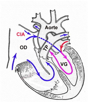
B
Operation decoder
Danielson / Carpentier repairs
Historical leaflet mobilization, annular reduction, atrialized-RV plication, and monocusp/bileaflet reconstruction. Residual TR/stenosis and RV geometry vary.
Historical leaflet mobilization, annular reduction, atrialized-RV plication, and monocusp/bileaflet reconstruction. Residual TR/stenosis and RV geometry vary.
Da Silva cone
Leaflets are extensively delaminated, rotated, and joined into a circumferential cone at the true annulus; often with ring and atrialized-RV/RA reduction.
Leaflets are extensively delaminated, rotated, and joined into a circumferential cone at the true annulus; often with ring and atrialized-RV/RA reduction.
1.5-ventricle repair
Bidirectional Glenn unloads a marginal RV while preserving RV–PA continuity; adds SVC/Glenn physiology.
Bidirectional Glenn unloads a marginal RV while preserving RV–PA continuity; adds SVC/Glenn physiology.
Starnes palliation
Severe neonatal pathway excludes/occludes tricuspid inflow and proceeds toward single-ventricle circulation; adult is a Fontan patient.
Severe neonatal pathway excludes/occludes tricuspid inflow and proceeds toward single-ventricle circulation; adult is a Fontan patient.
C
When to refer for surgery
Clinical triggerModerate-or-greater TR with HF symptoms or objective deterioration in exercise capacity.
Ventricular triggerProgressive RV enlargement or systolic dysfunction before end-stage RV disease.
Shunt triggerProgressive desaturation/right-to-left atrial shunt or paradoxical embolus after hemodynamic review.
Rhythm triggerRecurrent atrial/ventricular arrhythmia; congenital EP mapping/ablation is ideally completed before valve surgery.
D
Do not miss
- CMR functional-RV size/function and TR; echo leaflet/annulus anatomy.
- LV compression/dysfunction from the enlarged RV.
- Accessory pathways can be multiple and right-sided.
- ASD/PFO may be a pressure relief; do not close reflexively.
- Transvenous leads can worsen TR and complicate cone repair.
- Pregnancy tolerance depends on cyanosis, arrhythmia, RV and TR—not the label.
Timing principleSevere systemic venous congestion or advanced RV dysfunction can make a technically successful tricuspid repair too late. Refer while the functional RV and end organs remain recoverable.
E
Quantify the right heart correctly
| Measure | Why it matters |
|---|---|
| Apical displacement | Index septal-leaflet displacement to body size; confirms anatomy but does not alone set surgery timing |
| Functional RV | Exclude atrialized segment when judging effective chamber; CMR volumes/function and regional motion are valuable |
| TR mechanism/severity | Leaflet delamination, tethering, coaptation, annulus and jet; massive RA/RV can make standard measures discordant |
| Forward output | CPET, stroke volume and saturation can decline before classic LV-type EF thresholds |
| Atrial shunt | Direction at rest/exercise, RA pressure and PVR determine whether closure is safe |
| LV interaction | RV enlargement/septal shift can impair LV filling/function |
F
After cone or valve replacement
Early
RV adaptation
Low output/RV dysfunction, residual TR/stenosis, Glenn pressure if added, rhythm and AV block.Late
Durability
Recurrent TR, ring/valve gradient, functional-RV remodeling, RA size, ASD closure/residual shunt.Electrical
Scar and pathways
Recurrent AVRT/IART/AF, pacer/ICD need, lead interaction with repaired or prosthetic valve.Severe neonatal palliation in an adult chartA prior Starnes procedure means tricuspid exclusion and a single-ventricle/Fontan pathway; do not apply biventricular Ebstein algorithms.
Lesions that may reach this pathwayTricuspid atresia, double-inlet LV, unbalanced AVSD, HLHS, some DORV/heterotaxy, pulmonary atresia, and other hearts unable to support safe biventricular circulation.
A
The staged logic
Stage I
Balance flows
Shunt/Sano for low PBF; PA band for excess PBF; Norwood/DKS for systemic outflow; atrial septectomy for mixing
Balance flows
Shunt/Sano for low PBF; PA band for excess PBF; Norwood/DKS for systemic outflow; atrial septectomy for mixing
›
Stage II
Glenn/hemi-Fontan
SVC flow directly to PAs; ventricular volume load falls; IVC still returns to heart
Glenn/hemi-Fontan
SVC flow directly to PAs; ventricular volume load falls; IVC still returns to heart
›
Stage III
Fontan/TCPC
IVC/hepatic flow also routed to PAs; all systemic venous return becomes passive pulmonary flow
Fontan/TCPC
IVC/hepatic flow also routed to PAs; all systemic venous return becomes passive pulmonary flow
B
Norwood / DKS anatomy
- Atrial septectomy provides unrestricted pulmonary venous egress/mixing.
- Proximal PA is joined to diminutive native aorta (Damus–Kaye–Stansel / neo-aorta) and arch is reconstructed.
- Pulmonary flow comes from modified BTT shunt or RV–PA Sano conduit until Glenn.
- Coronary perfusion remains through the tiny native aortic root; arch/DKS/native-root obstruction can threaten myocardium.
Norwood chest pain/new RV dysfunctionUrgently assess reconstructed arch, DKS/neo-aorta, native aortic root, and coronary perfusion.
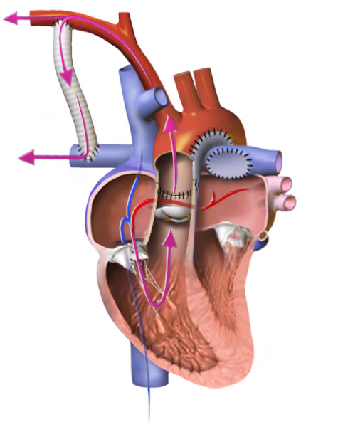
C
Before saying “Fontan patient”
| Systemic ventricle | LV, RV, or indeterminate? Systolic/diastolic function? Scar? Dyssynchrony? | Systemic AV valve | Tricuspid, mitral, common? Regurgitation/stenosis? |
| Systemic outflow | Native, DKS/neo-aorta, arch reconstruction? Obstruction or regurgitation? | Pulmonary circuit | Glenn/Fontan type, PA stenosis, fenestration, collaterals, thrombus? |
| Hepatic flow | Does it reach both lungs? PAVMs/cyanosis if maldistributed. | Prior palliation | Old shunts, PA bands, Sano, coils/stents, lymphatic procedures. |
D
Different diagnoses, same questions
| Birth lesion | Common systemic ventricle/valve | Extra adult issue |
|---|---|---|
| Tricuspid atresia | Usually systemic LV; mitral valve | Restrictive bulboventricular foramen/subaortic obstruction, old BTT/Fontan |
| Double-inlet LV | Dominant LV; common/paired AV valves | Rudimentary RV outlet chamber; DKS for systemic outflow |
| Unbalanced AVSD | Dominant LV or RV; common AV valve | Systemic AV-valve regurgitation and heterotaxy/venous anomalies |
| HLHS | Systemic morphologic RV; tricuspid valve | Norwood arch/DKS/native-root coronary pathway, systemic TR |
| PA/IVS or complex DORV | Variable | Coronary dependence or complex ventricular/outflow routing |
E
Stage-II circulation before Fontan
Sources of PBF
Glenn ± antegrade flow
Native/conduit antegrade pulmonary flow may remain; too much raises SVC pressure/volume load, too little worsens cyanosis.Cyanosis
Expected but explain change
Venovenous collaterals, PAVMs, PA stenosis, AV-valve/ventricular failure and pulmonary disease can worsen saturation.Intervention
Not all reach Fontan
Adults may remain Glenn-only because of PVR, ventricular/valve disease or preference; management protects low-PVR passive flow.F
Staged-operation procedural traps
- PA band or DKS can alter subaortic/systemic outflow.
- Old shunts/collaterals change perfusion and bleeding risk.
- Sano ventriculotomy contributes scar and RV dysfunction.
- Glenn pressure is central venous pressure from upper body.
- Hepatic-flow distribution determines PAVM risk.
- Atrial septectomy creates an embolic/air pathway until Fontan routing.
Systemic-outflow obstructionIn single-ventricle hearts, a restrictive VSD/bulboventricular foramen or arch/DKS obstruction can cause hypertrophy, AV-valve regurgitation, ischemia and sudden decline even when the resting gradient seems modest.
A
Which Fontan?
| Atriopulmonary | RA→PA; giant RA, energy loss, IART, thrombus. Selected conversion to TCPC + arrhythmia surgery. |
| Lateral tunnel | Intracardiac baffle routes IVC to PA; baffle leak/stenosis and atrial scar. |
| Extracardiac conduit | External tube IVC→PA; less atrial scar, but conduit/PA stenosis and no growth. |
| Fenestration | Fontan-to-atrium “pop-off”: lowers venous pressure and raises preload/output at the cost of cyanosis and paradoxical embolus. |


B
Name the failure phenotype—EF alone is not the diagnosis
Pump / valve / rhythm
High downstream pressure
Systolic or diastolic failure, systemic AV-valve regurgitation, outflow/arch obstruction, IART/AF, bradycardia or dyssynchrony.Circuit / pulmonary bed
Resistance before preload
Conduit/tunnel or branch-PA/pulmonary-vein narrowing, PVR, thrombus, collateral or fenestration flow, hepatic-flow streaming.Lymphatic / end organ
Failure with preserved EF
PLE, plastic bronchitis, effusions, FALD/HCC, renal decline, sarcopenia, bone and neurocognitive/mental-health burden.C
Minimum longitudinal surveillance
| Domain | Stable baseline | Escalate for change |
|---|---|---|
| ACHD + ECG + TTE | At least annually; stage C q6–12 mo, stage D visit q3–6 mo and TTE q6–12 mo | Symptoms, edema/ascites, saturation or murmur change, arrhythmia, falling function |
| CMR/CT · CPET · rhythm | Serial congenital imaging and exercise testing; 24-h Holter every 1–2 y in adults is the 2019 AHA Fontan expert-surveillance framework | Monitor sooner/longer for palpitations, syncope, exercise decline, new HF, bradycardia or suspected IART/AF; compare trajectory |
| Annual blood/urine bundle | CBC/platelets/lymphocytes; electrolytes/renal ± cystatin C and urine albumin; liver/GGT/bilirubin/albumin/INR; iron; BNP/NT-proBNP | Low muscle mass can hide renal decline; low albumin prompts stool α1-antitrypsin, Ig/lymphocyte assessment |
| Liver | At least annual imaging + laboratories including AFP; at least one Fontan-informed hepatology review | Nodule, splenomegaly, falling platelets, varices, ascites, jaundice or sarcopenia |
| Whole patient | Renal, lung/sleep, nutrition, muscle/bone/vitamin D, cognition, mood, contraception/pregnancy and vaccination review | Do not let preserved EF delay multiorgan or advanced-care evaluation |
D
Cardiovascular treatment anchors
| Target | Practical rule |
|---|---|
| Thromboprophylaxis | Anticoagulate known/suspected thrombus, prior TE/CVA, atriopulmonary Fontan or sustained AF/flutter. Without these, use aspirin or anticoagulation if no contraindication; individualize FALD/varices, platelets, renal function, bleeding and pregnancy. CHA₂DS₂-VASc is not validated here. |
| New AF/flutter/IART | Timely cardioversion, immediate anticoagulation/thrombus assessment, and imaging ± catheterization for obstruction or deterioration. Congenital EP early; atrial-based pacing and minimize ventricular pacing when feasible. |
| Congestion / HF drugs | Diurese carefully: underfilling can collapse output. Treat a defined ventricular/valve phenotype; acquired-HFrEF drugs have limited Fontan-specific outcome evidence and do not correct passive-flow resistance. |
| Exercise | Prescribe aerobic plus lower-limb resistance/respiratory training or cardiac rehabilitation; skeletal-muscle and respiratory pumps augment venous return. Avoid blanket inactivity. |
| Mechanical causes | Relieve pathway/PA/pulmonary-vein/outflow obstruction; treat AV-valve disease and rhythm. Use P22 for PVR, APCs, fenestration and collateral decisions. |
E
FALD and lymphatic failure: treat the circuit while treating the organ
| FALD | AST/ALT may be normal. Trend platelets, albumin, bilirubin/INR context, spleen/ascites and portal signs; stiffness reflects congestion + fibrosis. Characterize nodules with Fontan-expert multiphase CT/MRI. Biopsy is selective, especially for heart versus heart–liver planning. |
| PLE | Edema/effusions, diarrhea, low albumin/total protein, lymphopenia/Ig loss: confirm enteric α1-antitrypsin loss; correct obstruction/PVR/EDP/valve/rhythm; diet/protein/micronutrients, medical and lymphatic therapy through a Fontan program. |
| Plastic bronchitis | Branching airway casts, hypoxemia or obstruction = airway emergency. Stabilize airway/bronchoscopy as needed; correct hemodynamics and obtain dynamic lymphatic imaging for targeted embolization/decompression. |
| Lymphatic imaging | T2-weighted and dynamic-contrast MR lymphangiography can map hepatoduodenal, mesenteric or thoracic leaks; repeated drainage alone is not definitive management. |
Preserve passive flow—and refer before organs become irreversiblePositive-pressure ventilation/excess PEEP, hypovolemia, acidosis, hypoxia and hypercarbia raise impedance or reduce venous return. Progressive symptoms, recurrent admissions, active PLE/plastic bronchitis, refractory arrhythmia, falling CPET/CI, ascites/effusions, renal decline, sarcopenia or advanced FALD warrant early Fontan-experienced HF/transplant review; decide heart alone versus heart–liver with a multidisciplinary team.
A
No subpulmonary pump: tiny resistance changes matter
Qp ≈ (upstream Fontan pressure − pulmonary venous/atrial pressure)
÷
total cavopulmonary impedance
÷
total cavopulmonary impedance
The pressure head is small. A 1–2 mm Hg pathway gradient or modest PVR rise can reduce preload/output—or require higher systemic venous pressure to maintain flow. Separate conduit/branch-PA obstruction from true PVR with pressure pullbacks. A “normal” PA pressure does not exclude pulmonary vascular disease because low flow can mask it. In one adult cohort, PVRI >2 WU·m² + CI <2.5 L/min/m² identified higher risk; this is a prognostic signal, not a universal drug/intervention threshold.
B
Read the pressure-flow pattern, not one number
| Pattern | Interpretation / first targets |
|---|---|
| High Fontan + low/normal PAWP + ↑TPG | Conduit/tunnel or branch-PA resistance; pulmonary vascular disease after anatomy is cleared |
| High Fontan + high PAWP/EDP | Diastolic/pump or AV-valve disease, pulmonary-vein obstruction; not primarily a PVR-drug problem |
| “Acceptable” Fontan pressure + low CI | Advanced low-flow/underfilled physiology may conceal PVR; do not call the circuit reassuring |
| Ventricular dilation / high PV return | APCs, residual antegrade pulmonary flow, AV-valve regurgitation or excess volume |
| New cyanosis | Fenestration/baffle leak, VVC, PAVM/hepatic-flow maldistribution, thrombus/PE or lung disease |
C
Catheterization + CMR: make Qp credible before calculating PVR
- Document sedation, spontaneous vs positive-pressure ventilation, PEEP/FiO₂, pH/PaCO₂, hemoglobin and volume state.
- Measure SVC, IVC/conduit/tunnel and both branch-PAs with pullbacks across every narrowing.
- Obtain bilateral PAWP; direct pulmonary-vein/atrial pressure if discordant; systemic-ventricular EDP and arch/outflow gradient.
- Full saturation map with SVC and IVC separately; assess thrombus, fenestration, PAs/PVs, hepatic streaming, APCs/VVCs/PAVMs.
- Pulmonary TPG = mean PA distal to series obstruction − PAWP/LA; PVR = TPG ÷ measured total pulmonary flow. Mean Fontan pressure substitutes only when the pathway is unobstructed; use bilateral/flow-weighted assessment when lungs differ.
- Fenestration makes Qp<Qs; APC/residual antegrade flow adds distal Qp; streaming, assumed VO₂ and thermodilution can mislead.
2025 escalation gateNew/progressive symptoms, hypoxemia, functional decline or hepatic, renal, lymphatic, respiratory or neurologic dysfunction → ACHD hemodynamic + angiographic catheterization to guide therapy. Severe/progressive hypoxemia or hypotension → urgent TEE, CMR or Fontan-protocol CTA to exclude pathway/pulmonary thrombus or embolus; TTE cannot rule this out. Use dual-site delayed/multiphase CTA to avoid streaming artifact.
Hybrid flow checkPhase-contrast CMR can reconcile branch flow and collateral burden. In suitable anatomy, APC flow is estimated as ascending-aortic flow − (SVC + IVC), or pulmonary-venous flow − branch-PA flow; account for fenestration, valve regurgitation and other shunts.
D
Manage total impedance in the right order
1 · Remove series resistance
Stent significant Fontan/conduit or branch-PA narrowing; assess pulmonary veins and arch/systemic outflow
Stent significant Fontan/conduit or branch-PA narrowing; assess pulmonary veins and arch/systemic outflow
›
2 · Lower downstream pressure
Restore rhythm; address AV valve, ventricular EDP, pulmonary-vein and pump disease
Restore rhythm; address AV valve, ventricular EDP, pulmonary-vein and pump disease
›
3 · Reverse vasoconstriction
Treat hypoxia, hypercarbia/acidosis, atelectasis, OSA/lung disease, PE; avoid excess PEEP
Treat hypoxia, hypercarbia/acidosis, atelectasis, OSA/lung disease, PE; avoid excess PEEP
›
4 · Quantify connections
APC, VVC, fenestration, PAVM, residual antegrade flow; predict pressure/output trade-off
APC, VVC, fenestration, PAVM, residual antegrade flow; predict pressure/output trade-off
›
5 · Selected drug trial
Only for a precapillary/low-preload phenotype after anatomy and filling pressure are defined
Only for a precapillary/low-preload phenotype after anatomy and filling pressure are defined
E
“Pulmonary arterial contribution from the aorta” = APC flow
Aorta/systemic branch
Subclavian, bronchial, intercostal, internal mammary or other branch
Subclavian, bronchial, intercostal, internal mammary or other branch
›
Aortopulmonary collateral
Systemic arterial blood enters distal pulmonary bed
Systemic arterial blood enters distal pulmonary bed
›
Lung → pulmonary vein
Extra pulmonary venous return bypasses the passive Fontan inflow
Extra pulmonary venous return bypasses the passive Fontan inflow
›
Single ventricle → aorta
Recirculating left-to-right volume load; not extra effective systemic output
Recirculating left-to-right volume load; not extra effective systemic output
Consequences: systemic-ventricular dilation/EDP, higher pulmonary/Fontan pressure and energy loss, reduced effective systemic output, and major transplant bleeding. Smaller/stenotic native PAs may drive APC formation—fix the cause, not only the vessel count.
F
Do not confuse the four right-to-left/left-to-right pathways
| Connection | Direction / physiology | Management trap |
|---|---|---|
| APC | Aorta/systemic artery → pulmonary bed; left-to-right recirculation and ventricular volume load | Close selected important targets for symptoms/volume load; confirm native segmental PA supply and treat PA/arch obstruction |
| VVC | Fontan systemic vein → atrium/PV; right-to-left cyanotic pressure pop-off | Test occlude: blind closure may raise Fontan pressure, reduce output or worsen PLE/effusions |
| Fenestration | Deliberate Fontan → atrium pop-off; raises preload/output at cost of saturation/embolus | Saturation rise alone is not success; measure Fontan pressure, CI and oxygen delivery during test occlusion |
| PAVM | PA → PV intrapulmonary shunt; often absent/maldistributed hepatic effluent | Restore/balance hepatic flow to the affected lung; diffuse PAVMs are not an APC-coiling problem |
Separate accessory pulmonary blood flow: a residual native ventriculo-PA outlet, PDA or old systemic-to-PA shunt may add useful pulsatility but can also raise PA pressure and ventricular volume load; quantify it as its own circuit rather than calling it an APC.
G
Intervention and pulmonary-vasodilator gates
| Fontan/PA stenosis | Stent hemodynamically significant narrowing; in a pump-free circuit even a small gradient can matter. |
| Selected APC closure | Reasonable for symptoms or systemic-ventricular volume overload; consider pretransplant targets to reduce runoff/bleeding. No validated CMR percentage cutoff: embolize through congenital interventional review—not every visible vessel. |
| Fenestration | Consider closure for important cyanosis/embolus only after tolerated test occlusion; creation/enlargement can be an expert rescue/bridge. Fontan ≤15 mm Hg, CI ≥2 L/min/m² and CI fall ≤20% are one institution’s criteria—not guideline cutoffs. |
| VVC | Define origin, drainage and decompressive role. Embolize only a selected nonessential channel after test occlusion confirms acceptable Fontan pressure, CI, saturation and systemic oxygen delivery. A VVC may be a protective “natural fenestration”; no universal closure threshold exists. |
| Pulmonary vasodilator | May be considered to improve exercise capacity only after excluding obstruction and high PAWP/EDP. Use an objective ACHD-center trial (BP, saturation, symptoms/CPET, liver/hemoglobin as drug-appropriate), not routine empiric therapy. |
| Trial reality | FUEL: udenafil did not improve the primary peak-VO₂ endpoint, though several VAT measures improved. RUBATO: macitentan did not improve peak VO₂ or activity. No established reduction in Fontan failure, transplant, death or FALD. |
No blind occlusionEvery collateral or fenestration is both a lesion and a clue. Define why it formed, what territory it supplies or decompresses, and the effect of temporary occlusion on pressure, cardiac index, saturation and systemic oxygen delivery before permanent closure.
Use segmental descriptionState atrial situs/appendage morphology, bronchial/lung sidedness, AV and VA connections, systemic and pulmonary venous pathways, spleen status, and abdominal organ arrangement. “Situs ambiguus” alone is not a clinical map.
A
Patterns—not absolutes
| Pattern | Frequent cardiac anatomy | Noncardiac / rhythm implications |
|---|---|---|
| Right isomerism / asplenia | Complex AVSD, single ventricle, pulmonary atresia/stenosis, TAPVR | Infection risk from absent splenic function; paired sinus nodes/atrial tachyarrhythmia |
| Left isomerism / polysplenia | Interrupted IVC with azygos continuation, AVSD, systemic outflow lesions | Sinus-node dysfunction/AV block; splenic function variable, not guaranteed normal |
| Dextrocardia / mesocardia | Any connection pattern; mirror-image or discordant anatomy | ECG lead placement, imaging planes, defibrillation pads, and procedural access need adaptation |
B
Venous pathways that change procedures
Persistent left SVC
Usually to coronary sinus; can drain to LA with cyanosis. Matters for central lines, device leads, retrograde cardioplegia, and coronary-sinus dilation.
Usually to coronary sinus; can drain to LA with cyanosis. Matters for central lines, device leads, retrograde cardioplegia, and coronary-sinus dilation.
Interrupted IVC / azygos continuation
Femoral venous access may reach SVC rather than RA; transseptal/cath routes change. Often associated with left isomerism.
Femoral venous access may reach SVC rather than RA; transseptal/cath routes change. Often associated with left isomerism.
Kawashima circulation
Azygos/SVC flow goes to PAs while hepatic veins remain connected to atrium. Lack of hepatic effluent to lungs promotes pulmonary AVMs.
Azygos/SVC flow goes to PAs while hepatic veins remain connected to atrium. Lack of hepatic effluent to lungs promotes pulmonary AVMs.
Hepatic-vein completion
Routes hepatic venous blood to pulmonary arteries after Kawashima; maldistribution to one lung can leave unilateral PAVMs/cyanosis.
Routes hepatic venous blood to pulmonary arteries after Kawashima; maldistribution to one lung can leave unilateral PAVMs/cyanosis.
C
Whole-patient checklist
- Spleen: imaging plus functional assessment; vaccination and fever plan if asplenic/hyposplenic.
- Gut: malrotation history; abdominal pain/obstruction requires low threshold for evaluation.
- Lungs/airway: bronchial isomerism, ciliary dysfunction, recurrent infections in some patients.
- Liver: Fontan-related disease plus abnormal venous anatomy.
- Conduction: absent/displaced sinus node, twin AV nodes, AV block, complex macroreentry.
- Pregnancy/genetics: individualized recurrence counseling; review syndromic/extracardiac disease.
D
Rhythm anatomy in isomerism
| Pattern | Electrical implication | Planning point |
|---|---|---|
| Left isomerism | Absent/hypoplastic sinus node, atrial bradycardia, progressive AV block | Monitor chronotropic response and conduction; venous routes complicate pacing |
| Right isomerism | Paired sinus nodes and twin AV-node pathways can support complex reentry | Congenital EP mapping; do not assume usual triangle of Koch |
| Common AV junction | Displaced conduction axis; surgery/ablation carries AV-block risk | Use operative anatomy and 3-D mapping |
| Fontan/baffles | Atrial scar and inaccessible chambers compound native conduction anomalies | Plan access, surgical ablation and pacing together |
E
Cross-sectional map before a procedure
Venous
Every return
Bilateral SVC, bridging vein, azygos/hemiazygos, hepatic-vein entry, pulmonary veins and collaterals.Arterial
Arch and PAs
Right/left arch, aberrant branches, MAPCAs, PA confluence/stenosis, prior shunts and DKS.Abdominal
Access and organs
Aorta/IVC position, interrupted segments, liver sidedness, spleen, gut rotation and prior surgery.F
Asplenia fever plan
Confirm functional splenic status rather than relying on anatomy alone. Keep pneumococcal, meningococcal, Hib and influenza vaccination current; provide written urgent-fever instructions and antimicrobial prophylaxis when indicated by local asplenia guidance. Sepsis can destabilize complex circulation quickly.
G
Operations shaped by venous anatomy
| Bilateral Glenn | Both SVCs connected to PAs when bridging vein is absent/small; assess flow balance and each anastomosis. |
| Kawashima completion | Hepatic veins routed to PA/Fontan to restore hepatic effluent and treat PAVMs/cyanosis. |
| Complex Fontan conduit | Must incorporate anomalous IVC/hepatic returns without compression or streaming to one lung. |
Before any line, EP study, or surgeryCross-sectional venous mapping is not optional when interrupted IVC, bilateral SVCs, anomalous hepatic veins, baffles, or cavopulmonary connections are possible.
Do not stop at “anomalous coronary”Record artery and dominance; originating sinus and ostial height; separate/shared ostium; take-off angle and ostial shape; proximal caliber; presence, length and commissural relation of an intramural segment; subsequent course; myocardium supplied; scar; and coexisting CAD.
A
Interarterial and intramural are not synonyms
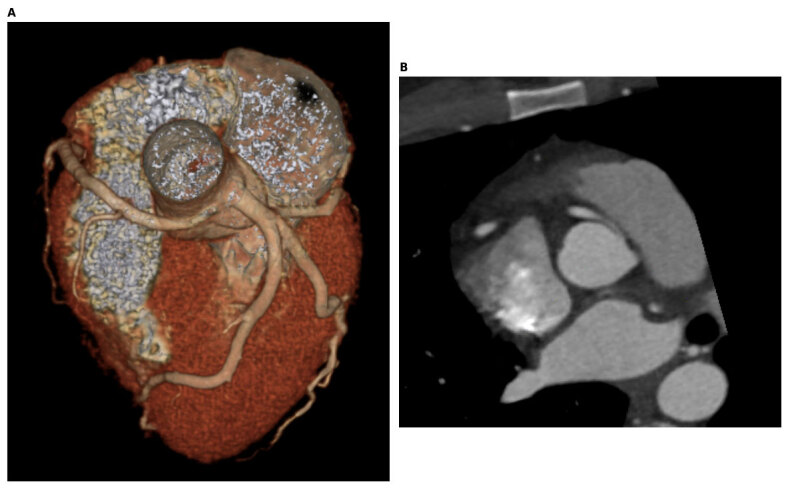
| Course | What it means |
|---|---|
| Interarterial | Between aorta and PA; risk depends strongly on associated ostial/intramural disease. |
| Intramural | Within the aortic wall; give length, proximal hypoplasia and relation to aortic commissure. |
| Intraseptal / subpulmonic | Through conal/septal myocardium; usually lower risk than intramural anatomy, not automatically benign if ischemic. |
| Prepulmonic | Anterior to PA; usually no dynamic ostial obstruction from the course itself. |
| Retroaortic LCx | Usually low SCD risk; critical before aortic/root/mitral surgery because injury or compression can occur. |
B
Risk is anatomy × physiology × patient
Higher-concern anatomy
Proximal obstruction
AAOLCA from right sinus; slit/stenotic ostium, acute take-off, proximal hypoplasia, long intramural segment and interarterial course—especially in combination.Higher-concern presentation
Exertional signal
Unexplained exertional syncope/angina, matched ischemia, ischemic VA/aborted arrest or scar; younger age and intense exercise increase concern.Attribution
A label is not causality
Negative stress does not erase risk; atypical pain in an older adult still requires evaluation for CAD, vasospasm and microvascular disease.C
Anatomic and physiologic work-up
CCTA first
ostium, proximal caliber, intramural length, take-off, great-vessel relation
ostium, proximal caliber, intramural length, take-off, great-vessel relation
›
CMR
ventricular function, perfusion, infarct/scar; coronary map if CT unsuitable
ventricular function, perfusion, infarct/scar; coronary map if CT unsuitable
›
Provocative imaging
maximal exercise preferred; dobutamine when exercise not feasible
maximal exercise preferred; dobutamine when exercise not feasible
›
Expert invasive study
angiography + IVUS/pressure-flow for discordant or selected high-risk cases
angiography + IVUS/pressure-flow for discordant or selected high-risk cases
Exercise or dobutamine better models dynamic obstruction than vasodilator-only stress. A normal study reduces demonstrated ischemic burden but does not prove zero SCD risk.
D
Repair versus surveillance
| Symptoms / matched ischemia | Repair recommended when attributable to AAOCA; confirm territory and exclude another dominant cause. |
| AAOLCA from right/opposite sinus, asymptomatic | Surgery is reasonable with high-risk anatomy—ostial stenosis/slit-like orifice, acute take-off, proximal hypoplasia or intramural/interarterial course. Prepulmonic or retroaortic variants without attributable ischemia generally do not require repair. |
| Related ventricular arrhythmia | Surgery is reasonable after scar assessment and review of competing arrhythmic causes. |
| AAORCA, no ischemia/complex VA | Observation with longitudinal ACHD follow-up and SDM is reasonable; integrate age, anatomy, activity and preference. |
| Middle-aged, no high-risk findings | No demonstrated survival benefit from prophylactic surgery; surveillance is often appropriate. |
E
Choose the operation from the target lesion
| Unroofing ± ostioplasty | Long, favorable intramural segment; watch residual distal narrowing, commissural injury and aortic regurgitation. |
| Neo-ostium / fenestration | Creates an exit from a long intramural segment when simple unroofing is incomplete or commissural relation is unfavorable. |
| Transection + reimplantation | Short/low/absent intramural segment or intrinsic origin/take-off not corrected by unroofing; avoid tension/kinking. |
| PA translocation | Selected interarterial anatomy without correctable intramural target; does not repair a slit-like or stenotic ostium. |
| CABG | Guideline default is to reserve CABG for coexisting obstructive CAD. If direct anatomic repair is exceptionally not feasible, any ligation-plus-bypass strategy requires expert congenital-coronary review because competitive native flow threatens graft patency. |
Exercise
Current SDM, not one blanket
Unrepaired interarterial AAOLCA generally requires correction before strenuous competition. Selected AAORCA without symptoms, ischemia or complex VA may participate through SDM and surveillance.After repair
Repair is not discharge
Baseline anatomy + maximal provocative testing; lifelong review for ostial restenosis, residual ischemia, AR, scar and VA. Recurrence and adverse events can occur.F
Routine AAOCA follow-up framework
| Physiologic stage | ACHD review + ECG | TTE | Individualize |
|---|---|---|---|
| A | Every 24–36 mo | Every 24–36 mo | CCTA/CMR for anatomy or scar; maximal exercise/dobutamine imaging for symptoms, activity decisions, changing findings and after repair. |
| B | Every 12 mo | Every 24 mo | |
| C–D | Every 12 mo | Every 12 mo |
Intervals are the current ACHD framework, not a substitute for earlier reassessment with new exertional symptoms, syncope, VA, ischemic ECG, ventricular change or postoperative concern.
Core physiologyAn epicardial coronary—usually mid-LAD—tunnels beneath myocardium. Systolic compression can persist into early diastole; tachycardia both intensifies compression and shortens filling time. Most bridges are incidental. Length, depth or angiographic “milking” alone does not prove the cause of pain, ischemia, arrhythmia or arrest.
CCTA report
artery/segment, entrance/exit, length, depth and complete/partial encasement
artery/segment, entrance/exit, length, depth and complete/partial encasement
·
Dynamic detail
systolic and diastolic caliber/compression when the acquisition permits
systolic and diastolic caliber/compression when the acquisition permits
·
Adjacent disease
proximal plaque/stenosis, distal bed and anomalous origin/course
proximal plaque/stenosis, distal bed and anomalous origin/course
·
Clinical phenotype
symptoms, exercise relation, scar/ischemia/VA and coexisting spasm/CMD
symptoms, exercise relation, scar/ischemia/VA and coexisting spasm/CMD
A
First establish causality
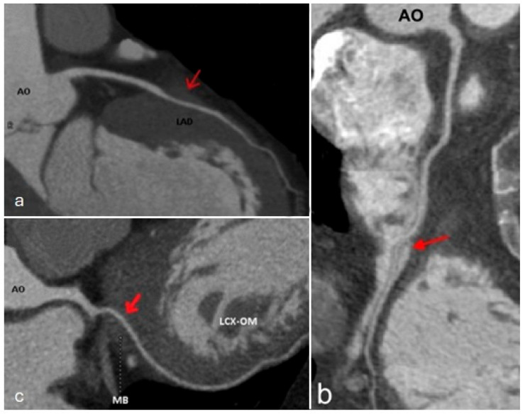
| Test | What it answers—and cannot |
|---|---|
| CCTA | Entrance/exit, length, depth, overlying muscle, proximal plaque; anatomy does not prove ischemia. |
| Maximal exercise + imaging | Reproduces symptoms, ischemia, BP response and complex VA under physiologic tachycardia/inotropy. |
| Angiography | Systolic milking and delayed diastolic relaxation; limited sensitivity and not causality by itself. |
| IVUS / OCT | Intramyocardial segment, compression and proximal plaque; an IVUS halo is not an intervention indication. |
| Provocative physiology | Dobutamine diastolic indices in selected refractory/discordant cases; phenotype vasospasm/CMD too. |
B
Dynamic lesion ≠ fixed stenosis
Pressure physiology
Adenosine mean FFR may miss it
Systolic distal-pressure overshoot can mask an important diastolic gradient. Dobutamine diastolic FFR is better studied; dobutamine iFR/RFR are expert-lab alternatives.Threshold caution
Do not turn a cutoff into prognosis
Dobutamine dFFR ≤0.76 is used in small referral cohorts, not validated as a stand-alone surgical or event-risk threshold.C
Treatment ladder
Incidental / asymptomatic
reassure; no bridge-specific drug; standard ASCVD prevention
reassure; no bridge-specific drug; standard ASCVD prevention
›
Concordant symptoms
β-blocker first: lower HR/contractility, lengthen diastole
β-blocker first: lower HR/contractility, lengthen diastole
›
Alternative
non-DHP CCB if β-blocker intolerant; useful when vasospasm documented
non-DHP CCB if β-blocker intolerant; useful when vasospasm documented
›
Before procedure
treat/exclude CAD, plaque, spasm, CMD, HCM, anemia and arrhythmia
treat/exclude CAD, plaque, spasm, CMD, HCM, anemia and arrhythmia
Avoid routine nitrates in an isolated bridgeVasodilation, reflex tachycardia and increased contractility can accentuate compression. Proven coexisting vasospasm requires individualized ANOCA expertise; aspirin/statin follow conventional plaque/ASCVD indications, not bridge anatomy alone.
D
Intervene only when all three gates are open
1 · Burden
life-limiting symptoms despite maximally tolerated medical therapy
life-limiting symptoms despite maximally tolerated medical therapy
+
2 · Concordance
objective matched ischemia or abnormal expert dynamic physiology
objective matched ischemia or abnormal expert dynamic physiology
+
3 · Attribution
multidisciplinary agreement bridge is dominant mechanism
multidisciplinary agreement bridge is dominant mechanism
| Surgical myotomy / unroofing | Preferred direct treatment for suitable anatomy; observational cohorts show symptom/QOL improvement, but randomized outcome data are absent. |
| CABG | Selected very long/deep bridge or unsafe myotomy; competitive flow can cause graft failure. Length/depth cutoffs are planning heuristics, not indications. |
| PCI | Not routine: repetitive external compression creates perforation, fracture, thrombosis and restenosis/reintervention risk. |
E
Exercise, arrest and the “do-not” list
Exercise
Test the activity being cleared
Incidental asymptomatic bridge permits sport. Symptomatic athletes need maximal provocative testing; persistent/residual ischemia weighs against competitive sport. Return after healing or medical control with no ischemia/complex VA.Do not
An image is not an endpoint
Do not stent an incidental bridge, treat an IVUS halo, use normal adenosine FFR as proof of harmlessness, reflexively give nitrates, or implant an ICD solely because a bridge is visible.F
Persistent symptoms after “successful” treatment
| After medical therapy | Confirm adequate HR control and adherence, then reassess CAD/proximal plaque, vasospasm, CMD, HCM, anemia and rhythm rather than escalating from anatomy alone. |
| After unroofing | Repeat anatomic and maximal provocative assessment for residual tunnel/compression or ischemia; spasm/CMD and neuropathic or noncardiac pain can persist despite complete myotomy. |
| After arrest / sustained VA | Use the full secondary-prevention evaluation. Bridge repair does not replace scar assessment, EP review or an ICD when otherwise indicated. |
A
ALCAPA physiology changes after birth
PVR falls
PA pressure and oxygen content fall; antegrade LCA perfusion becomes inadequate
PA pressure and oxygen content fall; antegrade LCA perfusion becomes inadequate
›
Collateral rescue
dilated RCA sends septal/epicardial collaterals into LCA territory
dilated RCA sends septal/epicardial collaterals into LCA territory
›
Coronary steal
collateral blood runs retrograde through LCA into low-pressure PA
collateral blood runs retrograde through LCA into low-pressure PA
›
Adult burden
scar/ischemia, LV dysfunction, papillary injury/MR, VA and SCD
scar/ischemia, LV dysfunction, papillary injury/MR, VA and SCD
ACAPA is an origin abnormality—not a fistula.The affected coronary lacks its normal aortic origin and begins in the PA; a coronary-to-PA fistula has a normal aortic coronary origin plus an abnormal terminal connection. Define both proximal origins and flow direction before any occlusion. Adult survival implies collaterals, not benign physiology; ALCAPA repair is recommended even when incidental.
B
Recognition and complete imaging
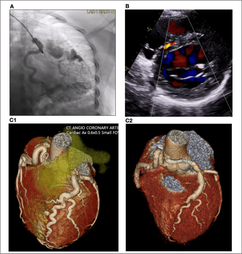
| Echo clues | Absent aortic LCA origin; enlarged/tortuous RCA; septal collaterals; retrograde coronary flow into PA; LV dilation/dysfunction and MR. |
| CCTA | Definitive origin, distance to aorta, course, collateral network, ostia and surgical relation; include adult CAD. |
| CMR | LV volumes/function, MR, perfusion/viability and ischemic/infarct scar; scar informs VA/SCD review. |
| Cath | When noninvasive anatomy/hemodynamics are incomplete, or coronary/bypass planning requires selective angiography. |
| ECG/rhythm | Inferolateral Q waves, ectopy/NSVT or sustained VA; monitor according to scar, symptoms and ventricular phenotype. |
C
ALCAPA versus ARCAPA
ALCAPA
Repair all patients
Large LV territory makes chronic ischemia/scar, MR and malignant VA central risks. Restore dual coronary origins and eliminate PA runoff; do not treat MR or cardiomyopathy without correcting the coronary lesion.ARCAPA
Dominance and physiology matter
Repair when ischemia, ventricular dysfunction or symptoms are attributable. Younger asymptomatic patients are often offered elective repair; in older asymptomatic adults individualize ischemia, dominance, operative risk/comorbidity and preference.Territory
dominance, nodal supply and all myocardium dependent on anomalous artery
dominance, nodal supply and all myocardium dependent on anomalous artery
›
Transfer
PA origin, distance, mobility and whether reimplantation can be tension-free
PA origin, distance, mobility and whether reimplantation can be tension-free
›
Myocardium
ischemia, viability, infarct scar, LV/RV function and rhythm substrate
ischemia, viability, infarct scar, LV/RV function and rhythm substrate
›
Associated work
MR mechanism, AP window/CHD and acquired CAD or bypass targets
MR mechanism, AP window/CHD and acquired CAD or bypass targets
D
Operations: create two coronary origins
| Direct reimplantation | Preferred when feasible: mobilize coronary button and implant into aorta; coronary-extension/interposition may bridge distance. Watch ostial stenosis, tension and kinking. |
| Takeuchi repair | Aortopulmonary window + intrapulmonary baffle directs aortic blood to anomalous ostium. Late baffle leak/stenosis and supravalvar PA stenosis are signature failures. |
| Ligation + CABG | Selected adult anatomy not reimplantable: close PA origin to abolish steal and graft the dependent territory, commonly LIMA–LAD. Verify all myocardium supplied and graft patency. |
| Simple ligation alone | Leaves a one-coronary circulation and is generally inadequate when a dual system can be constructed. |
| ARCAPA repair | Direct reimplantation when possible; otherwise PA-origin closure with bypass selected by anatomy and dominance. |
E
Mitral valve, scar and ventricular arrhythmia
Mitral regurgitation
Revascularize the cause
Functional/papillary ischemic MR may improve after coronary repair and LV recovery. Add mitral repair for severe structural or unlikely-to-recover MR after team review.LV recovery
Function may improve; scar remains
Trend LV/MR by echo and CMR. Persistent LGE can remain arrhythmogenic despite normalized EF and a patent two-coronary repair.SCD strategy
Phenotype, not acronym
Assess sustained VA/arrest, scar, residual ischemia and LV dysfunction using standard secondary-prevention and congenital EP principles.F
Lifelong post-repair surveillance
- Echo: LV size/function and MR; PA gradient after Takeuchi.
- CCTA/CMR: reimplantation ostium, tunnel/graft and coronary perfusion/scar.
- Rhythm monitoring for palpitations, syncope, scar or ventricular dysfunction.
- Takeuchi: baffle leak/stenosis and supravalvar PS; reimplantation: ostial stenosis/kink.
- CABG: graft patency plus native PA-origin closure/steal.
- Rarer ACXPA/ALADPA: apply the same territory, runoff and dual-circulation logic.
AP-window/PDA trap: before closing a PA-level shunt, document both coronary origins. Elevated PA pressure may be sustaining antegrade flow in ACAPA; closing the shunt without coronary repair can abruptly cause ischemia and steal.
Repair stops ongoing steal; it does not erase established scar.Normal postoperative EF does not exclude abnormal strain, residual perfusion defects or scar-mediated VA. Use symptoms, CMR, provocative testing and rhythm history—not anatomy alone—to define residual risk.
The recipient determines the shunt; the donor coronary determines closure risk.Prefer explicit donor→recipient naming (for example, RCA→RA or LAD→PA) over the inconsistent label “coronary arteriovenous fistula.” Map every normal myocardial branch before closing the shortcut.
Coronary-cameral
Coronary → chamber
Drainage into RA, RV, LA or LV. Right-sided termination usually creates a left-to-right shunt; left-sided termination causes runoff without Qp:Qs.Coronary-to-vascular
Coronary → vessel
PA, coronary sinus, SVC, pulmonary vein or other vascular bed. Coronary-sinus drainage is a particularly high-thrombosis phenotype.Acquired
Iatrogenic or traumatic
After biopsy, surgery, ablation, trauma or infarction; mechanism and timing alter the closure plan. Do not assume every fistula is congenital.A
Physiology follows the exit pressure and resistance
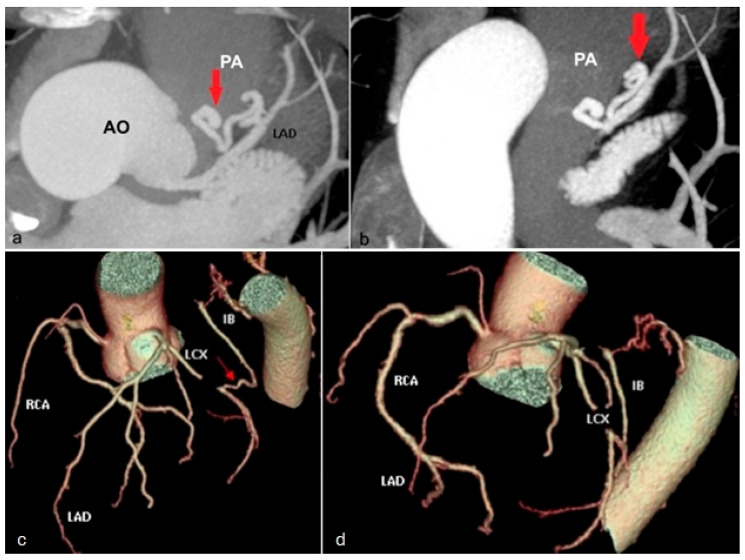
Low-resistance runoff
dilates donor and steals flow from myocardium
dilates donor and steals flow from myocardium
›
Volume/shunt
right/PA/CS drainage loads pulmonary circuit; left chamber causes runoff
right/PA/CS drainage loads pulmonary circuit; left chamber causes runoff
›
Late substrate
ectasia/aneurysm, stasis/thrombus, branch loss, ischemia/HF/VA
ectasia/aneurysm, stasis/thrombus, branch loss, ischemia/HF/VA
Potential complicationsIschemia/infarction, chamber overload/HF, atrial or ventricular arrhythmia, thrombosis/embolization, infective endarteritis/endocarditis and—rarely—rupture.
B
The pre-closure map
| Define | Why it changes the plan |
|---|---|
| Origin(s) and segment | Donor artery; proximal/mid/distal takeoff; single versus bilateral/multiple fistulas; dominant territory. |
| Course and branches | Tortuosity, length and every normal myocardial branch arising before/within/after the tract—these must be preserved. |
| Exit | Chamber/vessel, pressure, number of exits and narrowest safe occlusion site; distal closure minimizes residual blind pouch. |
| Donor remodeling | Maximum diameter, aneurysm, calcification, thrombus and expected stagnant segment after closure. |
| Hemodynamic burden | Qp:Qs/chamber enlargement for right/PA drainage; ischemia and distal perfusion; pressure/balloon test when dependence uncertain. |
| Proximal vs distal | Distal fistula can leave the entire ectatic donor as a low-flow cul-de-sac after exit closure; proximal discrete branch closure often preserves a near-normal distal coronary. |
| Diffuse microfistulas | Multiple tiny distal coronary-to-chamber channels—often to LV—may have no single safe closure target; quantify physiology and treat the phenotype. |
TTE assesses flow/chambers; gated CCTA maps the entire tract and branches; CMR adds Qp:Qs/function/perfusion; selective angiography + hemodynamics is often needed for closure planning.
C
Who should be considered for closure?
Closure-favoring
Harm is demonstrated or evolving
Attributable symptoms/ischemia; meaningful shunt or chamber enlargement; ventricular dysfunction; progressive donor dilation/aneurysm; IE, thrombosis/embolus, rupture or other complication.Observe
Small, asymptomatic, no consequence
Longitudinal clinical and anatomic follow-up. Fistula size alone is not an automatic closure rule; current guidance calls for knowledgeable multidisciplinary review.D
Catheter versus surgery
| Transcatheter favorable | Single accessible tract with a narrow distal exit, no indispensable branches near landing zone and acceptable residual pouch. Coils, plugs/occluders or selected covered stent; occlude as distally as safely possible. |
| Surgery favorable | Large/aneurysmal, multiple/tortuous or branch-rich anatomy; coronary-sinus drainage; need for aneurysm resection, distal reconstruction/bypass or concomitant cardiac surgery. |
| Before release/ligation | Confirm normal branch perfusion and exit occlusion; consider balloon test. Plan bailout for device migration, side-branch compromise, dissection or ischemia. |
E
Closure creates a new thrombosis problem
Immediate
Protect myocardium
Device migration/embolization, donor dissection, side-branch loss, acute thrombosis/MI and residual flow; ECG, echo and ischemia review before discharge.Late
Stagnant ectatic donor
Thrombosis/embolization and MI can occur after technically successful closure, especially with large distal or coronary-sinus fistulas; residual/recanalized flow is possible.Antithrombotic plan
No universal regimen
Antiplatelet versus anticoagulation and duration depend on residual ectasia, thrombus, flow, device and bleeding risk; document the expert plan rather than extrapolating routine PCI therapy.F
Follow-up and important mimics
| Post-closure | Early echo/ECG; gated CTA or angiography for device/tract/donor patency; symptom-driven ischemia testing. Lifelong surveillance when donor remains large/aneurysmal or flow persists. |
| Not a coronary fistula | Ruptured sinus of Valsalva/aorto-cameral tunnel begins in aortic root; PDA/AP window begins in aorta; unroofed coronary sinus is septal/venous; pseudoaneurysm may be acquired. |
| PA/IVS exception | RV–coronary sinusoids may support RV-dependent coronary circulation. Never decompress or occlude as an isolated cameral fistula before proving coronary dependence. |
| Endocarditis prevention | Meticulous dental care. Do not promise routine lifelong antibiotic prophylaxis; apply current highest-risk criteria and any repair/device-specific interval. |
Adult presentation can be late or mislabeledDysphagia, dyspnea/wheeze/stridor, cough, exercise limitation, recurrent pneumonia/aspiration or hemoptysis may reflect fixed compression, airway malacia or esophageal dysfunction. A normal echo does not exclude a ring or sling.
A
Name what completes—or approximates—the ring
| Anatomy | Compression / operation target |
|---|---|
| Double aortic arch | Complete ring from dominant + smaller/atretic arch around trachea/esophagus; divide nondominant/atretic segment and ligament after full branch mapping. |
| RAA + aberrant LSA | Left ligament to LPA completes ring; Kommerell diverticulum/retroesophageal segment may require resection and subclavian revascularization, not ligament division alone. |
| LAA + aberrant RSA | Usually incomplete ring but can cause dysphagia lusoria; define Kommerell diverticulum and airway/esophageal relation—see P29. |
| Circumflex arch | Arch crosses behind trachea/esophagus to opposite descending aorta; division alone may not decompress—selected arch translocation/uncrossing. |
| LPA sling | LPA from RPA passes between trachea and esophagus; often complete tracheal rings/long-segment stenosis. Reimplant LPA; assess need for slide tracheoplasty. |
| Innominate compression | Anterior pulsatile tracheal compression without complete ring; surgery reserved for clearly attributable severe symptoms. |
B
Diagnostic sequence
1 · Symptom phenotype
solids vs liquids, stridor/wheeze, cough, aspiration, recurrent infection, positional/exertional trigger
solids vs liquids, stridor/wheeze, cough, aspiration, recurrent infection, positional/exertional trigger
›
2 · Gated CTA / MRA
arch side/branches, ligament, diverticulum, trachea/bronchi, esophagus and PA anatomy
arch side/branches, ligament, diverticulum, trachea/bronchi, esophagus and PA anatomy
›
3 · Functional airway/GI
PFT, dynamic CT or bronchoscopy; barium swallow/endoscopy/manometry as indicated
PFT, dynamic CT or bronchoscopy; barium swallow/endoscopy/manometry as indicated
›
4 · Team synthesis
prove symptoms fit compression and plan vascular + airway + esophageal needs
prove symptoms fit compression and plan vascular + airway + esophageal needs
| Common miss: “incomplete” | An ARSA or innominate artery can still create important focal compression without a complete ligamentous ring. |
| Common miss: static airway | Resting inspiratory caliber may underestimate expiratory collapse/tracheobronchomalacia; use dynamic imaging or bronchoscopy when symptoms persist. |
| Common miss: one system | A barium impression does not define aortic risk; CTA alone may not explain dysmotility. Integrate vascular, airway and esophageal data. |
C
When to operate
Symptomatic
Decompression is reasonable
Surgery is reasonable for symptoms attributable to a vascular ring or PA sling. Progressive dysphagia, airway compromise, aspiration/recurrent infection or pulmonary injury strengthens the indication.Asymptomatic
No ring operation for anatomy alone
No benefit from surgery for an asymptomatic ring/sling without another indication. A Kommerell diverticulum may create a separate aortic indication—apply the weak-evidence thresholds on P29.D
Operation must match the compressive structure
| Double arch | Divide smaller/atretic arch and ligament; address diverticulum/fibrous remnant and free esophagus/airway while preserving dominant branches. |
| RAA + ALSA/KD | Divide ligament, eliminate retroesophageal compressive segment/KD when relevant and revascularize LSA; avoid a long residual stump. |
| ARSA | Carotid/subclavian transposition or bypass with division/occlusion of origin; KD/open/hybrid/TEVAR strategy depends on aorta and mass effect—see P29. |
| PA sling | Detach anomalous LPA and reimplant on main PA anterior to trachea; reconstruct long-segment complete tracheal stenosis when present. |
| Circumflex arch | Selected aortic translocation/uncrossing rather than simple ligament division when posterior arch remains the compressor. |
E
Persistent symptoms do not always mean recurrent ring
- Airway: tracheobronchomalacia or fixed stenosis can outlast vascular decompression.
- Esophagus: dysmotility/reflux may persist after relief of the anatomic impression.
- Residual anatomy: KD, long subclavian stump, dominant arch or scar adhesion can continue compression.
- Nerves/lymph: recurrent laryngeal/phrenic injury, Horner syndrome or chylothorax.
- Branch/PA: arm ischemia/steal, subclavian graft stenosis or LPA reimplantation stenosis.
- Surveillance: symptom-guided airway/GI evaluation plus CTA/MRA for residual aortic/branch/PA disease.
F
Post-repair review and reintervention triggers
| Domain | What to reassess | Escalate when |
|---|---|---|
| Vascular anatomy | Residual stump/KD, arch or branch aneurysm, graft/transposition and LPA reimplantation | Growth, endoleak/pseudoaneurysm, branch/PA stenosis or persistent compressive structure |
| Airway | Stridor/wheeze, cough, infection, dynamic caliber and fixed complete-ring stenosis | Critical narrowing, recurrent pneumonia, severe malacia or need for airway reconstruction |
| Esophagus | Solid/liquid dysphagia, aspiration, motility and reflux; exclude another luminal lesion | Progressive weight loss, aspiration, residual impression or treatable severe dysmotility |
| Neurovascular | Voice/diaphragm, bilateral arm BP/pulses, vertebral/subclavian flow | New hoarseness, phrenic deficit, steal, embolus or arm ischemia |
Urgent cross-sectional imagingNew severe chest/back pain, hemoptysis or GI bleeding, rapidly progressive dysphagia, stridor, aspiration/recurrent pneumonia, neurologic deficit or arm ischemia may signal aortic/branch complication or critical compression.
Arteria lusoria = aberrant subclavian arteryThe usual adult pattern is a left arch with an aberrant right subclavian arising last from the proximal descending aorta. A right arch with aberrant left subclavian often arises from a Kommerell diverticulum and may complete a true ring with a left ligamentum. Do not report either as a harmless “variant” until the airway, esophagus, diverticulum and entire aorta are assessed.
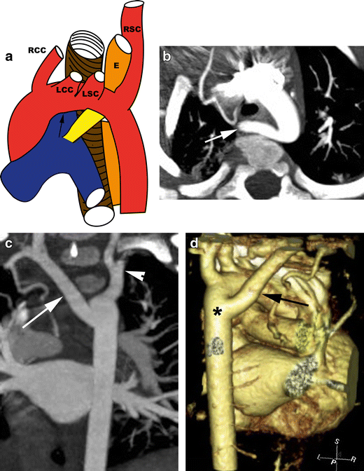
A
Decode the arch phenotype
| Pattern | Connection and compression | Adult consequence |
|---|---|---|
| LAA + ARSA | RCCA, LCCA, LSA, then ARSA; usually behind the esophagus | Dysphagia lusoria, difficult right-radial access, KD/aneurysm or dissection risk |
| RAA + ALSA | ALSA commonly arises from KD; left ligamentum to LPA can close the ring | Esophageal/tracheal compression plus KD aortic risk |
| Course | About 80% retroesophageal, 15% between trachea/esophagus, 5% anterior to trachea | The relationship, not the vessel name alone, predicts symptoms and approach |
B
CTA or MRA report: four non-negotiables
1 · Topology
Arch side, branch order, origin, retroesophageal/retrotracheal course, ligament/ductal remnant
Arch side, branch order, origin, retroesophageal/retrotracheal course, ligament/ductal remnant
›
2 · Compression
Minimum tracheal caliber, bronchomalacia, esophageal impression, aspiration or lung injury
Minimum tracheal caliber, bronchomalacia, esophageal impression, aspiration or lung injury
›
3 · Measure KD twice
Orifice/transverse vessel and apex-to-opposite aortic wall in orthogonal planes
Orifice/transverse vessel and apex-to-opposite aortic wall in orthogonal planes
›
4 · Plan repair
Whole-aorta disease, thrombus, vertebral origins, arm/cerebral flow and endograft landing zones
Whole-aorta disease, thrombus, vertebral origins, arm/cerebral flow and endograft landing zones
C
Intervention gates: symptoms are the clearest indication
| Gate | Current practical threshold | Interpretation |
|---|---|---|
| Acute | Rupture, dissection, malperfusion/embolization, rapid clinical change | Urgent multidisciplinary aortic treatment; do not apply elective size rules |
| Symptomatic | Attributable dysphagia, dyspnea/stridor/cough, aspiration/recurrent infection, arm ischemia or chest/back pain | Repair should be considered regardless of size; exclude another cause but do not wait for advanced dysmotility |
| Ring / PA sling | 2025 ACHD: surgery is reasonable when symptomatic; no benefit for an asymptomatic ring/sling without another indication | A KD may create a separate aneurysm indication even when compression is absent |
| ACC/AHA 2022 | KD orifice >3.0 cm or combined KD + adjacent descending aorta >5.0 cm | Repair may be reasonable; Class IIb, low-quality data |
| ESVS 2026 | Transverse aberrant vessel ≥3.0 cm or combined edge-to-edge distance ≥5.5 cm | Treatment may be considered; Class IIb-C. Measurement definitions and society thresholds differ |
| Incidental, normal caliber | ESVS: imaging every 5 years from age 40 may be considered | Shorten for KD/dilation, growth, aortopathy, symptoms or family/genetic risk |
D
What the operation must accomplish
| Transposition / bypass | Move flow to carotid/subclavian or ascending aorta, then divide/occlude the retroesophageal origin. A long patent stump can remain compressive or become aneurysmal/thromboembolic. |
| Open KD / DTA repair | Resect the diverticulum and replace involved descending aorta; offers direct decompression and is favored for mass effect, unsuitable landing zones or connective-tissue disease. |
| Arch / FET | Hemi- or total-arch reconstruction with branch revascularization, sometimes frozen elephant trunk, for extensive arch-to-descending disease or dissection. |
| Hybrid / TEVAR | Subclavian revascularization plus endograft exclusion and often origin plug/coil. Watch endoleak, residual sac compression, bypass patency and reintervention. |
| Ring release | Double arch: divide nondominant/atretic arch and ligament. RAA/ALSA: ligament division alone may be inadequate in adults when KD or a compressive retroesophageal segment remains. |
E
After repair
- Symptoms: dysphagia/dyspnea usually improve, but fixed esophageal dysmotility, reflux or tracheobronchomalacia can persist.
- Branch: bilateral arm BP/pulses; duplex/CTA for transposition or bypass stenosis, steal or arm ischemia.
- Nerves/lymph: hoarseness, Horner syndrome, phrenic injury and chylothorax are operation-specific complications.
- Open repair: CT/MR within 1 year, then about every 5 years if no residual aortopathy; annually when residual disease remains.
- TEVAR/hybrid: CTA at 1 month and 12 months, then annually if stable; MRI is an alternative when appropriate.
- Whole aorta: continue lifelong surveillance for adjacent aneurysm, dissection, pseudoaneurysm, sac growth or endoleak.
Act nowNew chest/back pain, hemoptysis or GI bleeding, hoarseness, neurologic deficit, arm ischemia/pulse change, rapidly progressive dysphagia, stridor, aspiration or recurrent pneumonia warrants prompt cross-sectional imaging and an aortic/ACHD team.
The connection auditFor every major vessel, state where it begins, what it passes behind or through, where it ends, whether it carries oxygenated or deoxygenated blood, and whether it is essential, obstructed, decompressing, or a source of shunt/collateral flow. This prevents closure of a necessary pathway and failed venous/catheter access.
A
Arch and pulmonary-artery variants
| Variant | What connects abnormally | Why the adult team cares |
|---|---|---|
| RAA, mirror-image | Left brachiocephalic first, then right carotid/subclavian; descending aorta on right | Often accompanies conotruncal CHD; usually not a ring by itself, but changes sternotomy, cannulation and arch access |
| Double / circumflex arch | Two arches encircle airway/esophagus, or an arch crosses behind them to the opposite descending aorta | Map dominant/atretic segments, ligament, bronchial compression and residual KD before division or uncrossing |
| Cervical / persistent 5th arch | High arch in neck or parallel systemic-to-systemic channel | May be incidental, aneurysmal, obstructed or mistaken for a carotid/subclavian mass; image the entire route |
| LPA sling | LPA arises from RPA and passes between trachea and esophagus | Airway compression/complete tracheal rings; repair reimplants LPA and may require tracheal reconstruction |
| Isolated/interrupted PA | One proximal branch PA is absent; distal intrapulmonary PA receives ductal/systemic collaterals | Unilateral small lung, hemoptysis, differential perfusion and PH; define every collateral before embolization/reconstruction |
| Hemitruncus | One branch PA arises directly from ascending aorta | High-pressure/high-flow lung with early pulmonary vascular disease; repaired adults need branch, valve, RV and PH surveillance |
B
Systemic venous connections
| Connection | Signature | Procedure / physiology consequence |
|---|---|---|
| Persistent LSVC → CS/RA | Dilated coronary sinus; left-sided vertical vein | Usually no shunt. Alters central line/lead route, cannulation and retrograde cardioplegia; report bridging vein and RSVC |
| LSVC → LA / unroofed CS | Left-arm agitated saline appears in LA; cyanosis or paradoxical embolus | Do not close until all systemic and pulmonary venous drainage and coronary-sinus anatomy are defined |
| Interrupted IVC → azygos | Absent hepatic/suprarenal IVC continuity; enlarged azygos/hemiazygos to SVC | Femoral venous wire tracks up the spine instead of directly to RA; hepatic veins often enter atrium separately |
| Retroaortic brachiocephalic | Left brachiocephalic vein passes behind ascending aorta | Can be injured or obstructed during arch/PA surgery and complicates lines/leads |
| IVC/hepatic vein → LA | Unexplained systemic desaturation with otherwise normal intracardiac anatomy | Right-to-left venous connection; map hepatic/IVC pathways and exclude post-Fontan streaming before redirection |
C
Pulmonary venous and decompression channels
PAPVR / TAPVR
Wrong atrial destination
One or more pulmonary veins reach systemic veins/RA. Map each vein, obstruction and Qp:Qs; see P7 for Warden, Scimitar and TAPVR pathways.Levoatriocardinal vein
LA/PV to cardinal vein
A decompression channel from LA or pulmonary vein to systemic venous circulation, often with left-sided obstruction. It can mimic PAPVR; correct the obstructive physiology before closure.Vertical vein / collaterals
Residual or intentional pop-off
After TAPVR/Fontan or with venous obstruction, a patent channel may decompress pressure. Test hemodynamics before occlusion; symptoms can worsen if the pathway is essential.D
Systemic-to-pulmonary and aortocardiac connections
| PDA / AP window | Continuous systemic-to-PA flow; quantify chamber load and PVR before closure. Distinguish from hemitruncus. |
| MAPCAs / bronchial arteries | May be essential pulmonary blood supply in PA/VSD or a bleeding/high-flow collateral after repair. Never embolize before proving distal PA supply. |
| Coronary-to-PA/cameral fistula | Coronary runoff can cause dilation, steal, shunt, thrombosis or IE; define origin, drainage and myocardial branches before device/surgery. |
| Aortoatrial tunnel / ruptured SOV | Continuous aortic-to-atrial/RV flow can mimic PDA/VSD; identify the aortic origin, receiving chamber and valve/coronary relationships. |
| Fontan venovenous collaterals | Systemic venous-to-pulmonary venous/atrial channels cause cyanosis but may decompress high Fontan pressure; cath/test occlusion before closure. |
E
Bedside and catheterization clues
1Right radial wire repeatedly enters descending aorta: suspect ARSA; stop forceful manipulation and define arch anatomy.
2Dilated coronary sinus or unusual left-arm line: inject left-arm saline and map LSVC/bridging vein before EP or surgery.
3Femoral venous wire ascends paraspinally: interrupted IVC with azygos continuation; plan jugular, transhepatic or alternate access.
4Unexplained cyanosis: seek systemic vein to LA, unroofed CS, baffle/fenestration leak, venovenous collateral or PAVM.
5Hemoptysis + small/unilateral PA: define MAPCAs/bronchial collaterals, PA interruption, aneurysm and pulmonary pressures.
6Dysphagia/stridor: image arch branches, ligament, airway and esophagus together - not an echo-only work-up.
F
Pre-procedure connection map
Venous return
SVCs, IVC/azygos, hepatic and pulmonary veins
SVCs, IVC/azygos, hepatic and pulmonary veins
›
Arterial supply
arch branches, coronaries, each lung and all collaterals
arch branches, coronaries, each lung and all collaterals
›
Created pathways
baffles, conduits, shunts, Fontan, fenestrations, grafts
baffles, conduits, shunts, Fontan, fenestrations, grafts
›
Occlusion test
is the channel harmful, incidental, or a necessary pop-off?
is the channel harmful, incidental, or a necessary pop-off?
Never close an unexplained vessel by appearance aloneFirst prove its origin, destination, saturation/flow, pressure gradient, dependent territory and what happens during test occlusion.
Mean PA pressure
>20 mmHg
current PH threshold
Wedge pressure
≤15 mmHg
precapillary profile
PVR
>2 WU
precapillary/PAH threshold
A
Eisenmenger essentials
Definition
Advanced pulmonary vascular disease
A prior systemic-to-pulmonary shunt becomes bidirectional or right-to-left, producing cyanosis. Shunt closure is harmful because it removes the RV/systemic pressure pop-off.Targeted therapy
Start in the guideline-defined group
In adults with Eisenmenger syndrome, LVEF >40%, and symptoms or reduced exercise capacity, initial monotherapy with PAH-directed therapy—an endothelin-receptor antagonist or PDE5 inhibitor—is recommended. If symptoms persist or exercise capacity worsens, add the complementary class with ACHD–PH specialists. BREATHE-5 specifically showed that bosentan improved exercise capacity and hemodynamics without worsening oxygen saturation.B
Cyanosis is multisystem disease
| Problem | Practical management |
|---|---|
| Secondary erythrocytosis | Adaptive. No routine phlebotomy. Check ferritin/iron at least annually; microcytosis worsens viscosity and exercise. |
| Hyperviscosity symptoms | Hydrate and correct iron first. Phlebotomy only for persistent symptoms with hematocrit >65%, using isovolumic replacement in expert care. |
| Bleeding + thrombosis | Hemoptysis, platelet/coagulation abnormalities coexist with PA thrombosis and embolic risk; anticoagulation is not routine and must be individualized. |
| Brain/air | Use meticulous air filters/de-airing for IV lines; evaluate headache/neurologic symptoms for abscess, stroke, or hyperviscosity. |
| Kidney/gout | Monitor renal function, proteinuria, urate; avoid dehydration and nephrotoxins. |
| Procedures | Avoid endocardial leads when possible; anesthesia and ventilation can destabilize SVR/PVR balance. |
C
Hemoptysis and acute decompensation
StabilizeACHD/PH critical-care team; oxygen if hypoxemic, correct reversible triggers, maintain SVR/preload, avoid abrupt systemic vasodilation and high airway pressures.
Find the sourceCTA when stable for PA aneurysm/thrombus, systemic collaterals, infection, infarct, or aortopulmonary source; coordinate bronchoscopy/interventional radiology cautiously.
Escalate earlyRefractory HF, syncope, recurrent hemoptysis, or falling functional status should trigger transplant discussion (bilateral lung + defect repair or heart–lung, anatomy dependent).
D
Shunt operability by PVR
| PVR | 2025 U.S. approach | Practical caution |
|---|---|---|
| ≤2 WU | Favorable zone when lesion-specific chamber/Qp:Qs indication is present | Still assess LV filling pressure and anatomy |
| >2 to <5 WU | ASD/VSD/PDA closure generally reasonable after expert evaluation | Confirm accurate output/wedge and absence of right-to-left physiology |
| 5–8 WU baseline | ASD may benefit if PAH therapy lowers PVR <5; PDA may be considered; VSD usefulness uncertain | Evidence limited; consider fenestration/test occlusion and long-term PAH plan |
| Persistent high PVR | Benefit uncertain or harmful; Eisenmenger is a no-closure state | ESC guidance is more conservative; multidisciplinary ACHD–PH decision |
| PVR >10 WU | No closure for PDA/AVSD and generally inoperable shunt physiology | Do not use “treat-to-close” as an open-ended promise |
E
Cyanotic blood and specimen handling
CBC
Interpret against saturation
A “normal” hemoglobin can represent relative anemia. Microcytosis/ferritin identify iron deficiency despite erythrocytosis.Coagulation
High Hct distorts tubes
With very high Hct, laboratory citrate adjustment may be needed for accurate coagulation testing; coordinate with lab.Pulse oximetry
Know the baseline
Trend rest/exercise; abrupt change suggests thrombosis, arrhythmia, infection, shunt/collateral or ventricular deterioration.F
Routine care protections
- Vaccinate against indicated respiratory pathogens.
- Avoid dehydration; give a clear illness/travel plan.
- Meticulous dental hygiene and IE risk review.
- Use IV air filters/de-airing; avoid unnecessary central lines.
- Individualize contraception and thrombosis risk.
- Plan altitude/air travel by baseline saturation and reserve.
PregnancyPAH/Eisenmenger is extremely high risk (mWHO IV). Provide expert preconception counseling and effective contraception; pregnancy requires immediate pregnancy-heart-team care.
A
Arrhythmia predicts anatomy
| Substrate | Common rhythm | Immediate parallel search |
|---|---|---|
| Mustard/Senning | Sinus-node disease, IART/flutter, AF | Baffle obstruction/leak, systemic RV/TR, thrombus; rhythm control preferred |
| Atriopulmonary Fontan | IART/AF, sinus-node disease | Fontan obstruction, thrombus, low output, FALD; prompt cardioversion |
| Repaired TOF | IART and scar-related monomorphic VT | PR/PS, RV/LV dysfunction, RVOT/VSD-patch isthmuses, SCD risk |
| Ebstein | AVRT/WPW, atrial arrhythmia | Accessory pathways, TR/RV load, ASD shunt; ablate before valve surgery |
| ccTGA/AVSD | Progressive AV block, atrial/ventricular arrhythmia | Systemic RV/valve, displaced conduction anatomy, pacing dyssynchrony |
B
Before a lead crosses anything
STOPAtrial switch
CMR/CT + venography for baffle stenosis/leak; treat defect before transvenous lead; plan embolic/anticoagulation strategy.
STOPFontan
Most pathways lack routine venous access to ventricle. Consider epicardial, coronary-sinus, transpulmonary/transbaffle, or leadless options at an expert center.
PLANIntracardiac shunt
Transvenous lead increases systemic embolic risk; close appropriate shunt first or use a non-endocardial system.
PLANSystemic RV
Avoid unnecessary dyssynchrony. High-grade block may favor CRT/conduction-system pacing; anatomy defines the target and access.
C
SCD risk is lesion-specific
Primary prevention
Not an EF-only decision
Integrate anatomy, ventricular function, scar, syncope, NSVT, QRS, inducibility, surgical history, and competing procedural risks.Secondary prevention
Standard indication, complex execution
Survived arrest or sustained hemodynamically important VT generally supports ICD; fix reversible hemodynamic lesions and plan access.Subcutaneous ICD
Avoids venous pathways
Useful when pacing is not needed; screen carefully for abnormal QRS/T waves. Extravascular/leadless systems may expand options.D
Acute tachycardia rules
- Unstable or poorly tolerated rhythm: synchronized cardioversion without delay.
- Wide-complex rhythm in repaired TOF/systemic RV: presume VT until proven otherwise.
- Fontan/atrial-switch IART may conduct 1:1 and collapse output despite modest ventricular dysfunction.
- Anticoagulation and thrombus imaging depend on rhythm duration and circuit.
- Correct hypoxemia, electrolyte disorder, infection, thyroid/drug triggers.
- After conversion, investigate the hemodynamic lesion that triggered the rhythm.
E
Ablation is an anatomic procedure
| Setting | Technical issue | Strategy |
|---|---|---|
| Atrial switch IART | Critical isthmus may lie on pulmonary-venous side of baffle | Transbaffle/retrograde access with imaging; treat baffle lesion first |
| Fontan IART | Extracardiac conduit blocks atrial access; AP Fontan has giant scarred RA | Transconduit/transbaffle, surgical/hybrid or conversion at expert center |
| TOF VT | Slow-conduction isthmuses between patch, annulus and scars | Electroanatomic mapping/ablation before PVR can preserve access |
| Ebstein AVRT | Multiple broad right-sided pathways and displaced true annulus | Map before cone/valve surgery; surgical ablation if needed |
| Heterotaxy/ccTGA | Displaced/twin AV nodes and unusual conduction axis | Segmental anatomy, prior maps and congenital EP technique |
F
Medication and anticoagulation pearls
Antiarrhythmic
Reserve and substrate
Ventricular dysfunction, cyanosis, renal/hepatic disease, bradycardia and QRS/QT alter drug choice; ablation often avoids chronic toxicity.Anticoagulation
Circuit-specific
Sustained IART/AF in atrial switch/Fontan generally warrants therapy; cyanotic bleeding and Fontan liver disease alter agent/dose.Rate control
Can worsen output
Fixed-stroke-volume systemic-RV/Fontan patients may depend on heart rate; excessive slowing can decompensate.G
Pacing follow-up
- Trend ventricular function/TR and paced QRS.
- Image venous/baffle/Fontan pathway with symptoms.
- Epicardial lead failure may be clinically silent.
- Plan extraction with congenital surgeon/extraction team.
- Reassess CRT/conduction-system/leadless options.
- Keep a clear diagram of lead course in the chart.
After ICD therapyInvestigate PR/PS, baffle/Fontan obstruction, ischemia, ventricular/valve failure and correctable scar substrate; a shock is not the endpoint.
Holter is one member of the ambulatory-ECG familyChoose the least burdensome system likely to record the clinical event. The target may be symptom–rhythm correlation, asymptomatic surveillance, arrhythmia-burden quantification, rate/chronotropy, or treatment efficacy—not merely “rule out arrhythmia.”
A
Match the recorder to how often the event occurs
Nuance: continuous 1–14-day monitoring is useful when quantifying ectopy/AF burden even if symptoms are absent. A 12-lead Holter is preferable when QRS/PVC morphology, ST pattern, or QT dynamics—not only rate—is the clinical question. For syncope or inability to trigger, choose continuous/auto-detect monitoring rather than a patient-activated event recorder. MCT = mobile cardiac telemetry; ILR/ICM = implantable loop recorder/insertable cardiac monitor.
B
Order it for a defined ACHD question
Symptoms / deterioration
Capture the event
Palpitations; syncope/presyncope; episodic dizziness; unexplained fatigue, desaturation or exercise decline; new/worsening HF; suspected tachycardia, pauses or intermittent AV block.Silent high-risk substrate
Do not wait for palpitations
Mustard/Senning, Fontan, ccTGA/conduction disease, repaired TOF with SCD-risk review, Ebstein/accessory pathway, atriotomy scars, ventricular scar or prior NSVT.Therapy / procedure
Verify efficacy and safety
After cardioversion/ablation; after drug or rate-slowing change; before/after valve or hemodynamic intervention; unexplained ICD/device alerts; quantify residual AF/IART, ectopy or pacing-related rhythm.Syncope is not a routine outpatient-Holter problem when risk is highExertional or supine syncope, injury, abnormal hemodynamics, severe ventricular dysfunction, documented high-grade block, sustained/wide-complex tachycardia or unstable symptoms require urgent monitored evaluation. Do not delay treatment merely to obtain symptom–rhythm correlation.
C
Lesion-specific surveillance: cadence is not one-size-fits-all
| Substrate | Stable surveillance anchor | What the monitor must find |
|---|---|---|
| Mustard / Senning | 2025 guideline: ambulatory monitoring for bradycardia/sinus-node disease; in an asymptomatic stable patient, Holter about every 3–5 y is reasonable. Shorten for rate-slowing therapy, symptoms or new HF. | Sinus/junctional rhythm, pauses, chronotropy, IART/flutter/AF, ventricular arrhythmia; pair a positive study with baffle, systemic-RV and TR review. |
| ccTGA | Without known conduction disease, ambulatory monitoring about every 5 y is reasonable; with PR/AV-conduction disease and no pacemaker, use symptoms and block severity to set a shorter interval. | Progressive PR prolongation, intermittent second-/third-degree AV block, pauses, atrial/ventricular arrhythmia; involve congenital EP before block becomes symptomatic. |
| Fontan | The 2025 guideline does not prescribe a fixed Holter cadence. An older 2019 AHA expert framework suggested 24-h Holter every 1–2 y in adolescents/adults; individualize because interval evidence is limited, and monitor promptly with symptoms or declining output. | Sinus-node disease, junctional rhythm, IART/flutter/AF, ectopy/VT. New burden triggers pathway/PA, valve/ventricle, thrombus and end-organ assessment. |
| Repaired TOF | Use ambulatory ECG when symptoms occur and when NSVT/ventricular ectopy will change SCD-risk or pre-PVR/ablation planning; extend duration if events are infrequent. | NSVT rate/duration/morphology, PVC burden/couplets, atrial tachyarrhythmia and symptom correlation; integrate with QRS, RV/LV function, scar, hemodynamics and CPET—not as an ICD test by itself. |
| Other ACHD | Ebstein/WPW, AVSD, post-atriotomy scar, ventricular dysfunction/scar and prior arrhythmia merit risk- and symptom-based monitoring. An uncomplicated simple repaired lesion does not require automatic annual Holter. | Ask the lesion-specific question: AVRT/pre-excitation, IART/AF, AV block, NSVT, chronotropic incompetence or treatment recurrence. |
D
Read the whole report—not “AF: yes/no”
| Report | Minimum interpretation | ACHD consequence |
|---|---|---|
| Recording quality | Analyzable time, leads, artifact, sleep/activity/medication diary; baseline rhythm and QRS morphology | A low-quality “negative” study is nondiagnostic; single-lead recordings may misclassify wide-complex rhythms |
| Rate / conduction | Minimum/mean/maximum HR; sinus versus junctional; pauses; AV block; rate trend during reported activity | Separate medication effect from sinus-node/AV-node disease; exertional chronotropy may require CPET |
| Atrial rhythm | PAC burden; SVT/IART/AF episode count, duration, rate and total burden; symptom relation | In Fontan/systemic RV, even a tolerated atrial rhythm may reduce output and change anticoagulation/rhythm strategy |
| Ventricular rhythm | PVC count/% burden, morphology, couplets; NSVT count, rate, length, timing and symptoms | New NSVT prompts lesion/scar/hemodynamic/SCD review; it is not an automatic ICD indication |
E
Convert the result into an action
Urgent / same-day ACHD-EP triage
Do not file and forget
Sustained or symptomatic wide-complex tachycardia; high-grade AV block or symptomatic pause; arrhythmia-correlated syncope; rapid IART/AF with Fontan or systemic RV; ventricular rhythm with ischemia or hemodynamic compromise.Accelerated outpatient review
Interrogate the substrate
New AF/IART, NSVT, rising ectopy, sinus-node disease/chronotropic incompetence or meaningful symptom correlation → ECG/echo ± CMR/CT/CPET, medications/electrolytes/thyroid/OSA review, and congenital EP/hemodynamic planning.A negative monitor answers only the window recordedIf the typical event did not occur, escalate duration rather than declaring “no arrhythmia.” If a clean recording captures the typical symptom in sinus rhythm, redirect the differential. Exercise-triggered symptoms need maximal exercise ECG/CPET; a consumer PPG alert is a screening signal, and a single-lead wearable ECG can capture an event, but clinical decisions should use clinician-reviewed ECG evidence. QT/JT, QRS morphology and broad-complex discrimination may require 12-lead recording.
A
HF treatment begins with the circulation
| Phenotype | First questions | Therapy principle |
|---|---|---|
| Systemic LV | Residual shunt/obstruction, valve disease, ischemia, dyssynchrony? | Apply standard HFrEF/HFpEF evidence when anatomy allows, plus lesion correction |
| Systemic RV | Systemic TR, baffle/LVOTO, pacing burden, IART? | Evidence for conventional GDMT is limited; selected HFrEF therapy may be reasonable, but anatomy/rhythm/valve treatment dominates |
| Fontan | Pathway/PA obstruction, PVR, collaterals, AV valve, rhythm, lymphatic/end-organ disease? | Careful diuresis; correct circuit lesions; rehab; selected pulmonary vasodilator/lymphatic therapy; avoid preload collapse |
| Cyanotic/PAH | Shunt direction, PVR/SVR, iron, thrombosis/bleeding, infection? | PAH-targeted therapy and physiology protection; generic systemic vasodilation can worsen shunting |
B
Refer before the window closes
Advanced HF signals
Trajectory beats one threshold
Repeated admissions, progressive NYHA limitation/CPET decline, escalating diuretics, hypotension, refractory arrhythmia, rising BNP, ventricular/valve decline, cyanosis, PLE/plastic bronchitis, worsening liver/kidney disease.Therapy options
Anatomy changes candidacy
Transplant; systemic-RV or selected single-ventricle VAD; heart–liver transplant in selected FALD; bilateral lung + shunt repair or heart–lung for Eisenmenger. Prior sternotomies, collaterals, sensitization, and venous anatomy raise risk.C
Pregnancy: preconception, not first trimester
Pregnancy heart teamDefine anatomy/repair, ventricular and valve function, aorta, saturation, PA pressure, rhythm, medications, anticoagulation, genetics, contraception, and maternal goals. Use the 2025 ESC mWHO 2.0 classification and an individualized delivery/postpartum plan.
Usually lower riskSuccessfully repaired simple shunts without sequelae; small VSD/PDA with normal LV and no PH. Unrepaired ASD adds arrhythmia/paradoxical-embolism risk; avoid air and venous stasis.
Very high riskPAH/Eisenmenger, severe ventricular dysfunction, severe symptomatic obstruction, or major aortopathy may place pregnancy in mWHO IV. Explain maternal and fetal risks with a Pregnancy Heart Team, support informed reproductive choices, and offer effective contraception.
DeliveryVaginal delivery is preferred for most ACHD. Routine cesarean offers no cardiac benefit and can cause harm unless an obstetric or defined high-risk cardiac indication exists. Monitor the early postpartum volume shift.
D
Evidence by HF phenotype
| Therapy | Systemic LV | Systemic RV | Fontan |
|---|---|---|---|
| Diuretic | Congestion, standard monitoring | Useful; avoid preload collapse | Narrow preload window |
| RAAS/ARNI | Standard indication | Limited trials; no assumed LV-equivalent benefit | No routine benefit absent indication |
| Beta-blocker | Standard HFrEF/arrhythmia use | Selected; bradycardia matters | Can reduce rate-dependent output |
| MRA/SGLT2i | Standard acquired-HF evidence | Emerging limited data | Limited; volume/renal effects matter |
| PAH therapy | Only indicated PH phenotype | Treat PAH when present | Selected elevated-PVR/low-output physiology |
E
Pregnancy lesion checkpoints
| Circulation | Main maternal risk | Preconception action |
|---|---|---|
| Aorta/CoA | HTN, aneurysm/dissection | Image entire aorta; all-limb/ambulatory BP |
| Systemic RV | HF, TR, IART | CMR/echo, rhythm plan, medication reset |
| Fontan | HF/arrhythmia, thrombosis/bleeding, fetal loss | Circuit, liver, saturation and anticoagulation review |
| PAH / Eisenmenger | Very high maternal risk | mWHO IV. Expert counseling and shared decisions |
| Cyanosis without PAH | HF, thrombosis and fetal risk | Unrepaired cyanotic CHD without Eisenmenger: mWHO III. Saturation and ventricular function refine risk |
| Valve/conduit | Obstruction, prosthesis/anticoagulation | Gradient/function, exercise and anticoagulation plan |
F
Postpartum is a cardiac stress test
Autotransfusion and rapid postpartum fluid and hemodynamic shifts can precipitate HF or arrhythmia after an apparently stable delivery. Define monitoring duration, fluid/diuretic strategy, anticoagulation restart, lactation-compatible drugs, contraception and early ACHD follow-up before discharge.
Medication resetReview teratogenic HF/PAH/anticoagulant drugs before conception; do not stop essential therapy without a substitute and coordinated plan.
How to use this glossaryThe expansion is only the first step. The context column states what the shorthand means in this guide and the final column points to the pages where the anatomy or decision is developed. Red terms have more than one clinically important meaning.
I · anatomy A-MII · anatomy N-ZIII · operationsIV · tests/flowV · clinical
| Term | Expansion | ACHD context or trap | See |
|---|---|---|---|
| AAOCA | Anomalous aortic origin of a coronary artery | Umbrella term; report ostium, proximal caliber, intramural segment and course rather than saying only "interarterial." | P24 |
| AAOLCA | Anomalous aortic origin of the left coronary artery | Usually left coronary from the right/opposite sinus; dominance and the entire proximal anatomy matter. | P24 |
| AAORCA | Anomalous aortic origin of the right coronary artery | Usually right coronary from the left/opposite sinus; symptoms, dynamic ischemia, anatomy and activity guide management. | P24 |
| ACAPA | Anomalous coronary artery from the pulmonary artery | Umbrella for ALCAPA, ARCAPA and rarer branch origins; distinguish from a coronary-to-PA fistula. | P26 |
| ACHD / CHD | Adult congenital heart disease / congenital heart disease | "Adult" changes the care setting, not the anatomy; repaired, palliated and unrepaired circulations all remain CHD. | P1 |
| ALCAPA | Anomalous left coronary artery from the pulmonary artery | Postnatal PA-pressure fall creates runoff/steal; adult repair restores a two-coronary circulation. | P26 |
| ALSA | Aberrant left subclavian artery | Often arises from a Kommerell diverticulum with a right aortic arch; may complete a ring with a left ligament. | P28-29 |
| AP | Anatomic-physiologic; aortopulmonary; or atriopulmonary | AP class on P1, AP window on shunt pages, and AP Fontan on single-ventricle pages are different concepts. | P1,8,21 |
| AP window | Aortopulmonary window | Direct aorta-to-PA communication with separate semilunar valves; not PDA, truncus or a coronary fistula. | P8,30 |
| APC | Aortopulmonary collateral | Systemic/aortic arterial contribution to the pulmonary bed; can recirculate volume and burden a Fontan. | P7,22 |
| APVC | Anomalous pulmonary venous connection | Umbrella term: partial connection is PAPVR; total connection is TAPVR. Map every pulmonary vein and obstruction. | P7 |
| ARCAPA | Anomalous right coronary artery from the pulmonary artery | Consequences vary with dominance, collateralization, ischemia and ventricular effect; not managed identically to ALCAPA. | P26 |
| ARSA | Aberrant right subclavian artery | Arteria lusoria, usually the last branch of a left arch with retroesophageal course; define KD and compression. | P28-29 |
| ASD | Atrial septal defect | Specify secundum, primum, sinus venosus or coronary-sinus type; PFO is not an ASD. | P5-6 |
| AV | Atrioventricular | Used for AV connection, AV valve and AV conduction/block; chamber morphology still must be named. | P1,9,12 |
| AVSD | Atrioventricular septal defect | Partial, transitional or complete common-AV-junction lesion; adult failure often centers on the left AV valve and LVOT. | P9 |
| BAV | Bicuspid aortic valve | Track valve dysfunction, ascending aortopathy and associated coarctation; repaired arch disease does not remove BAV risk. | P17-18 |
| ccTGA | Congenitally corrected transposition of the great arteries | AV and VA discordance are both present; the morphologic RV and tricuspid valve are usually systemic without anatomic repair. | P12 |
| CoA | Coarctation of the aorta | Hypertension and diffuse aortopathy may persist despite an acceptable resting repair gradient. | P17 |
| CS | Coronary sinus | Relevant to unroofed CS defects, persistent LSVC drainage and some coronary fistula exits. | P5,23,27 |
| DCRV | Double-chambered right ventricle | Progressive muscular RV obstruction, often with a VSD; distinguish the intracavitary level from valvar or infundibular PS. | P18 |
| DORV | Double-outlet right ventricle | Both great arteries arise predominantly from the RV; VSD relationship and outflow obstruction define routing options. | P16 |
| DTA | Descending thoracic aorta | Named separately from the KD orifice and aberrant vessel when measuring complex arch pathology. | P29 |
| d-TGA | Dextro-transposition of the great arteries | AV concordance with VA discordance; adult physiology depends on atrial switch, arterial switch or complex routing. | P10-11 |
| HLHS | Hypoplastic left heart syndrome | Adult survivors generally have Norwood-Glenn-Fontan palliation with a systemic morphologic RV. | P20-22 |
| KD | Kommerell diverticulum | Aneurysmal arch-branch origin associated with aberrant subclavian anatomy, compression and separate aortic risk. | P28-29 |
| LA / RA | Left atrium / right atrium | Name atrial morphology, situs and venous connections rather than assuming left-right position. | P1,23 |
| LPA / RPA | Left / right pulmonary artery | Branch size, distortion and flow split matter after shunts, ASO, TOF repair and Fontan. | P3,11,13 |
| LSVC / PLSVC | Left / persistent left superior vena cava | Drainage destination changes shunt, cannulation, lead-access and Fontan/Kawashima planning. | P23,30 |
| LV / RV | Left / right ventricle | In ACHD these mean morphology, not merely screen position; always name which ventricle supports the systemic circulation. | P1 |
| LVOT / LVOTO | Left ventricular outflow tract / obstruction | Prior tunnel, VSD routing, subaortic membrane or complex repair determines the obstructed segment. | P12,16,18 |
| MAPCA(s) | Major aortopulmonary collateral artery/arteries | May provide essential segmental pulmonary supply in PA/VSD; never embolize before native supply is mapped. | P15,30 |
I · anatomy A-MII · anatomy N-ZIII · operationsIV · tests/flowV · clinical
PVR WU = resistance; "underwent PVR" = pulmonary valve replacement.
RAA right aortic arch on ring pages; right atrial appendage in a Warden repair.
VA ventriculoarterial connection in anatomy; ventricular arrhythmia in EP.
PA pulmonary artery alone; pulmonary atresia in PA/IVS or PA/VSD.
PV pulmonary vein in venous-return pages; pulmonary valve in outflow pages.
SV single ventricle in Fontan care; sinus venosus when modifying an ASD.
| Term | Expansion | ACHD context or trap | See |
|---|---|---|---|
| LAA | Left aortic arch | Meaning on ring/lusoria pages; elsewhere LAA commonly means left atrial appendage, so spell it out. | P28-29 |
| LCA / LAD / LCx | Left coronary / left anterior descending / left circumflex | Name the exact anomalous donor, bridged segment or fistula branch; "left coronary" may be insufficient. | P24-27 |
| PA | Pulmonary artery | Main or branch PA in most text; in compound diagnoses such as PA/IVS, PA means pulmonary atresia. | P1,15 |
| PA/IVS | Pulmonary atresia with intact ventricular septum | Coronary sinusoids may create RV-dependent coronary circulation; do not decompress the RV before mapping. | P15,27 |
| PA/VSD | Pulmonary atresia with ventricular septal defect | Pulmonary supply may come from native PAs, duct, shunt or MAPCAs and must be mapped segment by segment. | P15 |
| PAPVR | Partial anomalous pulmonary venous return | One or more, but not all, pulmonary veins reach the systemic venous side; count each vein and its obstruction. | P7 |
| PAVM(s) | Pulmonary arteriovenous malformation(s) | Intrapulmonary right-to-left shunting may follow uneven hepatic flow after cavopulmonary connections. | P22-23 |
| PBF | Pulmonary blood flow | Early single-ventricle palliation balances inadequate oxygenation against pulmonary overcirculation and rising PVR. | P2-3,20 |
| PDA | Patent ductus arteriosus | Aortic-to-PA ductal connection; distinguish from AP window, MAPCA and coronary-to-PA fistula. | P8,30 |
| PFO | Patent foramen ovale | Dynamic flap/tunnel rather than tissue-deficiency ASD; stroke closure requires causal attribution. | P6 |
| PV | Pulmonary vein or pulmonary valve | Venous-routing pages use vein; TOF/outflow pages use valve. State the structure when the context is not obvious. | P7,13-18 |
| RCA | Right coronary artery | Specify anomalous origin, intramural course, dominance, fistula donor or compression risk rather than using RCA alone. | P24-27 |
| RAA | Right aortic arch or right atrial appendage | Arch meaning on ring/lusoria pages; atrial-appendage meaning at the cephalad Warden anastomosis. | P7,28-29 |
| RCCA / LCCA | Right / left common carotid artery | Used with LSA/RSA to report exact arch branch order before intervention or access. | P29-30 |
| RVDCC | Right-ventricle-dependent coronary circulation | Seen in selected PA/IVS; coronary perfusion depends on high RV pressure through sinusoids. | P15,27 |
| RVOT / RVOTO | Right ventricular outflow tract / obstruction | Differentiate native tract, transannular patch, conduit and branch-PA levels before calling it "RVOT disease." | P11,13-18 |
| SOV | Sinus of Valsalva | Rupture can create an aorto-cameral connection that mimics a coronary fistula or other continuous-flow lesion. | P27,30 |
| SV | Single ventricle; sometimes sinus venosus | Fontan pages use single ventricle; ASD shorthand such as SVASD uses sinus venosus. Do not assume stroke volume. | P5,20-23 |
| SVASD | Sinus venosus atrial septal defect | Usually SVC-type and commonly associated with PAPVR; not a device-suitable secundum ASD. | P5,7 |
| SVC / IVC | Superior / inferior vena cava | Map bilateral SVCs, interrupted IVC and azygos continuation before Fontan, cannulation, leads or venous intervention. | P3,23,30 |
| TAPVR | Total anomalous pulmonary venous return | All pulmonary veins drain anomalously; repaired adults require surveillance for confluence/individual-vein obstruction. | P7 |
| TGA | Transposition of the great arteries | Always specify d-TGA versus ccTGA and the actual operation; the abbreviation alone does not identify the circulation. | P10-12 |
| TOF | Tetralogy of Fallot | Adult physiology depends on RVOT repair type, PR, residual obstruction, ventricular remodeling and scar. | P13-14 |
| VA | Ventriculoarterial connection or ventricular arrhythmia | Anatomy pages describe VA concordance/discordance; EP/coronary pages use VA for ventricular arrhythmia. | P1,24,32 |
| VSD | Ventricular septal defect | Location and relationship to valves/outflows predict AR, conduction injury, LVOT routing and closure options. | P8-9,15-16 |
| VVC(s) | Venovenous collateral(s) | Fontan systemic-vein-to-atrium/PV decompression channel; may cause cyanosis yet remain pressure-protective. | P22,30 |
| AR | Aortic regurgitation | Seen with outlet VSD, BAV, Ross/ASO neo-aorta and LVOT disease; not ARSA. | P8,11,17-18 |
| AS | Aortic stenosis | Specify valvar, subvalvar or supravalvar obstruction; "subAS" is subaortic stenosis. | P17-18 |
| MR | Mitral regurgitation | Valve meaning in this guide unless paired as CT/MR imaging; use CMR/MRI for magnetic resonance when possible. | P18,25-26 |
| PR | Pulmonary regurgitation or PR interval | TOF/outflow pages mean valve regurgitation; rhythm pages may mean the ECG conduction interval. | P13-14,33 |
| PS | Pulmonary stenosis | May be valvar, subvalvar, supravalvar, conduit or branch-PA obstruction; name the level. | P13,15,18 |
| TR / TV | Tricuspid regurgitation / tricuspid valve | Especially consequential when the morphologic TV is systemic in ccTGA or after an atrial switch. | P10,12,19 |
I · anatomy A-MII · anatomy N-ZIII · operationsIV · tests/flowV · clinical
Operation language is circulation language"Repaired" is not a useful endpoint. Record the operation, the tissue or device used, the routes created, and what remains pressure- or volume-loaded.
| Term | Expansion | ACHD context or trap | See |
|---|---|---|---|
| AP Fontan | Atriopulmonary Fontan | Older RA-to-PA connection with marked atrial dilation and IART risk; different from modern TCPC pathways. | P20-21 |
| ASO | Arterial switch operation | Jatene repair with coronary transfer and Lecompte maneuver; not an atrial switch. | P4,11 |
| BDG | Bidirectional Glenn | SVC-to-both-PAs cavopulmonary connection; upper-body venous return reaches the lungs without a ventricle. | P3,20 |
| BTT / mBTT | Blalock-Taussig-Thomas / modified BTT shunt | Systemic-to-PA source of pulmonary flow; classic uses subclavian division, modified uses an interposition graft. | P2-3 |
| CABG | Coronary artery bypass grafting | May appear in acquired CAD, selected AAOCA/bridge repair, ALCAPA repair or fistula reconstruction. | P24-27 |
| CRT | Cardiac resynchronization therapy | Lead route, ventricular morphology and activation pattern are anatomy-dependent; standard LV criteria may not transfer directly. | P32 |
| DKS | Damus-Kaye-Stansel connection | Joins pulmonary root/outflow to the systemic aorta in selected single-ventricle or Yasui pathways. | P3-4,20 |
| EC Fontan | Extracardiac-conduit Fontan | IVC flow reaches PAs through an external conduit; fenestration and transvenous atrial access vary. | P20-21 |
| FET | Frozen elephant trunk | Hybrid arch plus proximal descending-aortic graft strategy for selected extensive arch/KD disease. | P29 |
| ICD | Implantable cardioverter-defibrillator | Indication and route require lesion-specific SCD risk, venous/baffle/shunt mapping and valve considerations. | P32 |
| LIMA-LAD | Left internal mammary artery to LAD bypass | Example arterial graft; competitive native flow can matter in dynamic or nonfixed coronary lesions. | P24-26 |
| LT Fontan | Lateral-tunnel Fontan | Intra-atrial baffle channels IVC flow to PAs; tunnel leak/stenosis and atrial access are operation-specific. | P20-21 |
| MCS / VAD | Mechanical circulatory support / ventricular assist device | Feasibility differs for systemic RV, Fontan and single-ventricle anatomy; define which chamber and failure phenotype. | P34 |
| PAB | Pulmonary artery band | Restricts pulmonary flow; residual PA distortion, supravalvar PS or altered ventricular loading may persist. | P2-3,20 |
| PCI | Percutaneous coronary intervention | Used for acquired fixed CAD; routine stenting is generally a poor solution for a myocardial bridge. | P24-25 |
| PTFE | Polytetrafluoroethylene | Common graft/patch material in shunts, conduits and repairs; record size, location and age. | P2-4 |
| PVR | Pulmonary valve replacement | Procedural meaning in TOF: surgical or transcatheter valve implantation. With WU or PVRI, PVR means resistance. | P14 |
| REV | Réparation à l'étage ventriculaire | Ventricular-level repair using LV-to-aorta routing and anterior RV-PA reconstruction, often without a prosthetic conduit. | P4,11 |
| RV-PA | Right ventricle-to-pulmonary artery | May describe a conduit or Sano connection; specify valve, material, size and whether it is the sole pulmonary outlet. | P3-4,15 |
| SAPT / DAPT | Single / dual antiplatelet therapy | Used after selected devices and vascular procedures; regimen and duration are indication- and device-specific. | P6,34 |
| SAVR / TAVR | Surgical / transcatheter aortic valve replacement | Congenital annulus, coronaries, prior root/arch work and lifetime strategy may limit standard acquired-valve pathways. | P17-18 |
| TAP | Transannular patch | TOF RVOT patch crossing the pulmonary annulus; relieves obstruction but creates chronic PR and scar substrate. | P13 |
| TCPC | Total cavopulmonary connection | Modern Fontan circuit using lateral-tunnel or extracardiac routing rather than atriopulmonary connection. | P20-22 |
| TEVAR | Thoracic endovascular aortic repair | Selected arch/DTA/KD strategy; requires landing-zone, branch revascularization and endoleak surveillance. | P29 |
| TPVR | Transcatheter pulmonary valve replacement | Requires conduit/RVOT landing-zone definition and coronary-compression testing; not interchangeable with all PVR. | P14 |
Shunt
Added connection
Creates or preserves flow between circulations; name source, recipient, direction and whether still patent.Baffle / tunnel
Internal routing
Channels blood within atria or ventricles; leaks and stenoses create operation-specific physiology.Conduit
Prosthetic outflow
Has a type, diameter and lifespan; obstruction, regurgitation, calcification and IE are expected surveillance targets.+
Named-operation decoder
eponyms are shorthand for flow| Name | Circulation created or adult clue | See |
|---|---|---|
| Mustard / Senning | Atrial switch: systemic and pulmonary venous baffles redirect flow while the morphologic RV remains systemic. | P4,10 |
| Jatene / Lecompte | Arterial switch plus coronary transfer; branch PAs are brought anterior to the aorta. | P4,11 |
| Rastelli / REV / Nikaidoh | Different ways to route LV to aorta and RV to PA in complex TGA/DORV; conduit and LVOT consequences differ. | P4,11,16 |
| Norwood / Sano | Pulmonary root becomes systemic outflow through DKS; pulmonary flow is supplied by a shunt or RV-PA Sano. | P3-4,20 |
| Warden | High PAPVR repair using PV baffling plus SVC division/reimplantation to the right atrial appendage. | P4,7 |
| Takeuchi | Intrapulmonary tunnel carries aortic blood from an AP window to an anomalous left coronary origin. | P4,26 |
| Ross / Ross-Konno / Yasui | Pulmonary autograft, enlarged LVOT, or LV-to-pulmonary-root/DKS routing; all create operation-specific root/conduit surveillance. | P4,18 |
| Cone | Ebstein repair mobilizes and rotates tricuspid leaflets into a functional cone around the true annulus. | P4,19 |
I · anatomy A-MII · anatomy N-ZIII · operationsIV · tests/flowV · clinical
Read every number with its methodA pressure without the measurement site, a flow without the route included, or a ventricular volume without morphology/indexing can be actively misleading in ACHD.
| Term | Expansion | ACHD context or trap | See |
|---|---|---|---|
| ABPM | Ambulatory blood-pressure monitoring | Detects masked, nocturnal or treated hypertension after coarctation repair. | P17 |
| BP / HR | Blood pressure / heart rate | Document baseline, limb/site and rhythm; arm-leg BP is essential in arch disease. | P1,17 |
| CCTA / CTA | Coronary CT angiography / CT angiography | CCTA targets coronary detail; CTA maps vessels, airways and extracardiac relationships. | P1,24-30 |
| CI | Cardiac index | Cardiac output indexed to body surface area; low flow can mask PVR in Fontan physiology. | P22 |
| CMR | Cardiovascular magnetic resonance | Reference method for many ACHD ventricular volumes, regurgitant fractions, flow splits, baffles and scar. | P1 |
| CPET | Cardiopulmonary exercise testing | Tracks peak VO2, ventilatory efficiency, desaturation, chronotropy and objective trajectory. | P1,33-34 |
| CT | Computed tomography | Fast high-resolution anatomy for coronaries, pulmonary veins, aorta, conduits, stents and airway relationships. | P1 |
| ECG | Electrocardiogram | Trend rhythm, PR/QRS/QT, conduction, hypertrophy and ischemic patterns in lesion context. | P1,32-33 |
| EDP | End-diastolic pressure | Ventricular filling pressure; elevation can drive Fontan failure or make shunt closure unsafe. | P5,22 |
| EF / LVEF / RVEF | Ejection fraction / LV EF / RV EF | Interpret with ventricular morphology, loading and CMR volumes; EF alone does not define ACHD failure. | P1,14,34 |
| FFR / dFFR | Fractional flow reserve / diastolic FFR | Dynamic bridge assessment may require dobutamine and diastolic analysis; adenosine mean FFR can miss it. | P25 |
| ICE | Intracardiac echocardiography | Procedural alternative to TEE for selected septal/device interventions; still requires complete preprocedure anatomy. | P5-6 |
| iFR / RFR | Instantaneous wave-free ratio / resting full-cycle ratio | Nonhyperemic coronary indices; bridge use is specialized and not a stand-alone intervention trigger. | P25 |
| IVUS / OCT | Intravascular ultrasound / optical coherence tomography | Invasive definition of coronary ostia, intramural segments, bridges and selected fistula anatomy. | P24-27 |
| LGE | Late gadolinium enhancement | CMR marker of myocardial scar; interpret with lesion, ventricular function, ischemia and arrhythmia history. | P13-14,24-26 |
| LVEDVi / RVEDVi | Indexed LV / RV end-diastolic volume | CMR volume normalized to body surface area; RVEDVi is central to repaired-TOF remodeling assessment. | P13-14 |
| LVESD | LV end-systolic dimension | Linear chamber dimension used in selected valve decisions; do not confuse with indexed end-systolic volume. | P18 |
| LVESVi / RVESVi | Indexed LV / RV end-systolic volume | CMR residual volume after ejection; RVESVi is an important TOF PVR-remodeling metric. | P14 |
| MAP | Mean arterial pressure | Perfusion pressure; unrelated to MAPCA despite the shared first three letters. | P34,40 |
| mPAP | Mean pulmonary artery pressure | One component of PH classification; pressure alone does not define precapillary PAH. | P5,31 |
| MRA | Magnetic resonance angiography or mineralocorticoid receptor antagonist | Imaging meaning on vascular pages; drug-class meaning in HF therapy. Spell it out when both are plausible. | P29,34 |
| PAWP / PCWP | PA wedge / pulmonary capillary wedge pressure | Estimate pulmonary venous/left-sided filling pressure; verify waveform and anatomy before using in PVR. | P5,22,31 |
| PEEP | Positive end-expiratory pressure | Excess airway pressure can impair passive Glenn/Fontan pulmonary flow and venous return. | P22,40 |
| PVR | Pulmonary vascular resistance | Hemodynamic meaning when paired with WU or PVRI; distinguish from pulmonary valve replacement. | P5,22,31 |
| PVRI | Pulmonary vascular resistance index | PVR indexed to body surface area; can identify adverse low-flow Fontan pulmonary vascular physiology. | P22 |
| Qp / Qs | Pulmonary / systemic blood flow | Know which routes were counted: fenestration, APCs, accessory flow and streaming can corrupt simple calculations. | P5,22 |
| Qp:Qs | Pulmonary-to-systemic flow ratio | Quantifies net shunt magnitude; does not replace chamber load, direction, filling pressure or PVR. | P5,7-9 |
| QRS / QT / JT | Depolarization / repolarization intervals | Trend width, morphology and correction method; JT may help when QRS is broad. | P13-14,32-33 |
| SpO2 | Peripheral oxygen saturation | Record baseline, site and conditions; a change may signal shunt/collateral, lung, rhythm or low-output deterioration. | P1,31,40 |
| SVR | Systemic vascular resistance | Abrupt changes can destabilize cyanotic, Fontan and shunt physiology; interpret alongside PVR and flow. | P20,31,40 |
| TCD | Transcranial Doppler | Detects right-to-left microbubble passage but does not localize PFO versus pulmonary passage. | P6 |
| TEE / TTE | Transesophageal / transthoracic echocardiography | Complementary tools for valves, septa, baffles and veins; TEE detail does not replace full extracardiac mapping. | P1,5-7 |
| TPG | Transpulmonary gradient | Mean PA pressure distal to pathway obstruction minus PAWP/LA pressure. Fontan pressure substitutes only when the pathway is unobstructed. | P22 |
| VO2 / VAT | Oxygen uptake / ventilatory anaerobic threshold | CPET endpoints for longitudinal function; compare serial tests with the same protocol and lesion context. | P1,22,34 |
| WU / WU·m2 | Wood units / indexed Wood units | Units for PVR / PVRI; always state whether resistance is indexed and how flow/oxygen consumption were derived. | P5,22,31 |
I · anatomy A-MII · anatomy N-ZIII · operationsIV · tests/flowV · clinical
Clinical shorthand is not a management planState the lesion, operation, systemic ventricle, hemodynamic substrate and clinical question. Common acquired-cardiology labels can conceal an anatomic cause in ACHD.
| Term | Expansion | ACHD context or trap | See |
|---|---|---|---|
| AF | Atrial fibrillation | May be poorly tolerated in Fontan, atrial-switch or systemic-RV physiology; restore rate/rhythm with hemodynamic review. | P21,32-33 |
| AFP / HCC | Alpha-fetoprotein / hepatocellular carcinoma | Components of Fontan liver surveillance; interpret with imaging and hepatology rather than AFP alone. | P21 |
| ANOCA / CMD | Angina with nonobstructive coronary arteries / coronary microvascular dysfunction | Alternative or coexisting causes of symptoms when a myocardial bridge or coronary variant is found. | P24-25 |
| APS | Antiphospholipid syndrome | Independent thrombotic disorder; a PFO should not replace evaluation and treatment of the actual embolic mechanism. | P6 |
| ARNI / RAAS | Angiotensin receptor-neprilysin inhibitor / renin-angiotensin-aldosterone system | Standard LV-HF evidence cannot automatically be extrapolated to every systemic RV or Fontan circulation. | P34 |
| ASA | Atrial septal aneurysm | PFO high-risk feature in this guide; not aspirin. Interpret with shunt and stroke phenotype. | P6 |
| BNP / NT-proBNP | B-type natriuretic peptide / N-terminal pro-BNP | Trend markers of ventricular or circulatory stress; absolute values vary by lesion and renal function. | P1,21,34 |
| CAD / ASCVD | Coronary artery disease / atherosclerotic cardiovascular disease | Acquired disease can coexist with congenital coronary anatomy and requires independent prevention/assessment. | P24-27 |
| CBC / INR | Complete blood count / international normalized ratio | Useful in cyanosis, iron status, thrombocytopenia, anticoagulation and Fontan liver/coagulation assessment. | P21,31 |
| CCB | Calcium-channel blocker | Non-dihydropyridine CCB is a selected myocardial-bridge alternative; rate slowing can worsen conduction disease. | P25,32-33 |
| DCI | Decompression illness | Association with PFO is not a routine stand-alone closure indication; syndrome and exposure context matter. | P6 |
| DVT / PE / VTE | Deep-vein thrombosis / pulmonary embolism / venous thromboembolism | May supply paradoxical emboli or complicate Fontan/shunt physiology; identify the source rather than blaming PFO alone. | P6,21,31 |
| FALD | Fontan-associated liver disease | Congestive/fibrotic liver phenotype requiring longitudinal portal-hypertension and malignancy surveillance. | P21 |
| GDMT | Guideline-directed medical therapy | Define the phenotype first: benefits differ for systemic LV, systemic RV, Fontan and cyanotic failure. | P34 |
| HCM | Hypertrophic cardiomyopathy | Potential competing cause of ischemia or symptoms when evaluating myocardial bridging. | P25 |
| HF | Heart failure | May reflect obstruction, valve/baffle/conduit failure, rhythm or Fontan/end-organ physiology despite preserved EF. | P21-22,34 |
| HTN | Hypertension | Particularly important after coarctation and in aortopathy; can alter shunt-closure filling physiology. | P5,17 |
| IART | Intra-atrial reentrant tachycardia | Scar-related macroreentry common after atrial switch and Fontan; do not assume typical cavotricuspid flutter. | P10,21,32 |
| IE | Infective endocarditis | Prosthetic valves, conduits, shunts, patches and residual jets create lesion-specific substrates and imaging targets. | P14,40 |
| ILR / ICM | Implantable loop recorder / insertable cardiac monitor | Synonymous long-term rhythm devices here; ICM does not mean ischemic cardiomyopathy on the monitor page. | P33 |
| MCT | Mobile cardiac telemetry | Auto-detect external monitoring for less-than-daily events or burden when near-real-time review is useful. | P33 |
| MI | Myocardial infarction | Possible presentation or complication of coronary anomaly, fistula, thrombosis or postclosure donor-vessel stasis. | P24-27 |
| mWHO | Modified World Health Organization maternal cardiovascular risk | Pregnancy risk framework; class IV denotes conditions in which pregnancy is extremely high risk. | P31,34 |
| NSVT / VT / VA | Nonsustained VT / ventricular tachycardia / ventricular arrhythmia | Interpret duration, morphology and lesion substrate; VA here means arrhythmia, not ventriculoarterial connection. | P24-26,32-33 |
| NYHA | New York Heart Association functional class | Symptom scale; pair with objective CPET, saturation, rhythm and imaging trajectory. | P1,34 |
| OSA | Obstructive sleep apnea | Reversible contributor to fatigue, arrhythmia, hypoxemia and pulmonary vascular load. | P22,32 |
| PAC / PVC | Premature atrial / ventricular complex | Report burden, morphology, complexity and symptom relation; APC means aortopulmonary collateral, not PAC. | P32-33 |
| PASCAL / RoPE | PFO-Associated Stroke Causal Likelihood / Risk of Paradoxical Embolism | Causal-attribution tools for PFO-associated stroke; RoPE is not a recurrence-risk score. | P6 |
| PDE5i | Phosphodiesterase type-5 inhibitor | PAH-targeted drug class; Fontan use is selected and monitored rather than routine empiric therapy. | P22,31 |
| PH / PAH | Pulmonary hypertension / pulmonary arterial hypertension | PH is the umbrella; PAH is a specific precapillary phenotype. Do not use the terms interchangeably. | P5,31 |
| PLE | Protein-losing enteropathy | Major Fontan lymphatic/end-organ failure phenotype; evaluate hemodynamics, pathway and lymphatic anatomy. | P21-22 |
| PPG | Photoplethysmography | Consumer wearable pulse signal; an alert is screening, not a definitive ECG rhythm diagnosis. | P33 |
| QOL / SDM | Quality of life / shared decision-making | Important when evidence is limited and exercise, surgery or surveillance tradeoffs are preference-sensitive. | P24-25,34 |
| SCD | Sudden cardiac death | Risk integrates lesion, scar, rhythm, ventricular status and hemodynamics rather than EF alone. | P14,24,32 |
| SVT / AVRT / WPW | Supraventricular tachycardia / AV reentrant tachycardia / Wolff-Parkinson-White | Accessory pathways are especially relevant in Ebstein; pre-excitation may require congenital-EP evaluation. | P19,32 |
| TIA / CVA | Transient ischemic attack / cerebrovascular accident | Define the actual neurologic syndrome and imaging; TIA alone is not the strongest PFO-closure evidence phenotype. | P6,21 |
A
Do-not-do list
NOShunt closure
Do not close ASD/VSD/PDA before PVR and LV filling-pressure assessment when indicated; never close Eisenmenger physiology.
NOBlind RVOT expansion
No TPVR or conduit stent without coronary-compression assessment and landing-zone planning.
NOBlind device access
No transvenous lead across atrial-switch baffle/Fontan/shunt without venous imaging, leak review, and embolic plan.
NOPA/IVS decompression
Do not lower RV pressure before defining coronary sinusoids and RV-dependent coronary circulation.
CAUTIONAir and filters
Use meticulous de-airing; consider in-line filters with right-to-left shunt/fenestration/cyanosis.
CAUTIONGeneric anesthesia
Preserve preload/SVR, avoid hypoxia/acidosis/high PEEP, and plan vascular access with ACHD anesthesia for complex physiology.
B
Endocarditis prevention and diagnosis
| Dental prophylaxis is for highest risk | When fever/bacteremia occurs |
|---|---|
| Prior IE; prosthetic cardiac valve/material; unrepaired cyanotic CHD; repaired CHD with prosthetic material for the first 6 months; or lifelong when residual shunt/regurgitation is adjacent to prosthetic material. | Obtain cultures before antibiotics when stable; image prosthetic valves, RV–PA conduits, shunts, devices, patches, and septic pulmonary embolic sources. Early ACHD–IE–surgery team involvement. |
| Native uncomplicated ASD/VSD/PDA/PAPVR alone is not an indication. Daily oral health is more important than episodic antibiotics. | New conduit/valve gradient, regurgitation, HF, embolus, abscess/conduction change, or persistent bacteremia is a complication signal. |
C
Exercise and noncardiac procedures
- Encourage activity: individualized aerobic + resistance prescription; use CPET when moderate/complex disease or symptoms.
- Restrict specifically: severe obstruction/aortopathy, active myocarditis, uncontrolled arrhythmia, severe PH/cyanosis, or high-risk coronary disease.
- Pre-op: anatomy/operation summary, saturation, ventricular/valve status, PA pressure, rhythm/device, anticoagulant, end organs, access.
- Fontan: avoid dehydration and excess airway pressure; anticipate FALD/coagulopathy and venous collaterals.
- Cyanosis: air filters, careful hemostasis, iron/platelet/coagulation review; avoid routine phlebotomy.
- CoA/aorta: BP control and avoidance of extreme pressure surges; ensure recent aortic imaging for major surgery.
D
Red flags: contact the ACHD center now
1Rhythm: syncope, sustained palpitations, new wide-complex tachycardia, device alert.
2Oxygen: new desaturation/cyanosis, hemoptysis, neurologic event, paradoxical embolus.
3Infection: fever/bacteremia with conduit, prosthetic valve, shunt, patch, or device.
4Fontan: new AF/IART, ascites, edema, falling albumin, PLE, plastic bronchitis.
5Ischemia: chest pain/ischemic ECG in ASO, Norwood, AAOCA, ALCAPA/Ross.
6Aorta: chest/back pain, hoarseness, arm–leg BP/pulse change after arch repair.
E
Typical ACHD-clinic follow-up intervals
| Lesion | Stage A | B | C | D |
|---|---|---|---|---|
| ASD | 36–60 mo | 12–24 | 6–12 | 3–6 |
| VSD | 36–60 mo | 12–60 | 6–12 | 3–6 |
| AVSD | 24–36 mo | 12–24 | 6–12 | 3–6 |
| APVC | 36–60 mo | 24 | 6–12 | 3–6 |
| PDA | 36–60 mo | 12–60 | 6–12 | 3–6 |
Illustrative 2025 intervals; shorten for symptoms, PH, ventricular/valve disease, arrhythmia, pregnancy, end-organ involvement or recent intervention. Moderate/complex lesions use lesion-specific schedules.
F
Emergency handoff in 30 seconds
Circulation
ventricle, shunt/baffle/Fontan, baseline SpO₂/BP
ventricle, shunt/baffle/Fontan, baseline SpO₂/BP
›
Threat
rhythm, obstruction/thrombus, valve/HF, ischemia/aorta, infection
rhythm, obstruction/thrombus, valve/HF, ischemia/aorta, infection
›
Hazard
air, access, PVR/SVR, preload/ventilation, anticoagulation
air, access, PVR/SVR, preload/ventilation, anticoagulation
›
Team
ACHD + congenital subspecialists now
ACHD + congenital subspecialists now
G
Every discharge summary
- Segmental anatomy and current circulation.
- Operations/devices/conduits/baffles and residual lesions.
- Baseline saturation, rhythm, ventricular/valve status.
- Anticoagulation and IE-prophylaxis indication.
- Access/air precautions and red flags.
- Named ACHD follow-up, interval and pending tests.
Reference, not protocolThis guide supports recognition and discussion; it does not replace lesion-specific imaging, invasive hemodynamics, multidisciplinary ACHD review, or local emergency/procedural protocols.