AKI is a trajectory, not one creatinine. Confirm the baseline and clock, trend urine output, examine volume/perfusion, review exposures, inspect urine, and exclude obstruction. Use the highest stage met by either criterion while determining cause. Estimated GFR is unreliable when creatinine is changing rapidly.
A
KDIGO detection & staging
either creatinine or urine output| Stage | Serum creatinine | Urine output |
|---|---|---|
| AKI definition | Rise ≥0.3 mg/dL in 48 h or ≥1.5× baseline within 7 d | <0.5 mL/kg/h for 6 h |
| 1 | 1.5–1.9× baseline or ≥0.3 mg/dL rise | <0.5 mL/kg/h for 6–12 h |
| 2 | 2.0–2.9× baseline | <0.5 mL/kg/h for ≥12 h |
| 3 | ≥3× baseline, creatinine ≥4 mg/dL with acute rise, or kidney-replacement therapy | <0.3 mL/kg/h for ≥24 h or anuria ≥12 h |
B
Phenotype before fluids
cause map| Pattern | Clues | Next move |
|---|---|---|
| Low effective perfusion | Bleeding/GI loss/sepsis. Hypotension, cool extremities, low JVP. HF with low output may coexist with congestion | Cause-specific resuscitation. Small reassessed boluses only when fluid responsive/appropriate. Restore perfusion without causing edema |
| Venous congestion | JVP/edema/ascites, RV failure, weight gain, pulmonary/systemic congestion. Rising creatinine during HF | Decongest and measure response. Do not give fluid solely because creatinine rose |
| Intrinsic kidney | Active sediment, protein/blood, systemic features, toxin/ischemia, prolonged course | Microscopy/protein quantification/serologies as indicated. Stop culprit. Early nephrology for glomerular/vascular/unclear disease |
| Obstruction | Retention, pelvic malignancy, stones, solitary kidney, anuria/oliguria, hydronephrosis risk | Bladder scan/catheter when appropriate. Kidney imaging. Urgent decompression if infected, bilateral, solitary kidney, or severe |
C
First-day data set
trend, do not snapshot- Current and prior creatinine/eGFR, urine output and fluid balance, weight, BP/perfusion, JVP/edema/lung findings.
- BMP, Mg/phos, CBC, urinalysis + microscopy. Urine protein/albumin when relevant.
- Medication/exposure timeline: contrast, NSAID, antibiotics, RAASi/MRA, diuretics, SGLT2i, metformin, anticoagulants, chemotherapy, supplements.
- Ultrasound/bladder assessment when obstruction or unclear volume phenotype remains plausible.
Practice boundary: verify baseline-selection rules, urine-output capture, microscopy interpretation, contrast safeguards, biopsy/serology triggers, and kidney-support escalation.
Reassess before stopping effective decongestion for a creatinine rise alone. In acute HF, kidney-function decline accompanied by improving congestion may not carry the same adverse signal as decline with persistent congestion. This is supported by secondary observational analyses, not proof that every creatinine rise is benign.
A
Interpret the rise
four questions1 · Is congestion improving?Symptoms, JVP/edema, weight/net balance, natriuretic peptide context, hemoconcentration, imaging/venous Doppler when used.
2 · Is perfusion adequate?BP/MAP trend, mentation, extremities, lactate if shock suspected, urine output, arrhythmia, low-output features.
3 · Is there kidney injury?Active sediment, nephrotoxin/ischemia, persistent oliguria, severe/progressive rise, hyperkalemia/acidosis, systemic disease.
4 · Is the diuretic response adequate?Document loop dose/time, urine output and/or spot urine sodium according to the current HF approach. Escalate dose/route or sequential nephron blockade thoughtfully. Avoid declaring “resistance” without an adequate loop dose and measurement.
B
Decision matrix
congestion × perfusion| Perfusion adequate | Hypoperfusion / shock | |
|---|---|---|
| Congested | Continue/intensify decongestion. Reassess response and electrolytes. Accept a context-appropriate functional creatinine change only with improving congestion and stability | Shock + congestion: urgent hemodynamic/critical-care evaluation. Treat cause, support perfusion, then decongest with close monitoring |
| Not congested | Look for intrinsic/obstructive/drug-related cause. Avoid unnecessary fluid and diuretic | Cause-specific resuscitation: hemorrhage, sepsis, overdiuresis, RV/LV failure. Small reassessed fluids only when appropriate |
C
Escalate now
do not wait for a number- Refractory hyperkalemia or severe acidemia. Pulmonary edema/hypoxemia refractory to medical therapy. Uremic pericarditis/encephalopathy/bleeding. Toxic dialyzable ingestion. Persistent oliguria/anuria with complications.
- Rapidly progressive AKI, active urine sediment/systemic disease, transplant/solitary kidney concern, suspected vascular catastrophe, or diagnosis uncertain after focused evaluation.
- Kidney-replacement therapy is initiated for clinical complications and trajectory, not a creatinine threshold alone.
D
Discharge is part of AKI treatment
close the loopRecord peak and discharge creatinine, presumed cause and confidence, unresolved findings, weight/volume target, medication changes and restart gates, BMP timing, nephrology/HF follow-up, and patient-facing sick-day/return precautions.
KDIGO emphasizes medication review at transitions and a clear restart plan whenever medications are stopped during acute illness.
A
Medication decisions in AKI / acute illness
indication + acute hazard| Class | Consider temporary hold / change when | Restart gate |
|---|---|---|
| NSAIDs / nephrotoxins | Stop nonessential exposure. Review antibiotics, supplements, contrast, chemotherapy and interacting drugs | Prefer avoidance/substitute. If essential, use explicit indication, dose, monitoring |
| ACEi / ARB / ARNI | Hypotension/shock, severe progressive AKI, clinically important hyperkalemia. Not automatically for a modest creatinine rise during effective decongestion | Hemodynamics stable, K acceptable, creatinine trajectory stable/improving. Restart for HF/CKD/HTN benefit with BMP clock |
| MRA | Hyperkalemia, severe AKI or inability to monitor. In HFrEF, discontinue spironolactone/eplerenone if K cannot be maintained <5.5 mmol/L | For HFrEF spironolactone/eplerenone, initiation requires K <5.0 and eGFR >30. Reassess after restart at about 1 week and 4 weeks. Other agents/indications have separate criteria |
| SGLT2 inhibitor | Critical illness, ketoacidosis/ketosis, prolonged fasting, major surgery, severe volume depletion. Use label-specific pre-op hold | Stable, eating/drinking, no ketosis, volume/perfusion acceptable. Name restart owner |
| Metformin | Severe AKI or tissue hypoxia/hypoperfusion. Follow contrast and eGFR policy | Kidney function and perfusion stable, oral intake resumed, no acidosis risk |
| Diuretics | Reduce/hold when truly depleted or hypoperfused. continue/escalate when congested with monitoring | Set discharge dose to volume target, daily weights, action threshold, BMP date |
| DOAC / LMWH / digoxin | Recalculate kidney-dependent exposure. Hold or adjust for bleeding, accumulation, procedure, or toxicity | CrCl/eGFR equations assume stable creatinine. In evolving AKI, use trajectory, pharmacy review, drug levels when available and a more controllable alternative when needed |
ACEi/ARB after initiation or dose increase: check BP, K and creatinine within 2–4 weeks, sooner after unstable AKI. A creatinine rise >30% within 4 weeks prompts evaluation for reversible causes and dose reassessment. This is not a universal “acceptable AKI” threshold. An expected early SGLT2 eGFR dip alone is usually not a reason to stop.
B
Four-part order
make it executable- Action: hold/reduce/continue ___.
- Why: acute hazard ___, while chronic indication remains ___.
- Restart gate: objective conditions ___ and earliest reassessment ___.
- Ownership: clinician/service ___. Labs ___ on ___. Patient told ___.
Copy-ready ledger: “___ held on ___ for ___. Indication ___ remains. Restart ___ dose when ___. BMP/K/Cr on ___. Owner ___. If gate fails, ___.”
Practice boundary: verify agent-specific eGFR/CrCl criteria, contrast safeguards, SGLT2 perioperative windows, RAASi/MRA potassium algorithms, and laboratory timing.
Inspect, quantify, and contextualize. A urinalysis without sediment review, protein quantification, medication timeline, or baseline comparison may miss actionable intrinsic kidney disease.
A
Sediment pattern map
pattern is not pathology| Finding | Raises concern for | Immediate next step |
|---|---|---|
| Bland / hyaline casts | Low effective perfusion, hemodynamic/drug effect. May also occur in CKD | Reassess congestion, perfusion, intake/losses, medications, and response to treatment |
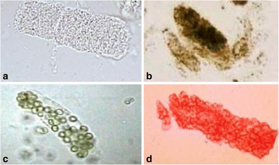
| Granular “muddy brown” casts | Acute tubular injury from ischemia, sepsis, pigment, or toxin | Remove insult, optimize perfusion/volume, dose medications to current function, trend complications |
| RBC casts / dysmorphic RBCs | Glomerular disease | Quantify protein, focused serologies, urgent nephrology. Consider biopsy pathway |
| WBC casts / pyuria | Interstitial nephritis or pyelonephritis. Pyuria alone is nonspecific | Exposure and infection review. Culture/imaging when indicated. Avoid reflex diagnosis from eosinophils |
| Crystals / pigment | Drug, urate/oxalate, tumor lysis, rhabdomyolysis, hemolysis | Identify morphology and exposure. CK/hemolysis/toxin/metabolic workup as the context directs |
B
Quantify what matters
do not stop at “protein positive”- Urine albumin-to-creatinine ratio for albumin-predominant disease. Protein-to-creatinine ratio when total protein matters.
- Confirm persistent hematuria/proteinuria after the acute episode when appropriate. Compare with prior values.
- Serologies are phenotype-driven: nephritic/nephrotic sediment, pulmonary-renal or systemic inflammatory features, complement pattern, monoclonal concern.
- FeNa is supportive, not a verdict. The classic <1% teaching came from 17 oliguric patients and excluded recent diuretic exposure. Loop diuretics can raise FeNa without proving tubular necrosis. CKD, sepsis, contrast, pigment, glomerular disease, and mixed physiology further weaken simple labels.
- Kidney/bladder ultrasound for obstruction risk, unexplained AKI, anuria/oliguria, or structural concern. Bladder scan/catheter when retention is plausible.
- Escalate vascular imaging for abrupt severe pain, embolic context, refractory hypertension, infarct/thrombosis concern.
Practice boundary: microscopy and protein-assay availability, focused serologies, ultrasound triggers, and biopsy/vascular escalation vary by setting.
A
Kidney-support activation
complication + trajectory| Threat | Act while consulting nephrology/critical care |
|---|---|
| Electrolyte / acid–base | Refractory hyperkalemia, severe acidemia, or rapidly worsening metabolic complication despite appropriate medical treatment |
| Volume / oxygenation | Pulmonary edema or hypoxemia refractory to diuretic/vasodilator strategy. Shock may require hemodynamic support first |
| Uremia / toxin | Pericarditis, encephalopathy, bleeding or other symptomatic uremia. Dialyzable toxic ingestion. Severe tumor lysis/pigment burden by context |
| Trajectory | Persistent anuria/oliguria with accumulating complications, or expected inability to maintain metabolic/volume control |
B
Imaging with contrast
diagnostic benefit × kidney risk- Do not defer time-critical, outcome-changing imaging solely because AKI/CKD exists. State the diagnostic consequence of delay and consider a noncontrast/alternate test only if it answers the same question.
- For IV iodinated CT contrast, use a diagnostic dose. Prophylactic isotonic saline is considered for AKI or eGFR <30 in patients not on maintenance dialysis, only when volume expansion is safe. Individualize selected high-risk eGFR 30–44 cases. These thresholds do not automatically apply to intra-arterial angiography.
- Review nephrotoxins and kidney-dependent medications. Distinguish contrast-associated AKI from injury proven to be contrast-induced. Do not start or alter dialysis solely to remove contrast.
C
AKI → recovery / AKD / CKD
close every transition| Before discharge | After discharge | Patient-facing |
|---|---|---|
| Peak/current creatinine and urine output. Cause + confidence. Unresolved protein/blood/imaging. Volume target. Complete hold/restart ledger | Named BMP/K date, albuminuria/urine reassessment when relevant, medication restart/titration owner, HF/nephrology/primary-care follow-up. Reassess at 3 months for resolution or new/worse CKD, with earlier labs based on discharge risk | Hydration/weight plan individualized to HF, medications to avoid/hold, return thresholds for low urine, dyspnea/edema, weakness/palpitations, vomiting, confusion |
Copy-ready recovery line: “AKI peak Cr ___, current ___, likely ___ (confidence ___). Unresolved ___. Medication restart gates ___. Labs ___. Owner ___. Return precautions reviewed.”