AF requires an ECG-quality tracing and clinician visual confirmation. An irregular pulse, photoplethysmography alert, or device notification alone is not the diagnosis. Once AF is confirmed, make four parallel decisions: stability, stroke prevention, symptom/rhythm strategy, and risk-factor treatment.
First 10 min
Confirm12-lead: irregular R–R, no repeating P waves. Compare old ECG.
MonitorBP · rhythm · SpO₂. IV access. Oxygen only if hypoxemic.
Find driverSepsis · bleeding · ACS · PE · thyroid · alcohol · K/Mg.
Clock + clotOnset certainty, OAC adherence, stroke risk, last dose.
1
Is the patient unstable because of AF?
causality mattersRate-driven instability
- Shock or severe hypotension
- Ongoing ischemic chest discomfort
- Acute pulmonary edema / HF
- Acutely altered mental status
Immediate synchronized biphasic cardioversion. Start ≥200 J. Sedate if feasible without delaying shock, escalate if unsuccessful, and re-enable/confirm SYNC before every shock.
AF may be a marker, not the cause
- Hemorrhage or severe hypovolemia
- Sepsis, hypoxemia, PE, ACS
- Thyrotoxicosis or withdrawal
- Postoperative adrenergic stress
Correct the driver while treating AF. Reflex rate suppression can worsen compensatory tachycardia in shock. Emergency cardioversion is never delayed for anticoagulation. Give OAC as soon as safely feasible afterward.
2
Wide + irregular: pause before AV-nodal blockade
separate WPW from polymorphic VTIf pre-excited AF is suspected/confirmed, do not give adenosine, any β-blocker, diltiazem, verapamil, digoxin, or IV amiodarone. Unstable → synchronized cardioversion. Stable → monitored IV procainamide or ibutilide with urgent EP input. If the diagnosis is uncertain, use an expert/ACLS wide-complex pathway and withhold AV-nodal blockers until pre-excitation is excluded. Polymorphic VT or a rhythm that cannot synchronize → unsynchronized defibrillation.
Mechanism secure?Rhythm is truly AF and pre-excitation has been excluded.
Perfusion independent?Tachycardia is not compensating for shock, bleeding, or low output.
EF gate passed?LVEF is >40% before diltiazem or verapamil.
Conduction safe?No high-grade AV block, profound brady risk, or unsafe IV nodal-blocker stacking.
3
If stable: define the immediate lane
rate, rhythm, or bothTreat the driverReplete K/Mg, address infection/volume/ischemia/thyroid/respiratory stress, stop stimulants, review QT and interacting drugs.
Control rateIf hemodynamically stable: IV β-blocker. Diltiazem/verapamil only if EF >40%. If ineffective/contraindicated: digoxin. Magnesium adjunct. See P.5.
Restore rhythmFavor with important symptoms, first/recent AF, HF/tachycardia-mediated CM, inadequate rate control, or patient preference. Check the cardioversion clock (P.6).
4
AF-CARE at every encounter
ESC 2024 frameworkC · ComorbidityWhich drivers and modifiable risks can be treated today?
A · Avoid strokeIs OAC indicated, safe, adherent, and correctly dosed?
R · Reduce symptomsRate, rhythm, or both—and what outcome matters to the patient?
E · EvaluateRecheck burden, symptoms, toxicity, stroke/bleeding risk, and substrate.
5
Fast rhythm discrimination
treat the tracing, not the monitor labelNarrow + irregular AF is likely, but compare atrial activity for flutter with variable block, multifocal atrial tachycardia, frequent ectopy, or artifact.
Wide + irregular Consider AF with aberrancy, pre-excited AF, or polymorphic VT. Withhold empiric AV-nodal blockade until the mechanism is secure.
Regular tachycardia Search for flutter with fixed block, SVT, or VT. A regular ventricular rhythm suggests another mechanism, but AF can be regular with complete AV block or ventricular pacing.
Device alert only Obtain and visually review an ECG-quality tracing. Do not commit to lifelong therapy from pulse irregularity or photoplethysmography alone.
6
Choose a safe destination
disposition is part of treatmentAdmit / monitor when risk persists
- Instability, ischemia, acute HF/hypoxemia, syncope, major bleeding, or a serious untreated driver.
- Wide-complex/pre-excited rhythm, persistent symptomatic RVR, significant brady–tachy pauses, or uncertain diagnosis.
- Need for monitored AAD initiation, urgent cardioversion/thrombus imaging, or an anticoagulation decision that cannot be made safely now.
Outpatient only when the loop is closed
- Hemodynamics and exertional symptoms are acceptable. Rate/rhythm plan is coherent and tolerated.
- AF is confirmed. Onset and likely driver are documented. Stroke risk, OAC dose, and first/next dose are settled.
- Patient understands red flags and missed-dose instructions, has medication access, and has time-defined follow-up.
Minimum handoff: ECG source + rate/QRS/QTc. Onset certainty. Stability and suspected driver. EF/structural substrate. Stroke score and OAC last/next dose. Drugs/shocks given and response. Unresolved tasks and named follow-up owner.
Name the phenotype, not just the rhythm. Document the confirming tracing, temporal pattern, symptom burden, onset certainty, substrate, and whether rhythm restoration remains a goal. Avoid vague terms such as “chronic,” “lone,” or routine “valvular/nonvalvular” AF.
1
Confirm and characterize
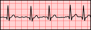
source → pattern → burdenAtrial fibrillationillustrative strip
Sinus rhythm3-second illustration
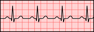
Recognition in three passes
- Rhythm: truly irregularly irregular?
- Atria: no repeating P morphology/PR?
- Safety: QRS width, pre-excitation, QTc, ischemia?
Three-second drawings teach morphology, not the duration needed to confirm a diagnosis. Review the complete diagnostic tracing. ECG vectors: Rocuronium Bromide, Wikimedia Commons, CC0 · AF source · sinus source.


History that changes care
- Exact/estimated onset. Spontaneous termination. Prior CV/ablation.
- Symptoms: dyspnea, fatigue, palpitations, angina, syncope. Functional effect.
- OAC drug, dose, adherence, last dose. Bleeding and stroke history.
Burden and substrate
- Clinical vs device-detected AF/AHRE. Longest episode and cumulative burden.
- LA size, LV/RV function, valves, HF, CAD, HCM/amyloid.
- Rhythm-control goal, candidacy, preference, access, pregnancy potential. Reconsider flutter, MAT, ectopy, or artifact when the pattern does not fit.
2
Temporal classification
do not infer severity from label| Pattern | Definition | Clinical note |
|---|---|---|
| First diagnosed | First documented AF episode | May already be persistent · does not mean “new onset.” |
| Paroxysmal | Terminates within 7 days (usually <48 h) | Stroke prevention is based on risk, not pattern. |
| Persistent | Continuous >7 days, including episodes terminated by cardioversion after ≥7 days | Duration, LA remodeling, and prior failures affect rhythm success. |
| Longstanding persistent | Continuous ≥12 months while rhythm control is still pursued | Shared expectations and earlier EP discussion are important. |
| Permanent | Patient + clinician decide no further rhythm-restoration attempts | A therapeutic decision—not a biological endpoint · revisit if circumstances change. |
U.S. disease-stage lens: Stage 1 at risk → Stage 2 pre-AF → Stage 3A paroxysmal / 3B persistent / 3C longstanding persistent / 3D successful ablation → Stage 4 permanent. The stage emphasizes prevention and progression. The temporal label still helps select therapy.
3
Initial evaluation
minimum useful dataset| Domain | Obtain | What it changes |
|---|---|---|
| ECG | Rate, QRS, QTc, ischemia, pre-excitation, prior infarct/conduction disease | Acute drug safety, rhythm diagnosis, pacing/ablation planning |
| Labs | CBC · BMP/CMP with K/Mg, renal/liver function · TSH · glucose/HbA1c | Driver, anemia/bleeding, DOAC/AAD dosing, thyroid disease |
| TTE | LV/RV function, LA size, valves, pulmonary pressure, structural disease | Rate/AAD choice, HF therapy, rhythm feasibility · do not delay indicated OAC |
| Targeted only | Troponin/ischemia, PE testing, infection studies, sleep testing, ambulatory monitor | Order when symptoms or probability support it—AF alone is not an indication |
4
Treat the atrial substrate
prevents progression and recurrenceWeight≥10%If BMI >27 and overweight/obese. Avoid weight cycling.
Exercise~210 min/wkModerate–vigorous, individualized. Avoid extreme endurance excess.
Alcohol0–3/wkMinimize/eliminate when rhythm control is desired.
Pressure + sleepControlOptimize BP. Assess OSA, tobacco, diabetes, HF and thyroid. OSA treatment has other benefits, but its effect on AF recurrence remains uncertain.
Caffeine: routine abstinence has no proven AF benefit unless a reproducible trigger. Smoking: offer behavioral + pharmacologic cessation support. ESC commonly targets treated BP 120–129/70–79 mm Hg when tolerated. Individualize for frailty/very advanced age.
Stroke prevention is independent of AF pattern and apparent rhythm success. Use annual thromboembolic risk, correct modifiable bleeding risks, and revisit at least yearly—and whenever age, kidney function, comorbidity, bleeding, or treatment changes.
1
Calculate the score correctly
U.S. vs ESCCHF/LV dysfunction +1
HHypertension +1
A₂Age ≥75 +2
DDiabetes +1
S₂Stroke/TIA/embolism +2
VVascular disease +1
AAge 65–74 +1
ScFemale sex +1 (U.S. modifier)
| Framework | Recommend OAC | Consider / reasonable | No OAC by score |
|---|---|---|---|
| ACC/AHA 2023 | Annual risk ≥2%: approximately CHA₂DS₂-VASc ≥2 men / ≥3 women | 1%–<2%: approximately 1 men / 2 women | No non-sex risk factors · aspirin has no benefit |
| ESC 2024 | CHA₂DS₂-VA ≥2 | CHA₂DS₂-VA 1 | CHA₂DS₂-VA 0 |
2
Choose the stroke-prevention lane
never substitute aspirinMechanical valve or clinically important mitral stenosisMechanical valve → VKA. With AF, ACC/AHA recommends warfarin over DOACs for rheumatic MS or MS of moderate-or-greater severity. ESC 2025 advises VKA (INR 2–3) and avoiding DOACs in rheumatic MS when MVA ≤2.0 cm². If etiology/severity falls between these definitions, clarify with valve/anticoagulation expertise before choosing a DOAC.
Eligible AF without a VKA-only indicationDOAC preferred over warfarin. Use the labeled dose. Verify CrCl, age/weight/SCr criteria, interactions, adherence, cost, and pregnancy status (P.4).
Already on antiplatelet therapyAspirin ± clopidogrel is not an OAC substitute. Beyond 1 year after stable coronary revascularization, OAC alone is usually preferred if there is no other antiplatelet indication.
Bleeding riskUse HAS-BLED to expose modifiable hazards and follow-up intensity—never as a stand-alone reason to deny OAC. Repair the risks, then reassess.
HHypertension
AAbnormal renal/liver +1 each
SStroke history
BBleeding history/predisposition
LLabile INR
EAge >65
DDrugs/alcohol +1 each
3
When the score is borderline
shared decision modifiersAF burden Persistent/permanent pattern, longer or more frequent episodes
Comorbidity Obesity, HCM, poorly controlled hypertension
Kidney eGFR <45 or proteinuria >150 mg/24 h
Atrium LA volume ≥73 mL or diameter ≥4.7 cm
Score-independent indication: clinical AF with hypertrophic cardiomyopathy or cardiac amyloidosis warrants OAC regardless of the conventional score when the patient is eligible. This removes the score as a gate—not the contraindication/bleeding review: correct modifiable bleeding risks, select the appropriate agent/dose, and use shared decision-making. Triple-positive antiphospholipid syndrome favors warfarin, not a DOAC.
4
Device-detected AF / atrial high-rate episodes
confirm the stored tracing| Confirmed episode | Stroke score | ACC/AHA action |
|---|---|---|
| ≥24 h | CHA₂DS₂-VASc ≥2 | OAC is reasonable within shared decision-making. |
| 5 min–24 h | CHA₂DS₂-VASc ≥3 | OAC may be reasonable · weigh episode duration and individual risk. |
| <5 min | Any | No OAC without another indication. |
ESC 2024: less prescriptive. A DOAC may be considered for selected high-stroke-risk patients with device-detected subclinical AF and no major bleeding risks. The actionable duration threshold remains uncertain. Continue surveillance for clinical AF.
ARTESIA: 4,012 patients with device-detected AF lasting 6 min–24 h. Apixaban vs aspirin reduced stroke/systemic embolism (0.78 vs 1.24 per 100 patient-years) but increased on-treatment major bleeding (1.71 vs 0.94). The efficacy and safety populations and follow-up windows differ, so do not subtract these rates into a single “net benefit.” NOAH-AFNET 6: edoxaban vs placebo (aspirin when indicated) did not significantly reduce CV death/stroke/embolism and increased death/major bleeding. It stopped early. Neither trial justifies treating an unconfirmed wearable alert.
5
Left atrial appendage occlusion (LAAO)
alternative for selected patientsPercutaneous
ACC/AHA: reasonable when CHA₂DS₂-VASc ≥2 and a nonreversible contraindication to long-term OAC. May be considered for high bleeding risk/patient preference after discussing procedural risk and the stronger evidence base for OAC. ESC is more conservative (“may be considered”).
Surgical
During cardiac surgery, LAA exclusion is an adjunct to continued anticoagulation, not a replacement. Target no residual flow and stump <1 cm by intraoperative TEE. Do not stop OAC solely because the LAA was surgically closed.
Stroke/TIA despite prescribed OAC is not automatic “DOAC failure.” Confirm the dose, adherence/last doses, CrCl, interacting drugs or food requirements, and competing noncardioembolic mechanisms. Do not routinely add an antiplatelet or switch DOAC↔DOAC/VKA solely to prevent recurrence without a clear indication.
6
Make anticoagulation durable
remove hazards. Do not create treatment gapsAct on HAS-BLED A score ≥3 means high bleeding risk: correct BP, interacting antiplatelets/NSAIDs, alcohol, anemia, renal/liver disease, and labile INR. Increase review frequency—do not deny OAC by score alone.
Separate temporary from permanent Severe nonreversible GI/GU/pulmonary or spontaneous intracranial/intraspinal bleeding may truly preclude OAC. Treatable-source, isolated-trauma, or procedure-related bleeding—and fall risk alone—usually do not.
Verify the exact dose Use Cockcroft–Gault CrCl and the product label. Recheck age, weight, SCr, liver disease, P-gp/CYP interactions, food requirements, adherence, and affordability.
Build surveillance CBC, renal/liver function, bleeding, adherence, and interacting drugs at least yearly—earlier with frailty, renal impairment, intercurrent illness, anemia, or a medication change.
Patient teach-back: purpose and exact schedule. Product-specific missed-dose instructions. Bleeding and neurologic red flags. NSAID/supplement cautions. What to do before procedures. And why a normal pulse, cardioversion, or ablation does not authorize self-discontinuation.
1
U.S. labeled dosing for stroke prevention in AF
do not underdose off-label| Drug | Dose / renal criteria | High-yield interactions and traps |
|---|---|---|
| Apixaban | 5 mg BID. Use 2.5 mg BID only if ≥2: age ≥80, weight ≤60 kg, SCr ≥1.5 mg/dL. | Combined P-gp + strong CYP3A4 inhibitor: reduce 5 mg BID by 50% · if already 2.5 mg BID, avoid. Avoid strong combined inducers. |
| Rivaroxaban | CrCl >50: 20 mg daily · CrCl 15–50: 15 mg daily · CrCl <15: avoid. Take with evening meal. | Avoid combined P-gp/strong CYP3A inhibitors or inducers. CrCl 15–<80 plus combined P-gp/moderate CYP3A inhibitor: generally avoid unless benefit justifies risk. |
| Dabigatran | CrCl >30: 150 mg BID · CrCl 15–30: 75 mg BID (U.S. dose based on pharmacokinetic modeling) · <15/dialysis: avoid/no recommendation. | CrCl 30–50 + dronedarone or systemic ketoconazole: 75 mg BID. CrCl 15–30: avoid P-gp inhibitors. Avoid P-gp inducers. Swallow capsule intact. |
| Edoxaban | CrCl >50 to ≤95: 60 mg daily · 15–50: 30 mg daily. Do not use if CrCl >95 · no labeled dose <15. | No P-gp-inhibitor dose reduction for U.S. NVAF dosing · avoid rifampin. Weight ≤60 kg reduction is a VTE—not U.S. AF—criterion. |
| Warfarin | For AF with a VKA-only mitral-stenosis indication, target INR 2.0–3.0 unless a separate indication changes the target. | Required for mechanical valves, but the INR target depends on valve type/location and patient risk. Review diet/drug interactions and time in therapeutic range. |
Severe kidney disease: CrCl <15/dialysis requires individualized specialist review. The U.S. guideline says warfarin or an evidence-based apixaban dose might be reasonable. Randomized outcome evidence is limited. Dabigatran and edoxaban have no labeled dose / are not recommended. Do not infer class-wide safety.
2
Major bleeding: stabilize, reverse, and plan the restart
life-threatening / critical-site bleedingStabilizeStop anticoagulant. ABCs, large-bore access, CBC/CMP, type/cross, drug + last dose, renal function, appropriate coagulation tests. Control the source and involve the procedural team early.
Reverse when indicatedUse the agent-specific pathway below. Follow local massive-bleeding and transfusion protocols. Reversal restores thrombotic risk.
Restart deliberatelyReassess the indication and source. Resume OAC when hemostasis and clinical safety permit. Document the date/decision owner rather than allowing an accidental indefinite hold.
| Anticoagulant | Reversal | Critical notes |
|---|---|---|
| Dabigatran | Idarucizumab 5 g IV as two consecutive 2.5-g doses | If unavailable: activated PCC 50 U/kg once is less established · hemodialysis can remove dabigatran. |
| Apixaban / rivaroxaban / edoxaban | U.S. 2026: 4-factor PCC using current reversal guidance · guideline table: 2,000 U once or 25–50 U/kg | Off-label DOAC reversal. Andexanet alfa is no longer marketed in the U.S. U.S. sales ended Dec 22, 2025 after FDA concluded that serious risks outweighed benefits. |
| Warfarin | 4F-PCC + vitamin K 10 mg IV. INR 2–<4: 25 U/kg (max 2,500) · 4–6: 35 U/kg (max 3,500) · >6: 50 U/kg (max 5,000) | Give vitamin K with PCC for durable reversal · recheck INR and clinical hemostasis. |
DOAC assay trap: normal PT/INR or aPTT does not reliably exclude active drug. Dabigatran → thrombin time/dilute TT or ecarin assay. Factor Xa inhibitors → drug-calibrated chromogenic anti-Xa. Interpret with last dose + CrCl. Do not delay source control or lifesaving reversal for a level.
3
Periprocedure interruption when a hold is required
full drug-free days| Agent / renal floor | Low bleed risk | High bleed risk |
|---|---|---|
| Apixaban CrCl >25 · edoxaban >15 · rivaroxaban >30 | 1 day | 2 days |
| Dabigatran CrCl >50 | 1 day | 2 days |
| Dabigatran CrCl 30–50 | 2 days | 4 days |
Timing: these are full days with no dose before surgery, plus no dose on procedure day. When hemostasis is secure, resume the next day after low-risk procedures or evening of day 2–3 after high-risk procedures. No heparin bridge for most AF. Individualize mechanical valves, recent stroke/TIA, neuraxial surgery, and CrCl below the table floors.
4
Routine safety loop
at every handoffCorrect drug AF vs VTE/CAD dose. No duplicate anticoagulant or unintended antiplatelet.
Correct patient CrCl, age, weight, SCr, liver disease, valve disease, APS, pregnancy.
Correct exposure Adherence, last dose, affordability, P-gp/CYP3A interactions, NSAIDs/alcohol.
Correct follow-up CBC/renal/liver interval, bleeding education, procedure plan, refill ownership.
Rate control buys time. It does not replace stroke prevention or a rhythm strategy. Treat the precipitant, verify EF/hemodynamics and pre-excitation, and choose a target from symptoms, ventricular function, exertional response, and pacing needs.
1
Stable AF with rapid ventricular response
choose by physiologyExclude the stop signsInstability caused by AF → synchronized cardioversion. Wide-irregular/pre-excited AF → no AV-nodal blocker (P.1). Correct hypoxemia, volume/bleeding, sepsis, ischemia, thyroid and K/Mg.
Hemodynamically stableIV β-blocker. Use diltiazem/verapamil only if EF >40%. Avoid acute β-blockade in decompensated HF/low-output states. Choose based on BP, bronchospasm, concurrent drugs, and catecholamine state.
Usual agents ineffective or contraindicatedConsider digoxin. IV magnesium is a reasonable adjunct. Digoxin is slower and less effective in high-catecholamine states.
Critical illness / decompensated HFIf β-blocker and non-DHP CCB are unsuitable, IV amiodarone may be considered. It can cardiovert—apply cardioversion anticoagulation rules. Avoid negative inotropy in low-output shock.
Usual stable targetWithout HF, resting <100–110 bpm is generally reasonable.
AF-CM / refractory HFConsider <80 rest and <110 with moderate exercise.
Verify the targetCheck symptoms, BP and rate with ambulation or ambulatory monitoring—not resting telemetry alone.
2
Acute and maintenance doses
adult reference—titrate to response| Agent | Acute IV | Oral | Main cautions |
|---|---|---|---|
| Metoprolol tartrate | 2.5–5 mg over 2 min · up to 3 doses | 25–200 mg BID | Hypotension/shock, brady/AV block, decomp HF, bronchospasm |
| Esmolol | 500 mcg/kg over 1 min, then 50–300 mcg/kg/min | — | Short acting · same β-blocker cautions |
| Diltiazem | 0.25 mg/kg actual wt over 2 min · may repeat 0.35 mg/kg · then 5–15 mg/h | ER 120–360 mg/d | Harm in moderate/severe LV systolic dysfunction · shock, WPW, AV block · avoid near IV β-blocker |
| Verapamil | 5–10 mg over ≥2 min · may repeat twice · then 5 mg/h (max 20 mg/h) | ER 180–480 mg/d | Potent negative inotrope · same non-DHP CCB cautions |
| Digoxin | 0.25–0.5 mg, then 0.25 mg q6h · max 1.5 mg/24 h | 0.0625–0.25 mg/d | Individualize load by lean weight, kidney function and prior use. The listed maximum is not a target. Brady/AV block and interactions. Aim level <1.2 ng/mL |
| Amiodarone | 150–300 mg over 1 h, then 10–50 mg/h over 24 h | Not preferred solely for rate | Hypotension, brady/QT, interactions/toxicity · may convert rhythm |
EF safety: do not give IV diltiazem or verapamil with known moderate/severe LV systolic dysfunction. Long-term non-DHP CCB therapy is harmful with LVEF <40%. In decompensated HF/low output, stabilize perfusion before aggressively suppressing the ventricular response.
3
When rate control is failing
reassess before stacking drugsRecheck the diagnosis and driver
- Atrial flutter may resist rate control. Rhythm strategy often works better.
- Persistent high catecholamines suggest pain, sepsis, hypovolemia, PE, withdrawal, thyrotoxicosis.
- Check administration, absorption, renal clearance, drug interactions, K/Mg.
Escalate the strategy
- Cardioversion when symptoms/hemodynamics demand and thrombus precautions are satisfied.
- Early EP input for recurrent symptomatic AF/flutter, HF, suspected AF-induced cardiomyopathy, or drug intolerance.
- Refractory long-term rates: AV-node ablation + pacing can improve symptoms/QOL. Creates pacemaker dependence.
After recovery from AF-induced cardiomyopathy: continue long-term rhythm surveillance. Recurrence may be silent and can re-induce LV dysfunction.
4
Discharge after acute rate control
close the loopVitals + function Rest and ambulation rate, BP, symptoms, congestion, oxygen need.
Medication One coherent regimen. Avoid unintended β-blocker + non-DHP CCB stacking.
Stroke plan OAC indication/dose/adherence, first/next dose, bleeding education.
Rhythm plan Onset, monitoring, EP/cardiology follow-up, cardioversion/ablation decision owner.
5
Long-term rate-control follow-through
measure function, not only a resting pulseChoose by EF β-blocker is broadly useful. Diltiazem/verapamil only when LVEF is >40%. Digoxin is reasonable with HF symptoms or when other agents are unsuitable. Do not use dronedarone in permanent AF solely for rate control.
Test the target Record the intended HR range and reassess symptoms, BP, rest and ambulation/exercise rate. Use ambulatory monitoring when the clinic pulse does not explain symptoms.
Monitor digoxin Renal function, K/Mg, interacting drugs, nausea/confusion/visual change, bradycardia, and AV block. Sample just before a dose or ≥6 h after the last dose. Interpret with symptoms and kidney function. Target <1.2 ng/mL, often 0.5–0.9 in HFrEF.
Look for tachy–brady Syncope, fatigue, pauses after AF termination, or rate-drug intolerance → ECG/monitoring and pacing evaluation rather than progressively stacking nodal blockers.
Rate-control failure is clinical: persistent symptoms, HF, exercise intolerance, recurrent RVR, or drug-limited bradycardia should reopen the rhythm-control/ablation discussion even when the resting number appears acceptable.
Electrical and pharmacologic cardioversion carry the same thromboembolic precautions. The safe path depends on hemodynamics, onset certainty, prior anticoagulation, thrombus imaging, and the post-cardioversion plan—not simply whether AF is labeled “acute.”
1
Cardioversion anticoagulation pathway
U.S. default. ESC difference shownUnstable because of AFShock now: synchronized biphasic ≥200 J, escalate. Anticoagulate as soon as safely feasible. Do not delay life-saving cardioversion.
AF ≥48 h or onset unknown — ACC/AHABefore elective electrical or drug cardioversion: ≥3 weeks uninterrupted therapeutic OAC OR imaging (TEE or delayed-contrast cardiac CT with an appropriate protocol) excluding intracardiac thrombus.
AF <48 h, not anticoagulated — ACC/AHAIf CHA₂DS₂-VASc ≥2, precardioversion imaging may be considered. If definite <12 h and score 0–1, benefit of imaging/periprocedural OAC is uncertain.
After cardioversion — defaultEstablish therapeutic OAC before elective cardioversion and continue uninterrupted for ≥4 weeks. Continue long term by stroke risk, not maintained sinus rhythm. ACC/AHA notes that in definite AF <12 h with CHA₂DS₂-VASc 0–1, benefit of pericardioversion anticoagulation remains uncertain—individualize.
ESC 2024 is more conservative: if AF is definitely >24 hours, do not cardiovert without ≥3 weeks therapeutic OAC or TOE excluding thrombus. Post-CV OAC may be omitted only with no thromboembolic risk factors and sinus rhythm restored within 24 hours of definite onset. If onset is uncertain, use the longer-duration pathway.
LAA thrombus: cancel elective cardioversion. ACC/AHA: therapeutic OAC for 3–6 weeks, then repeat imaging. ESC: at least 4 weeks and repeat TOE. Investigate adherence, dose, absorption, and interactions if thrombus develops despite prescribed OAC.
2
Electrical cardioversion technique
maximize first-shock success| Step | Action |
|---|---|
| Prepare | Consent if feasible · airway/suction/monitor/resuscitation ready · correct K/Mg · remove patches · sedation/analgesia by an appropriate team. |
| Pads + sync | Anterior–posterior vector is reasonable, especially persistent AF · select SYNC and verify markers on every R wave. |
| Energy | Initial synchronized biphasic ≥200 J · escalate energy/vector/pressure as appropriate. Reconfirm SYNC before every shock. If synchronization delays shock delivery and the condition is critical, follow ALS and proceed immediately to an unsynchronized shock. High-energy (maximum manufacturer setting) is the separate recommendation for polymorphic VT. |
| After | 12-lead, rhythm/rate/QTc, neurologic and airway check, medication plan, ≥4-week OAC plan, recurrence strategy. |
3
Pharmacologic cardioversion
monitored setting| Agent | Regimen | Use / restrictions |
|---|---|---|
| Ibutilide | ≥60 kg: 1 mg IV/10 min · <60 kg: 0.01 mg/kg. May repeat once after 10 min. | Avoid LVEF ≤40% · correct K/Mg · baseline QT · continuous ECG during and ≥4 h after or until QTc baseline · torsades risk. |
| Amiodarone IV | 5–7 mg/kg or 300 mg, then total 1,200–3,000 mg infusion/24 h | Conversion often 8–12 h · hypotension, brady/QT, phlebitis, interactions. Useful with structural disease/HF when appropriate. |
| Procainamide IV | 1 g over 30 min, then 2 mg/min for 1 h | Monitor BP/QRS/QTc · hypotension/TdP, renal elimination, can worsen HFrEF. Avoid after QT-prolonging agents. |
| Flecainide PITP | <70 kg: 200 mg once · ≥70 kg: 300 mg once | No prior MI/significant structural disease/scar. AV-nodal blocker ≥30 min first · first dose tested in monitored setting. |
| Propafenone PITP | <70 kg: 450 mg once · ≥70 kg: 600 mg once | Same structural and monitored-first-dose restrictions · β-blocking/bronchospasm concern. |
Pill-in-the-pocket is a selected-patient plan, not an unsupervised first dose. Dofetilide, oral amiodarone, and oral sotalol take days and are not practical acute conversion drugs. Never use a pharmacologic conversion strategy to bypass thrombus precautions.
4
If the shock fails or AF returns immediately
optimize before repeating the same attemptReconfirm the system Verify AF, SYNC markers on every R wave, pad contact/position, device mode, delivered energy, airway/sedation readiness, and hemodynamics.
Improve delivery Escalate energy using current device guidance. Optimize contact and vector, especially with obesity or high transthoracic impedance. Reconfirm SYNC before each attempt.
Correct the substrate Replete K/Mg. Treat hypoxemia, ischemia, decompensated HF, thyroid disease, infection, volume disturbance, withdrawal, and stimulant exposure.
Change the strategy Selected patients may benefit from monitored AAD pretreatment and repeat CV. Early recurrence should prompt a durable AAD/ablation/risk-factor plan—not serial unplanned shocks.
5
Close the loop after conversion
success is more than sinus rhythmSafety check 12-lead ECG with rate/QRS/QTc, BP, oxygenation, neurologic and airway recovery, and any drug-related bradycardia or hypotension.
Anticoagulation clock Document pre-CV doses/imaging, first/next dose, uninterrupted ≥4-week plan when indicated, and the long-term decision by stroke risk.
Medication plan State which rate/AAD drugs continue, stop, or change. Include renal/QT/laboratory surveillance and an exact pill-in-the-pocket plan if used.
Recurrence plan Define symptom and rate thresholds, when to seek urgent care, rhythm-monitor strategy, and time-defined cardiology/EP follow-up.
Two cardioversion traps: uncertain or missed OAC doses are not “uninterrupted” therapy—delay or use thrombus imaging as appropriate. Prior surgical/percutaneous LAA closure does not automatically remove cardioversion precautions. Review closure completeness, residual leak/device thrombus, current OAC, and specialist imaging strategy.
Discuss rhythm control early—not only after years of failed rate control. Symptoms/QOL matter. EAST-AFNET 4 also supports early rhythm control in selected recent AF with cardiovascular risk. It tested a strategy, largely using drugs, and does not mean every patient needs immediate ablation. Continue anticoagulation according to stroke risk.
1
Who should prompt an early rhythm discussion?
shared decision + substrateSymptoms/QOL Palpitations, dyspnea, fatigue, exercise intolerance, angina, patient preference. Record baseline mEHRA 1–4.
Heart failure HFrEF, worsening HF, or suspected AF-mediated/tachycardia-induced cardiomyopathy.
Recent/progressive AF First diagnosed or within ~1 year. Increasing burden or progression despite risk-factor care.
Rate strategy failing Inadequate control, brady–tachy limitation, medication intolerance, recurrent admissions.
2
Choose an antiarrhythmic by cardiac substrate
safety before efficacy| Substrate | Reasonable choices | Avoid / key note |
|---|---|---|
| HFrEF · LVEF ≤40% | Dofetilide or amiodarone | Avoid flecainide/propafenone, dronedarone · sotalol generally avoided. |
| No prior MI, structural disease, scar/fibrosis | Flecainide or propafenone + AV-nodal blocker · dronedarone · dofetilide/sotalol selected | Class IC drugs can organize flutter with 1:1 conduction without nodal blockade. |
| Prior MI / CAD scar / significant structural disease | Dronedarone only without severe LV dysfunction or recent HF decompensation · dofetilide or sotalol selected · amiodarone | Do not use flecainide or propafenone. |
| Normal LV function · other choices fail/unsuitable | Low-dose amiodarone 100–200 mg/day | Reserve because of cumulative organ toxicity and interactions. |
3
Maintenance dosing and monitoring
specialist-led initiation where indicated| Drug | Dose | Safety essentials |
|---|---|---|
| Dofetilide | CrCl >60: 500 mcg BID · 40–60: 250 mcg BID · 20–<40: 125 mcg BID · <20 contraindicated | Admit ≥3 days. Do not start if QTc >440 ms (>500 with conduction delay). After dose 1: reduce if QTc rises >15% or exceeds 500/550 · after dose ≥2, >500/550 → stop. Never confuse mcg with mg · major prohibited interactions include verapamil, HCTZ, trimethoprim, ketoconazole, cimetidine. |
| Dronedarone | 400 mg BID with meals | Avoid NYHA III–IV HF, HF decompensation within 4 weeks, or severe LV dysfunction. Also avoid permanent AF, brady <50, QTc >500, severe liver disease, or strong CYP3A inhibitors. |
| Flecainide | 50–300 mg/day divided q8–12 h | No MI/significant structural disease/HFrEF · AV-nodal blocker · follow QRS/AV conduction and renal/hepatic function. QRS widening >25% from baseline warrants prompt dose reduction/cessation review. |
| Propafenone | IR 150–300 mg q8h · ER 225–425 mg q12h | Same structural restriction · AV-nodal blocker · CYP2D6 interactions · raises digoxin/warfarin · bronchospasm. |
| Sotalol AF | Usually 80 mg q12h initially · extend the interval by CrCl using the exact product label and local formulary. | Admit ≥3 days. Baseline QT >450 ms → do not start · QT ≥500 → reduce dose, extend interval, or stop. Current U.S. products differ in renal cutoffs, so do not treat CrCl <40 as a universal class rule. Follow HR, K/Mg, CrCl, and bronchospasm/HF risk. |
| Amiodarone | Load total 6–10 g · usually 100–200 mg/day | Baseline ECG, TSH, LFT, chest X-ray · TSH/LFT 3–6 mo then q6 mo, annual ECG. Lung/thyroid/liver/eye/skin/neuro toxicity · increases warfarin/digoxin. |
QT-drug surveillance: after monitored initiation/reinitiation, repeat ECG, K/Mg, and CrCl every 3–6 months—and sooner after AKI, diarrhea/diuretic intensification, bradycardia, a dose change, or a new QT-prolonging drug. Name the monitoring owner and stop/escalation rule.
4
Vaughan Williams: the fast AAD map
dominant action ≠ whole drugWhat it does: groups antiarrhythmic actions by their predominant electrophysiologic effect. It is a memory map—not a license to choose by class alone.
What selects the drug: AF role plus MI/scar/structural disease, EF/HF, QRS/QTc/HR, CrCl/K/Mg, interactions, organ toxicity, and pregnancy.
IAINa + K · intermediate
Quinidine · procainamide · disopyramide → conduction↓. APD/ERP/QT↑.
AF role: procainamide IV in selected acute/pre-excited pathways. Not routine chronic therapy.
Trap: QRS/QT/TdP, hypotension/negative inotropy.
IBINa · rapid
Lidocaine · mexiletine → preferential diseased/ventricular tissue effect. APD little changed or shorter.
AF role: no routine AF conversion or maintenance role.
Trap: these are principally ventricular agents.
ICINa · slow/use-dependent
Flecainide · propafenone → marked conduction/QRS slowing. Little APD change.
AF role: conversion/maintenance only in a selected clean substrate.
Trap: no MI/scar/SHD/HFrEF. Add AV-nodal blocker. Follow QRS.
IIβ-receptor block
Metoprolol · esmolol · propranolol → automaticity and sinus/AV-node conduction↓.
AF role: rate/adrenergic lane—not reliable conversion.
Trap: brady/AV block/hypotension. Sotalol is II + III.
IIIK currents · APD/ERP↑
Dofetilide · sotalol · ibutilide. Amiodarone/dronedarone are multichannel.
AF role: drug-specific conversion/maintenance. Ibutilide is acute.
Trap: QT/TdP/renal gates. Amiodarone organ toxicity.
IVICaL block
Diltiazem · verapamil → sinus/AV-node conduction and refractoriness.
AF role: ventricular rate control—not AF conversion/maintenance.
Trap: avoid LVEF <40% and pre-excited AF.
Outside the six tiles: digoxin is a vagal AV-nodal rate drug—not an AF converter—and is weaker in adrenergic stress. Never use it in pre-excited AF. Amiodarone/dronedarone span multiple classes, and adenosine is not AF therapy.
5
Sicilian Gambit: mechanism → vulnerable parameter → target
reasoning framework, not a prescription1 · PhenotypeDefine the clinical rhythm, tissue, trigger, and treatment aim.
2 · Mechanism(s)Automaticity · EAD/DAD triggered activity · reentry.
3 · Vulnerable parameterExcitability · conduction · ERP/APD · Ca handling · nodal dependence.
4 · Target / profileINa · IKr/K · ICaL · β/vagal receptors · multichannel · ablation.
5 · Net clinical effectVerify desired response, adverse effects, and new proarrhythmia.
AF reentry example: vulnerable conduction velocity + refractory period (wavelength ≈ CV × ERP) may be modified through Na/K-current effects or removal of a trigger/circuit. The same change can also organize flutter, facilitate ventricular reentry, or provoke EAD/TdP.
Bedside limit: AF mechanisms are often mixed or uncertain. Mechanism explains the target. Substrate and drug-specific safety determine treatment. Cardioversion anticoagulation rules still apply.
The Sicilian Gambit asks what the drug actually touches—not only which class name it carries. Selected rows from the JCS/JHRS presentation of the Sicilian Gambit illustrate multiaction pharmacology. Dot intensity is not a measured potency, dose-response estimate, or comparison of clinical efficacy. Use it to explain mechanisms, not to select current AF treatment.
1
Drug → channel · receptor · pump → patient signal
qualitative relative action| Drug | Channels | Receptors | Pump | Clinical effects | ECG effects | VW | ||||||||||||
|---|---|---|---|---|---|---|---|---|---|---|---|---|---|---|---|---|---|---|
| Na | Ca | K | If | α | β | M2 | A1 | LV fxn | Sinus rate | Extra- cardiac |
PR | QRS | JT | |||||
| fast | med | slow | Na/K ATPase |
|||||||||||||||
| Lidocaine | ● | → | → | ● | ↓ | IB | ||||||||||||
| Mexiletine | ● | → | → | ● | ↓ | IB | ||||||||||||
| Procainamide | A | ● | ↓ | → | ● | ↑ | ↑ | ↑ | IA | |||||||||
| Disopyramide | A | ● | ● | ↓ | → | ● | ↕ | ↑ | ↑ | IA | ||||||||
| Quinidine† | A | ● | ● | ● | → | ↑ | ● | ↕ | ↑ | ↑ | IA | |||||||
| Propafenone | A | ● | ↓ | ↓ | ● | ↑ | ↑ | IC | ||||||||||
| Flecainide | A | ● | ↓ | → | ● | ↑ | ↑ | IC | ||||||||||
| Bepridil† | ● | ● | ● | ? | ↓ | ● | ↑ | IV* | ||||||||||
| Verapamil | ● | ● | ● | ↓ | ↓ | ● | ↑ | IV | ||||||||||
| Diltiazem | ● | ↓ | ↓ | ● | ↑ | IV | ||||||||||||
| Sotalol | ● | ● | ↓ | ↓ | ● | ↑ | ↑ | III/II | ||||||||||
| Amiodarone | ● | ● | ● | ● | ● | → | ↓ | ● | ↑ | ↑ | III* | |||||||
| Nadolol | ● | ↓ | ↓ | ● | ↑ | II | ||||||||||||
| Propranolol | ● | ● | ↓ | ↓ | ● | ↑ | II | |||||||||||
| Atropine | ● | → | ↑ | ● | ↓ | V/other | ||||||||||||
| Adenosine | ■ | ? | ↓ | ● | ↑ | V/other | ||||||||||||
| Digoxin | ■ | ● | ↑ | ↓ | ● | ↑ | ↓ | V/other | ||||||||||
Relative action: ● low · ● moderate · ● high · blank = not emphasized in the historical map, not proof of no modern action.
State / mode: A activated-state Na blocker · I inactivated-state · ■ agonist · ◆ mixed.
Signals: ↑ increase/prolong · ↓ decrease/shorten · → little change · JT is the QRS-end→T-end interval.
Read across, then apply a safety gate. A drug can hit several targets, and a target can produce both desired and proarrhythmic effects. VW = predominant Vaughan Williams class. *amiodarone is multichannel. Page 7—not this matrix—contains current AF substrate and monitoring choices.
Scope: selected entries checked against JCS/JHRS Table 6. †Quinidine and bepridil illustrate historical actions, not routine AF choices. Adenosine names the A1 agonist. The digoxin M2 effect is indirect through vagal activity. Verify current labeling.
2
Translate the matrix at the bedside
ECG is the visible pharmacologyQRS · INaQRS widening signals conduction slowing. Compare with the pretreatment ECG and reassess at steady state and with exertion when appropriate. Class IC appeal never overrides prior MI, scar, significant structural disease, or HFrEF restrictions.
JT/QT · K currentsRepolarization prolongation can increase ERP but also EAD/TdP risk. Recheck QT, K/Mg, CrCl, bradycardia, dose and interacting drugs.
PR · nodal targetsβ, M2/vagal, A1, and ICaL effects can slow AV conduction. That does not make nodal blockade safe in pre-excited AF.
Beyond the ECGLV depression, hypotension, organ accumulation, neurologic/pulmonary/thyroid toxicity and drug interactions may dominate even when intervals look acceptable.
Do not turn dots into a prescription: class IC agents remain restricted by MI/scar/structural disease/HFrEF. K-current/QT drugs require QT, K/Mg, renal, bradycardia and interaction surveillance. AV-nodal blockers are dangerous in pre-excited AF. Adenosine is not AF therapy.
Start with tissue and mechanism before naming a current. Working myocardium, nodal tissue, and Purkinje tissue use different phase-0 and phase-4 currents. APD and ERP overlap but are not interchangeable. Sodium-channel recovery can extend refractoriness beyond repolarization.
1
Cardiac action potential: two tissues, different levers
original animated vector
Sequence only · not to scale
Working myocardium0 INa · 1 Ito · 2 ICaL↔K · 3 IKr/IKs→IK1 · 4 IK1. Phase 4 is a stable resting potential—not spontaneous diastolic depolarization.
Nodal pacemaker4 If/ICaT/Ca clock · 0 ICaL · 3 IK. β1 steepens phase 4. M2/IKACh slows it. There is no fast INa phase 0 or prominent phase 1–2 plateau.
AfterdepolarizationsEAD occurs before repolarization finishes (phase 2/3): long APD/QT and dispersion matter. DAD follows repolarization: Ca overload, SR release and transient inward INCX matter.
2
Arrhythmia mechanism → vulnerable parameter → likely target
hypothesis map| Mechanism | Vulnerable parameter / desired effect | Likely current or target |
|---|---|---|
| Automaticity | ||
| Enhanced normal automaticity | Decrease phase-4 diastolic-depolarization slope. | HCN/If · ICaT · β1–cAMP · M2/A1 modulation where relevant. |
| Abnormal automaticity | Hyperpolarize a partially depolarized maximum diastolic potential and/or slow phase 4 or raise threshold. | Increase outward K · block ICaL or INa by tissue/voltage. |
| Triggered activity | ||
| Early afterdepolarization · EAD | Shorten excessive APD/repolarization dispersion or suppress the reactivated inward current. | Restore repolarizing K · reduce ICaL · late INa · INCX. |
| Delayed afterdepolarization · DAD | Reduce intracellular Ca overload/SR Ca release or stop the DAD reaching threshold. | β–cAMP · ICaL · RyR2 · INCX · INa block may suppress propagation, not unload Ca. |
| Reentry | ||
| Na-dependent · long excitable gap | Depress excitability/conduction enough to turn unidirectional into bidirectional block. | Use-dependent peak INa block in fast-response tissue. |
| Na-dependent · short excitable gap | Prolong ERP/wavelength enough to close the gap · post-repolarization refractoriness may help. | Repolarizing K-current block, commonly IKr · selected Na effects. |
| Ca-dependent | Depress slow-response conduction/excitability or increase refractoriness—often an AV-nodal limb. | ICaL · β1 · A1 modulation. |
| Other proposed mechanisms | ||
| Reflection | Prevent delayed electrotonic excitation across an inexcitable gap from returning retrogradely. | INa or ICaL, depending on responding tissue. |
| Parasystole | Slow firing or remove the protected automatic focus/exit. | If only if pacemaker-current dependent · focus-specific therapy or ablation may be more relevant. |
Na paradox: incomplete conduction slowing can shorten wavelength or enlarge an excitable gap and worsen reentry. Retain QRS and substrate gates.
K/QT paradox: IKr/APD prolongation can provoke EAD/TdP—especially with bradycardia, low K/Mg, renal accumulation, or interacting QT drugs.
Nodal limit: AV-nodal participation does not make an AV-nodal blocker safe in pre-excited AF. Adenosine is not AF treatment.
3
Worked mechanistic checks
cause before currentAF / reentryLonger ERP can close an excitable gap. Slower conduction can instead shorten wavelength. Confirm the atrial substrate and watch for organized flutter, QRS widening, and new proarrhythmia.
Pause-dependent EAD/TdPStop offending QT drugs, correct K/Mg, and give IV magnesium per the acute pathway. Overdrive pacing/isoproterenol is context-specific and must not be generalized to congenital long-QT syndromes.
DAD / Ca overloadTreat the cause: use digoxin immune Fab for life-threatening or potentially life-threatening digoxin toxicity/overdose per the current label. CPVT and focal outflow-tract arrhythmias have different targeted strategies. INa block may stop propagation without unloading Ca.
Bedside translation: wavelength ≈ conduction velocity × ERP is a useful tissue-level approximation, not a drug-selection equation. Mixed/uncertain mechanisms are common. Substrate, organ function, ECG, interactions, adverse effects, evidence and patient goals decide the intervention.
Context changes the safest AF plan. Use this page as a trigger for specialist or multidisciplinary input. Pregnancy, congenital disease, transplant, complex valve disease, and inherited arrhythmia syndromes require dedicated pathways.
1
Special situations
high-yield deviationsHCM / amyloidosisClinical AF → OAC regardless of conventional score when eligible. Favor early rhythm/EP discussion.Do not miss: score-independent does not mean contraindication-independent—review bleeding risk, modifiable hazards, agent, and dose.
Pre-excitation / WPWWide-irregular AF: synchronized CV or procainamide/ibutilide. Definitive accessory-pathway ablation.Never give: adenosine, β-blocker, diltiazem/verapamil, digoxin, or IV amiodarone.
Acute illness / sepsisTreat triggers and reassess stroke/bleeding risk. “Secondary” AF often recurs. Arrange surveillance rather than assuming cure.Do not miss: OAC benefit/timing during critical illness remains uncertain—individualize.
Post-cardiac surgeryRate and/or rhythm control by symptoms. When surgical bleeding risk permits, OAC for about 60 days is reasonable, then reassess.Do not miss: balance early bleeding. Review rhythm and OAC at 30–60 days.
AF + PCIUse a DOAC when eligible. Default: stop aspirin after 1–4 weeks, then OAC + a P2Y12 inhibitor—usually clopidogrel. Extend triple therapy to 30 days only when stent-thrombosis risk is high.Beyond 1 year: without prior stent thrombosis or another antiplatelet indication, use OAC alone.
HyperthyroidismTreat thyroid disease and control rate. Anticoagulate by the standard stroke-risk framework until euthyroid and sinus rhythm stable.Do not miss: rhythm restoration may fail until euthyroid.
PregnancyUrgent cardio-obstetrics/EP input. Synchronized cardioversion is used for instability. Drug and anticoagulant choices are gestation-specific.Do not miss: DOACs are not recommended. Avoid casual use of teratogenic/toxic AADs.
Athlete / enduranceIndividualize rate/rhythm treatment and return to exercise. Address triggers and detraining when appropriate.Do not miss: extreme endurance load may promote AF. Routine moderate exercise remains beneficial.
2
Ablation follow-through and pace-and-ablate
refer before substrate progressesAF catheter ablation
- Useful when AADs fail, are contraindicated/not preferred, and symptoms persist.
- Reasonable first-line option in selected symptomatic paroxysmal—and some persistent—AF.
- Recommended in appropriate HFrEF patients. Improves symptoms and may improve outcomes.
After ablation / AV-node ablation
- Use uninterrupted warfarin or continuous/minimally interrupted DOAC per EP protocol. Continue OAC at least 3 months U.S. / 2 months ESC, then by stroke risk—not apparent rhythm success.
- Recurrence can be silent. Monitor symptoms and rhythm. After ablation, fever, painful swallowing, chest pain or neurologic symptoms need urgent evaluation, including for delayed esophageal injury.
- AV-node ablation: implant/confirm pacing first. Program an initial lower rate 80–90 bpm. Choose RV/CSP/CRT strategy by EF and HF substrate. Counsel lifelong device dependence and follow-up.
3
Follow-up cadence
AF-CARE is longitudinalEarly after a changeWithin days–weeks after discharge, cardioversion, new rate/AAD/OAC therapy: symptoms, HR/BP, ECG/QTc, labs, adherence, bleeding.
Structured reassessmentAt least annually—and sooner with clinical change—recalculate stroke/bleeding risk. Review renal/liver function, weight, AF burden, function and goals.
Escalation triggersSyncope, ischemia/HF, persistent RVR, recurrent CV/admission, drug toxicity/QT, thromboembolism or major bleeding, suspected cardiomyopathy.
4
Minimum handoff / discharge dataset
make the next decision possibleRhythm proof Confirming ECG date/source. Pattern, onset certainty, longest/burden, discharge rhythm/rate.
Cardiac substrate EF, LA/valve findings, HF/CAD/HCM/amyloid, reversible drivers and pending tests.
Stroke plan Score/framework, OAC drug/dose/first or next dose, CrCl/weight/SCr, adherence, bleed plan.
Rhythm plan Rate target, AAD/monitoring, cardioversion/ablation status, recurrence instructions, decision owner.
Risk-factor Rx Weight/exercise/alcohol/tobacco/BP/OSA/diabetes goals—not “lifestyle discussed.”
Follow-up Cardiology/EP timing, ECG/monitor/labs, post-CV OAC stop-review date, red flags and contact route.
5
Patient-facing safety script
teach-back before leavingMedication teach-back
- Name/purpose/timing of OAC and rate/rhythm drugs.
- What to do for missed doses—use the specific label, never double by assumption.
- Call before starting NSAIDs, interacting prescriptions, supplements, or stopping OAC for a procedure.
Seek urgent help
- Stroke symptoms, syncope, chest pain, severe dyspnea, shock, or persistent rapid rate with symptoms.
- Major/ongoing bleeding, head injury while anticoagulated, melena/hematemesis, sudden severe headache.
- Do not self-stop anticoagulation after a normal pulse, cardioversion, or ablation.
Clinical reference only: verify current prescribing information, contraindications, renal/hepatic dosing, pregnancy status, drug interactions, and local cardioversion/reversal protocols. When U.S. and ESC recommendations differ, document which framework is being used.