Don't
Miss
Miss
AV block < age 60
Unexplained Mobitz II or complete heart block in an adult <60 → evaluate for CS while arranging necessary pacing. Do not delay emergency pacing. Up to a third have occult sarcoid.
Unexplained VT
New sustained VT or frequent ectopy without CAD — image for inflammation. CS is a great arrhythmic mimic of ARVC / idiopathic VT.
SCD can be first sign
Granulomas infiltrate the conduction system & myocardium → sudden death may precede any HF. Don't under-stage.
Treatable + arrhythmogenic
Two parallel jobs: suppress inflammation (immunosuppression) and protect from arrhythmia (device). One does not replace the other.
01
When to Suspect
Cardiac presentationsThree classic phenotypes
- Conduction disease AV block (esp. <60 yr), bundle branch block, sinus node dysfunction.
- Ventricular arrhythmia sustained VT, frequent PVCs, palpitations, aborted SCD.
- Heart failure / CM unexplained LV (or RV) dysfunction, regional wall-motion abnormality in a non-coronary distribution.
Other clues
- Basal septal thinning / aneurysm; RV involvement (mimics ARVC).
- Mild LV dilation with patchy dysfunction out of proportion to risk factors.
- Atrial arrhythmia (AF/flutter) from atrial infiltration.
Extracardiac sarcoid (biopsy targets)
- Lung / hilar nodes (most common — CXR/CT, EBUS-TBNA).
- Skin, lacrimal/parotid, peripheral lymph node, liver.
- Eye (uveitis), CNS, bone, spleen.
Isolated cardiac sarcoid exists
Isolated-CS estimates vary with definition and systemic screening. Some patients have no overt extracardiac disease at presentation. Absence of systemic sarcoid does not exclude CS — rely on cardiac imaging + criteria.
02
Diagnostic Criteria
HRS 2014 · two pathwaysPathway 1 — Histologic
- CS confirmed if endomyocardial biopsy shows non-caseating granuloma with no alternative cause.
- EMB sensitivity is low (~20–30%, patchy) — a negative biopsy does not exclude CS.
Pathway 2 — Clinical (probable)
Requires Histologic dx of extracardiac sarcoid AND ≥1 criterion below AND other causes excluded.
Functional Steroid ± immunosuppressant-responsive cardiomyopathy or AV block.
Structural Unexplained LVEF <40%; or unexplained sustained VT; or Mobitz II / 3° AV block.
Imaging Patchy FDG-PET uptake; or LGE on CMR; or positive gallium uptake.
"Probable" cardiac involvement is accepted as sufficient for a clinical CS diagnosis. JCS 2016 (Japan) offers a parallel scheme incl. an isolated-CS pathway.
Always exclude mimics
Rule out CAD (angiography/CTA), giant-cell myocarditis, ARVC, and other infiltrative disease before committing to CS.03
Initial Workup
First-line testsBaseline cardiac
- ECG AV block, BBB, Q waves, fragmented QRS, VT.
- Echo LV/RV function, basal septal thinning, WMA, aneurysm.
- Ambulatory monitor Holter / extended — block & VT burden.
- Biomarkers BNP/NT-proBNP, troponin; ACE non-specific.
Systemic survey
- CT chest (hilar/mediastinal nodes), exam of skin/eyes.
- FDG-PET can map both cardiac & extracardiac targets for biopsy.
- Serum/urine Ca, ACE, sIL-2R (adjuncts, not diagnostic).
Screen every systemic-sarcoid patient
History (palpitations/syncope) + ECG in all; if any cardiac symptom, abnormal ECG, or abnormal echo → advanced imaging (CMR and/or FDG-PET).
KEY
Suspect → confirm extracardiac tissue → image the heart → exclude mimics. Imaging (CMR + PET) is the diagnostic and staging backbone when tissue is from a non-cardiac site.
▶
One-line pathophysiology
Granulomatous infiltration → scar + arrhythmiaWhy the heart fails & fibrillates
T-cell–driven non-caseating granulomas infiltrate the myocardium — predilection for the basal septum, free wall & conduction system. Active inflammation (FDG-avid) causes AV block and an arrhythmogenic substrate; healing leaves patchy fibrosis (LGE) that anchors re-entrant VT and, when extensive, produces a dilated/restrictive cardiomyopathy. The two therapeutic targets — active inflammation (immunosuppression-responsive) and established scar (device + ablation) — coexist and must both be addressed.
Exclude — CADIschemic scar in a coronary territory; rule out with angiography / coronary CTA before attributing dysfunction to CS.
Exclude — giant-cell myocarditisFulminant course, refractory VT; histology shows giant cells without granulomas — different prognosis & therapy.
Exclude — ARVCRV-predominant CS mimics ARVC; favor CS with septal/LV LGE, FDG activity, or extracardiac sarcoid.
Exclude — other infiltrativeAmyloid, hemochromatosis, other infiltrative CM; pattern on CMR/PET and tissue distinguish them.
The
Rule
Rule
CMR = scar & riskLGE identifies myocardial injury/scar and predicts VT and death — even with preserved EF.
PET = activityFDG-PET supports active inflammation with adequate preparation. Interpret with clinical findings, perfusion, CMR and tissue.
Prep makes or breaks PETInadequate diet prep → false myocardial uptake. Suppress physiologic glucose uptake or the scan is uninterpretable.
01
Cardiac MRI
Structure · scar · prognosisFindings
- LGE pattern multifocal mid-wall / epicardial, classically basal septum & inferolateral; non-coronary distribution.
- Function regional WMA, basal septal thinning, aneurysm, LV/RV dysfunction.
- Active edema ↑ T2 / native T1 / ECV suggests inflammation (complement PET).
Why it matters
- Presence & extent of LGE independently predict VT, appropriate ICD therapy & mortality.
- Useful even when EF is normal — drives ICD discussion.
- Limitations: implanted devices (artifact), renal/gadolinium considerations.
LGE changes management Significant LGE raises arrhythmic risk independent of EF and can justify an ICD / EP study even in mildly symptomatic patients.
02
FDG-PET
Inflammation · staging · responsePreparation (critical)
- Diet high-fat, very-low-carbohydrate ≥1–2 meals (≥24 h), then fast 12–18 h.
- ± Heparin Optional IV unfractionated heparin in validated protocols. Incremental suppression benefit is uncertain.
- Protocol pair rest perfusion (e.g. ammonia/rubidium/SPECT) with FDG.
Interpretation
- Focal uptake can support inflammation. Focal-on-diffuse uptake may reflect incomplete suppression. No pattern alone proves CS.
- Perfusion defect + FDG uptake = active disease in scarred territory (highest-risk pattern).
- Quantify with SUVmax / cardiac metabolic activity to follow over time.
PET guides & monitors therapy
PET contributes to treatment selection and monitoring. Compare preparation, perfusion, uptake extent and intensity alongside symptoms, rhythm and ventricular function.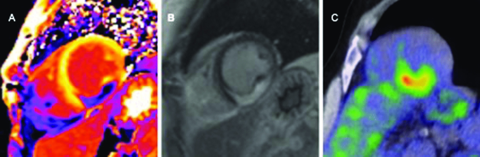
Resolve discordance before escalation
Review PET preparation and image quality, coronary disease and competing cardiomyopathies. A positive PET with an atypical phenotype deserves reassessment.
Biopsy an accessible extracardiac target when feasible. If cardiac tissue is needed, image/electroanatomic guidance may improve yield.
Noncaseating granulomas require exclusion of alternative granulomatous causes, especially infection.
▶
Reading the combined study
Perfusion + FDG patterns → meaning| Rest perfusion | FDG uptake | Interpretation |
|---|---|---|
| Normal | None (well suppressed) | No abnormal activity detected. This does not exclude prior scar, treated disease or all early CS. |
| Normal | Focal / focal-on-diffuse | Possible inflammation or incomplete suppression. Reconcile the pattern and preparation before diagnosing or treating CS. |
| Defect (scar) | Focal uptake in the defect | Highest-risk pattern — active disease within scarred myocardium; treat inflammation and protect from arrhythmia. |
| Defect (scar) | None | Fibrosis / burnt-out disease without active inflammation — unlikely to respond to steroids. |
| Any | Diffuse only | Usually inadequate suppression — repeat with strict high-fat/low-carb prep ± heparin before calling it disease. |
CMR LGE is classically mid-wall / epicardial at the basal septum & inferolateral wall (non-coronary); its presence & extent predict VT and mortality even at preserved EF. CMR & PET are complementary — CMR for scar/structure/prognosis, PET for activity/response. Many programs obtain both at baseline.
Who to treat Treat patients with active inflammation (FDG-avid) and clinically significant disease — AV block, VT, or LV/RV dysfunction. Published treatment evidence is mainly observational. Do not extrapolate pulmonary-sarcoid trials directly to cardiac disease. Goal: quiet inflammation, prevent progression of conduction/myocardial damage. Burnt-out scar without active uptake is unlikely to respond to immunosuppression.
A
Pharmacologic ladder
First-line steroids → steroid-sparing → biologicsCorticosteroidsFIRST-LINE
DrugPrednisone
DoseCommonly 0.5 mg/kg/day (≈30–40 mg) start; some use lower. Taper over 6–12 mo to ~5–10 mg maintenance.
MonitorGlucose, BP, bone, weight; PCP/GI prophylaxis as indicated.
NoteRe-image (PET) at ~3–6 mo to confirm response.
Steroid-sparingADD EARLY
OptionsMethotrexate 10–15 mg/wk (+folate); azathioprine; mycophenolate; leflunomide.
WhyLimit cumulative steroid toxicity; many add at the outset or by 3–6 mo.
MonitorCBC, LFTs; MTX: avoid in significant renal/liver disease, alcohol.
BiologicsREFRACTORY
DrugInfliximab (anti-TNF-α); adalimumab alternative.
UsePersistent FDG activity / progression despite steroid + sparing agent.
CautionScreen TB & hepatitis; HF warning: >5 mg/kg contraindicated in moderate/severe HF. Any use requires specialist review; infection risk.
B
Response, duration & monitoring
Integrate imaging, rhythm and functionAssessing response
- Serial FDG-PET at 3–6 mo: compare uptake, preparation and perfusion alongside symptoms, rhythm and function.
- Function recovery of LVEF & conduction can occur if treated while active.
- Arrhythmia VT burden may fall with inflammation control — but scar-related VT persists.
Duration
- Often ≥12 months; many require low-dose maintenance given relapse risk on withdrawal.
- Taper guided by imaging activity + clinical course, not a fixed calendar.
Caveats & pitfalls
- Immunosuppression ≠ arrhythmia protection it does not remove the need for a device in high-risk patients.
- Steroid toxicity anticipate infection, hyperglycemia, osteoporosis and fluid retention.
- Relapse common after discontinuation → re-image if symptoms/arrhythmia recur.
- No FDA-approved drug for CS — all use is guideline/consensus-based, off-label.
Parallel tracks Run immunosuppression and the device/arrhythmia plan (P.4) together — neither substitutes for the other.
C
Representative prednisone course & agent roles
Consensus regimens — individualize| Phase | Typical dose | Action & checkpoint |
|---|---|---|
| Induction | ~0.5 mg/kg/d prednisone (≈30–40 mg); high-dose pulse reserved for fulminant disease / GCM overlap | Start at diagnosis of active CS; add a steroid-sparing agent at or near outset to cap cumulative steroid. |
| Re-stage | Adjust to response and toxicity | Repeat FDG-PET at ~3–6 mo. Do not wait for PET to address steroid toxicity. Confirm prep and clinical activity before escalating. |
| Taper | Step down over 6–12 mo toward ~5–10 mg/d | Reduce as activity falls; do not taper on calendar alone. Steroid-sparing agent carries the load as prednisone comes down. |
| Maintenance | ≤10 mg/d ± steroid-sparing agent | Many need low-dose maintenance given relapse on withdrawal; reassess by serial imaging + rhythm. |
1stMethotrexate
10–15 mg weekly PO/SC + folate. Most-used steroid-sparing agent; avoid in significant renal/liver disease & alcohol use.
AltAzathioprine
Assess TPMT/NUDT15 risk before use; alternative when MTX not tolerated. Monitor CBC/LFTs for myelosuppression.
AltMycophenolate
Option for intolerance/refractoriness; teratogenic, monitor CBC. Leflunomide another alternative.
LastInfliximab
Anti-TNF-α for refractory activity; screen TB/hepatitis, doses >5 mg/kg contraindicated in moderate/severe HF. Lower doses also require specialist risk review and close monitoring.
A
ICD indications
HRS 2014 · AHA/ACC/HRS 2017| Class | Recommendation |
|---|---|
| I | ICD if spontaneous sustained VT/VF, prior cardiac arrest, or LVEF ≤35% despite optimal medical therapy + immunosuppression. |
| IIa | For LVEF >35%: ICD reasonable with an indication for permanent pacing, arrhythmic syncope, myocardial scar or inducible sustained VA. |
| IIb | HRS 2014: ICD may be considered for LVEF 36–49% or RVEF <40% despite therapy. Intermediate EF alone is not Class IIa. |
| EPS | Programmed stimulation is useful for risk stratification when LVEF >35% and the ICD decision is uncertain. |
Apply ICD recommendations when meaningful survival >1 year is expected and consistent with patient goals. Scar matters even with preserved EF. Consider CRT when pacing burden will be high or with LBBB + low EF.
01
Conduction Disease
Block & pacing- AV block may partially reverse with steroids, but recurrence/SCD risk persists → device with ICD capability preferred over bare PPM in most.
- Work up before PPM unexplained high-grade block <60 yr → evaluate for CS promptly. Do not delay emergency pacing, but results can change permanent device choice.
- CRT for high RV-pacing burden or LBBB + reduced EF.
Don't default to a pacemaker A patient getting "just a pacemaker" for unexplained block may need an ICD — evaluate promptly and select the permanent device with EP.
02
Ventricular Arrhythmia
VT management- Dual mechanism active inflammation and fixed scar → treat both: immunosuppression + antiarrhythmics ± ablation.
- Drugs β-blockers; amiodarone or sotalol for VT suppression; treat HF.
- Catheter ablation for recurrent/refractory VT or storm — often scar-related re-entry; best after inflammation controlled. Frequently needs repeat / epicardial approach.
- VT storm escalate immunosuppression, antiarrhythmics, sedation, ablation; consider steroids acutely.
B
Device selection & VT escalation
Match hardware to substrate| Scenario | Device / step | Rationale |
|---|---|---|
| Sustained VT/VF, arrest, or LVEF ≤35% | ICD (Class I) | Secondary prevention or severe LV dysfunction despite optimal therapy. |
| Mobitz II / complete heart block | Pacing with ICD capability | Block may not reverse with steroids; co-located arrhythmic risk favors defibrillator over bare PPM. |
| LVEF 36–49% or RVEF <40% | ICD may be considered (HRS IIb) | Intermediate EF alone. Scar, syncope, pacing need or inducibility may establish a stronger indication. |
| EF >35%, decision uncertain | EP study | Inducible sustained VT supports ICD; extensive LGE also shifts toward implant. |
| High pacing burden or LBBB + low EF | CRT | Avoid pacing-induced dyssynchrony; resynchronize when criteria met. |
| Recurrent VT / storm | AAD + catheter ablation | Escalate immunosuppression for active inflammation; ablate scar-related re-entry once quiet — often needs repeat / epicardial mapping. |
VT-storm bundle: amiodarone/β-blocker, sedation, treat HF, escalate steroids acutely, then ablation. Wearable defibrillator can bridge while CS work-up or steroid response is pending.
REMEMBER
Inflammation drives some arrhythmia; scar drives the rest. Steroids may reverse acute block and quiet VT, but established granulomatous scar remains a permanent arrhythmic substrate — which is why device protection and ablation stay on the table regardless of immunosuppression response.
Treat HF as HFrEF
Standard GDMT for reduced EF — ARNI/ACEi/ARB, β-blocker, MRA, SGLT2i — alongside immunosuppression.
Protect from SCD
Arrhythmic death can precede pump failure — device decisions (P.4) come early, not late.
Follow the activity
Serial FDG-PET + EF + rhythm monitoring guide immunosuppression titration.
Team sport
Cardiology · EP · rheumatology/pulmonary · advanced HF — manage in a dedicated program.
01
Heart Failure & Advanced
Pump managementMedical
- Full GDMT for LVEF <40% as in any HFrEF; decongest with diuretics.
- Atrial arrhythmia common — rate/rhythm control; anticoagulate per usual stroke-risk assessment.
- Anti-TNF caution infliximab can worsen HF. >5 mg/kg is contraindicated in moderate/severe HF.
Advanced / end-stage
- Refractory HF or VT → advanced therapies: LVAD or heart transplant — CS has excellent post-transplant outcomes (low, manageable recurrence).
- Palliative integrate for symptom & goals-of-care in end-stage disease.
Transplant is a good option Unlike many infiltrative diseases, CS patients do well after heart transplant — refer early when refractory.
02
Monitoring & Follow-up
What to trackCadence
- Imaging FDG-PET at ~3–6 mo after starting/changing therapy, then periodically; echo for EF.
- Rhythm device interrogation / ambulatory monitoring for block & VT.
- Labs immunosuppression toxicity panel (CBC, LFTs, glucose), BNP.
Prognostic markers
- LV systolic dysfunction, extensive LGE, sustained VT, and RV involvement portend worse outcome.
- Early treatment of active inflammation can recover function & conduction.
Extracardiac sarcoid
Co-manage lung/eye/skin/CNS disease with pulmonary & rheumatology.
Family / genetics
No clear Mendelian inheritance; counsel per systemic-sarcoid context.
Vaccination
On immunosuppression — keep vaccines current; infection vigilance.
Reassess goals
Re-evaluate device, drug & transplant candidacy at each visit.
03
Follow-up cadence & prognostic drivers
What to track and what it means| Domain | What & when | Why it matters |
|---|---|---|
| Disease activity | FDG-PET ~3–6 mo after starting/changing therapy, then periodically | Interpret uptake extent/intensity with preparation, symptoms, rhythm and function. SUV change alone is insufficient. |
| LV / RV function | Echo ± CMR for EF and remodeling | LV systolic dysfunction & RV involvement portend worse outcome; recovery possible if treated while active. |
| Rhythm | Device interrogation / ambulatory monitor for block & VT | Sustained VT and high-grade block mark arrhythmic risk; can occur before pump failure. |
| Scar burden | Extent of LGE on CMR (baseline, as needed) | Extensive LGE independently predicts VT & mortality even with preserved EF. |
| Drug safety | CBC, LFTs, glucose; BNP/NT-proBNP | Match monitoring to agent toxicity (P.6); keep vaccines current on immunosuppression. |
| Advanced options | Refer for LVAD / transplant when HF or VT refractory | CS has excellent post-transplant survival with low, manageable recurrence — refer early. |
SUMMARY
Diagnose early, image to stage activity, treat inflammation, and protect from arrhythmia — in parallel. CMR shows the scar (risk); PET shows the fire (treat & follow); the device guards against sudden death while immunosuppression cools the disease. Transplant is a legitimate endgame.
Principle Cardiac treatment evidence is mainly observational and consensus-based. Steroids are first-line; add a steroid-sparing agent early to limit cumulative toxicity. Titrate using clinical status, rhythm, function and serial PET, and anticipate relapse on withdrawal.
A
Drug-specific monitoring
Screen before, monitor during| Agent | Before starting | On therapy |
|---|---|---|
| Prednisone | Glucose, BP, bone-health risk | Glucose/BP/weight, bone protection; PJP prophylaxis if high-dose + second agent. |
| Methotrexate | CBC, LFTs, renal; avoid in significant liver/renal disease, alcohol | CBC + LFTs periodically; folate; pneumonitis is rare but watch. |
| Azathioprine | TPMT/NUDT15 assessment, CBC, LFTs | CBC + LFTs (myelosuppression). |
| Mycophenolate | CBC; pregnancy status | CBC; GI tolerance; teratogenic. |
| Infliximab | TB & hepatitis screen; HF risk review. >5 mg/kg contraindicated in moderate/severe HF | Infection vigilance; infusion reactions. |
Keep vaccinations current before/early in immunosuppression (avoid live vaccines once immunosuppressed). Anti-TNF (infliximab) can worsen HFrEF — screen LV function first.
01
Response & Taper
Follow the activity- Serial FDG-PET at 3–6 mo: compare uptake and preparation with the clinical trajectory.
- Taper by integrated response reduce steroid as activity falls; many need low-dose maintenance.
- Function EF & conduction can recover if treated while active; fixed scar will not.
- No fixed endpoint duration individualized — often ≥12 mo.
02
Relapse & Pitfalls
Expect recurrence- Relapse is common on withdrawal → re-image if symptoms, arrhythmia, or EF change.
- Immunosuppression ≠ device it does not remove the need for an ICD in high-risk patients.
- Burnt-out scar without FDG activity is unlikely to respond — don't escalate immunosuppression blindly.
- Off-label no FDA-approved drug for CS; all use is consensus-based.
Two treatment targets can coexist
Active inflammation and scar can coexist.
SUMMARY
Treat active inflammation and reassess it with the whole clinical picture, taper to the lowest effective dose, and re-image at the first hint of relapse. Match the agent's monitoring to its toxicity, keep the device plan running in parallel, and remember that established scar is a permanent substrate immunosuppression cannot reverse.