START
CV
Emergencies
Emergencies
ICI Myocarditis
hold ICI IV methylprednisolone 1 g/d × 3 d then taper after objective response. Urgent specialist escalation for deterioration or inadequate response. Adjunctive immunosuppression is individualized. 25–50% mortality in fulminant cases. Permanent discontinuation.
Anthracycline acute HF
Acute symptomatic HF → hold, urgent assessment + phenotype-directed GDMT. Anthracycline restart is an MDT decision after recovery. Consider dexrazoxane / liposomal options.
Hypertensive crisis
VEGF/TKI: SBP ≥180 or DBP ≥110 → hold, assess end-organ injury. IV therapy for hypertensive emergency. Reassess restart below 160/100, subject to product-label restrictions.
QTc ≥500 ms / Torsades
hold the agent. For torsades: IV magnesium and urgent rhythm care. Correct K⁺/Mg²⁺ and stop QT drugs. Pace if pause-dependent. Permanently d/c after Torsades (esp. ribociclib).
5-FU / Capecitabine ACS
stop exposure, ECG/troponin + ACS evaluation. Nitrates/CCB for vasospasm. Exclude obstructive CAD. Rechallenge only after MDT review, prophylaxis, and a monitored plan.
01
Baseline CV risk assessment
Before cancer therapyHFA-ICOS pre-treatment workup
- History prior HF / CAD / valve · cumulative anthracycline · chest RT · smoking · DM · HTN · CKD · pregnancy / late effects of childhood Ca.
- Exam + vitals BP both arms · HR · weight · JVP · edema · murmur.
- ECG if pre-existing CVD / CV risk, or the planned therapy is associated with arrhythmia or high CV-toxicity risk.
- LV imaging for drugs associated with LV dysfunction: TTE + GLS if available. CMR if echo quality is poor. MUGA if TTE is unavailable or infeasible.
- Biomarkers consider baseline hs-troponin · BNP for HIGH / V·HIGH risk rather than as a universal bundle.
- Labs lipids + A1c within 6 mo for systemic therapy. CBC · BMP · Mg / K · TSH according to regimen and comorbidity.
Stratify before cycle 1
Combine the therapy-specific HFA-ICOS proforma with patient factors to assign LOW MED HIGH V·HIGH. The tier informs monitoring, HIGH / V·HIGH referral, and multidisciplinary risk–benefit review. It is not itself a drug-hold rule.
Primary prevention candidates
- No routine prophylactic CV drug bundle Evidence does not support starting an ACEi/ARB, β-blocker, statin, or SGLT2i solely for primary CTR-CVT prevention. Treat standard CV indications. A specialist may select neurohormonal therapy or a statin in HIGH / V·HIGH risk.
- Anthracycline mitigation in HIGH-risk patients needing further therapy, consider dexrazoxane or a liposomal formulation, particularly near/above cumulative doxorubicin 250 mg/m² or with pre-existing cardiomyopathy.
- Lifestyle + risk factors exercise · smoking cessation · healthy diet · weight. CPS targets: A1c <7 · BP <130/80 · LDL <70. Individualize during active therapy.
02
Surveillance principles
Echo · biomarkers · ECG cadenceEcho / GLS cadence
- Anthracycline baseline LV imaging when dysfunction risk is present. Follow the drug/risk schedule. At 12 mo post-Tx, echo if early-stage/curative intent and cumulative doxorubicin ≥ 250 mg/m² or equivalent.
- HER2 baseline LV imaging + drug-specific on-Tx surveillance. Echo at 12 mo post-Tx.
- VEGF / TKI baseline · q3–6 mo if CV risk factors.
- BRAF/MEK baseline · 1 mo · then q3 mo.
- Survivorship after adult-onset cancer therapy: annual symptoms + CV risk-factor review. Re-stratify at 5 y. Childhood-cancer survivors follow their separate exposure-based pathway.
- Long-term echo MODERATE risk: q5y may be considered (IIb-C). HIGH / V·HIGH: years 1, 3, 5, then q5y (IIa-C). Evidence is limited.
Biomarkers
- hs-Tn / ECG ICI: baseline and early-cycle surveillance per risk pathway. New symptoms, troponin rise or conduction change → urgent clinical assessment, then targeted TTE/CMR. Do not wait for imaging if unstable.
- BNP echo cadence + at any new dyspnea / volume change.
- Trend > absolute — the patient is their own control only while modality, acquisition, loading, rhythm, vendor/software, and analysis remain comparable. Exclude technically invalid trend points.
ECG / electrolytes
- Pre-dose for any QT-prolonger. Review interacting drugs.
- Ribociclib ECG before therapy and ~day 14 of cycle 1, then as indicated. Start only if QTcF <450 ms. Correct electrolytes.
- Vandetanib / arsenic follow distinct label ECG and electrolyte schedules. Increase monitoring if unstable.
03
Therapy-specific decision rules
When to hold · who to callEF drop trigger (asymptomatic CTRCD)
Confirm LVEF/GLS and symptoms. Asymptomatic EF 40–49%: interrupt anthracycline. Selected HER2 therapy may continue with ACEi/ARB + BB and close monitoring. Follow the exact agent label and MDT plan.
Symptomatic HF (any cause)
New HF symptoms → hold diurese, GDMT (ARNI/ACE/ARB + BB + MRA + SGLT2 as tolerated). CV consult. Echo + biomarkers. Rule out ischemia.
QTc thresholds
Use QTcF: 480–500 ms → correct causes + closer monitoring. ≥500 ms → hold. Δ >60 ms alone below 500 is not an automatic hold. Stricter agent-label rules apply, including ribociclib and arsenic.
Hypertension (any agent)
Treat elevated BP. ≥160/100 generally needs ACEi/ARB + DHP-CCB. ≥180 systolic or ≥110 diastolic → hold culprit therapy pending control. Emergency = end-organ injury. Product labels may require earlier holds.
When to call cardio-onc
New CVD · LVEF <53% and/or relative GLS drop >15% · new arrhythmia · uncontrolled CV risk factors · important DDI concern. These CPS examples prompt evaluation/referral, not an automatic cancer-therapy hold.
B
Biomarker thresholds at a glance
Assay · context · serial changehs-Troponinng/L
≥ 99th percentile ULN + symptoms → CMR + cardiac consult. Interpret assay variation, renal disease, ischemia and myositis. During ICI therapy, a new rise requires urgent myocarditis assessment, not an automatic diagnosis.
BNPpg/mL
BNP > 35 (NT-proBNP > 125 equiv.) abnormal in non-acute setting. Rising trend → echo. Consider diuresis if volume-overloaded.
LVEF% by 3D / GLS
New EF <40%: severe asymptomatic CTRCD. EF 40–49%: moderate if fall ≥10 points, or a smaller fall plus new GLS/biomarker abnormality. EF ≥50%: mild only with new GLS/biomarker abnormality.
GLS% relative Δ
Subclinical injury signal: Δ > 15% relative drop from baseline. Confirm comparability and refer/evaluate. This signal alone is not an automatic hold criterion.
R
Baseline risk domains — not a scoring substitute
Use the therapy-specific proformaCardiac history
HF · CAD · MI · valvular · AF · prior cardiotoxic Tx
Imaging
EF < 50% · GLS reduced · ↑ LV mass · diastolic dysfn
Biomarkers
↑ hs-Tn · ↑ BNP at baseline
Demographics
Age ≥ 65 · pediatric · pregnancy · ethnicity (S Asian, AA)
CV risk factors
HTN · DM · CKD · obesity · smoking · dyslipidemia · OSA
Prior exposure
Anthracycline > 250 mg/m² · chest RT · prior HER2 / VEGF
START
L
How to score — count point-bearing rows, apply ceiling factors
Six point-based tools · ADT uses ASCVD riskLOWNo risk factors or a single Medium point. 0–1 med pts
MEDIUM2–4 Medium points. No High / Very-High factor. 2–4 med pts
HIGH≥5 Medium pts or any High-tier factor. ≥5 med · or any HIGH
V·HIGHAny Very-High factor (HF / cardiomyopathy etc). any VERY HIGH
A
Risk tier → pre-treatment action
Each drug card carries its own HFA-ICOS mini-scoreLOW
Proceed with routine class-based surveillance (echo / Tn per card).
MEDIUM
Closer monitoring. manage CV risk factors. Consider cardiology referral.
HIGH
Discuss risk / benefit. Refer to cardio-onc. Consider baseline Tn / BNP. Do not auto-start prophylactic CV drugs solely from the tier.
V·HIGH
MDT on alternative regimen / ↓ cum-dose / liposomal / dexrazoxane. Optimize established CVD. Specialist-selected prevention may be reasonable.
G
CTCAE / ASTCT grade criteria — what the grades actually mean
Versioned reference · use the trial-specified CTCAE edition| Toxicity | Grade 1 | Grade 2 | Grade 3 | Grade 4 |
|---|---|---|---|---|
| HypertensionCTCAE v5 | SBP 120–139 or DBP 80–89 | SBP 140–159 / DBP 90–99. Symptomatic. Monotherapy indicated | SBP ≥160 / DBP ≥100. >1 agent or more intensive Tx | Life-threatening — malignant HTN, crisis, neuro deficit. Urgent IV Tx |
| Heart failureCTCAE v5 · LVSD is a separate term | Asymptomatic. Abnormal imaging / biomarker only | Symptoms with moderate activity / exertion | Symptoms at rest / minimal exertion. Intervention indicated | Life-threatening. Urgent Tx — inotropes / mechanical support |
| MyocarditisCTCAE v5 | Not defined | Symptoms on moderate exertion | Symptoms at rest/minimal activity, intervention needed | Life-threatening, urgent support needed |
| Pericardial effusionCTCAE v5 | Not defined | Asymptomatic small-to-moderate effusion | Physiologic consequences | Life-threatening, urgent intervention |
| QTc prolongationCTCAE v5 | 450–480 ms | 481–500 ms | ≥501 ms or Δ >60 ms from baseline | Torsades / polymorphic VT / signs of serious arrhythmia |
| Atrial fibrillationCTCAE v5 | Asymptomatic. No intervention | Non-urgent medical intervention | Symptomatic, urgent Tx (rate/rhythm control, cardioversion) | Life-threatening. Hemodynamic compromise. Urgent intervention |
| Cytokine release (CRS)ASTCT 2019 | Fever ≥38°C. no hypotension / hypoxia | ↓BP not needing pressors and/or hypoxia on low-flow O₂ ≤6 L/min | ↓BP needing 1 pressor (±vasopressin) and/or O₂ >6 L/min / mask | ↓BP needing multiple pressors excluding vasopressin and/or positive-pressure ventilation |
| Peripheral sensory neuropathyCTCAE v5 | Asymptomatic. Loss of DTRs / paresthesia | Moderate. Limiting instrumental ADL | Severe. Limiting self-care ADL | Life-threatening. Urgent intervention |
| ProteinuriaCTCAE v5 | 1+. <1.0 g/24h | 2+. 1.0–3.4 g/24h | ≥3.5 g/24h (nephrotic-range) | — |
| DiarrheaCTCAE v5 | <4 stools/day over baseline | 4–6 stools/day over baseline | ≥7/day. Incontinence. Hospitalization | Life-threatening. Urgent intervention |
How to read the cards that follow
Each drug class is one block: agents (US brand + generic) · use / benefit · CV & non-cardiac toxicity · its HFA-ICOS mini-score (or a note where no proforma exists) · surveillance · and a granular, grade-tiered management plan with doses and hold / resume / rechallenge thresholds. CRS uses ASTCT. Grade by the worse hypotension/hypoxia domain. Fever need not persist after antipyretic or anti-cytokine therapy. A troponin rise alone is not a CTCAE v5 myocarditis grade. The 2026 ASTCT consensus retains the CRS framework with clarifications. ICANS assessment for developmental limitations or abnormal baseline cognition follows that updated consensus.
W
Worked example — patient → HFA-ICOS tier → action
New to cardio-onc? This is the workflow every card supports1 · The patient
68 F · HER2+ breast Ca · h/o HTN · prior doxorubicin 240 mg/m² · baseline EF 53%, GLS −16% (borderline). Now starting trastuzumab.
→
2 · Count points
Age 68 2 + HTN 1 + EF 53% 2 = 5 medium points → HIGH already. Remote anthracycline adds 2, or current sequential anthracycline adds 1. Borderline GLS is not a separate High-tier factor.
→
3 · Tier
HIGH
→
4 · Action
Refer cardio-onc · baseline echo+GLS. Consider hs-Tn/BNP · optimize standard CV indications. Discuss rather than automatically start prophylactic ACEi/BB · follow the trastuzumab surveillance plan.
DRUGS
CLASS MAP
Anthracyclines
Dose-dependent · cumulative · partly reversibleAnthracyclinesDNA topo-II inhibition · ROS / iron-mediated injury
CLASS
AgentsDoxorubicin (Adriamycin) · liposomal doxorubicin (Doxil) · Daunorubicin (Cerubidine) · Epirubicin (Ellence) · Idarubicin (Idamycin) · Mitoxantrone (Novantrone) · Valrubicin (Valstar).
UseBreast · HL / NHL · AML / ALL · sarcoma · gastric. Curative-intent backbone.Benefit≈ 33% ↓ breast-Ca mortality. > 80% cure in Hodgkin (ABVD). Core of AML 7+3 & pediatric ALL — often irreplaceable.
CV toxicity
- HFrEF / CTRCD: cumulative-dose injury with variable recovery. Historical doxorubicin trials estimated CHF in 5% at 400 and 26% at 550 mg/m². These are cohort estimates, not individual thresholds.
- Cumulative exposure increases risk. ≥250 mg/m² doxorubicin flags higher surveillance risk. There is no universally safe dose or absolute 550 mg/m² prohibition.
- A new GLS decline or troponin rise may precede LVEF decline. Neither appears in every case, and alternative causes must be assessed.
- Acute arrhythmia / myopericarditis during infusion (rare).
Non-cardiac toxicity
- Myelosuppression — neutropenia dose-limiting. Febrile neutropenia.
- Vesicant — extravasation necrosis. Mucositis · alopecia · N/V.
- Secondary leukemia (t-AML / MDS). Benign red urine.
HFA-ICOS · Anthracyclinepre-Tx
V·HIGHHF / cardiomyopathy
HIGHSevere valvular dz · MI / prior PCI / CABG · Stable angina · LVEF <50% · Prior anthracycline · Prior L-chest / mediastinal RT · Age ≥80
MEDLVEF 50–54%2 · Age 65–792 · ↑ baseline Tn1 · ↑ baseline BNP1 · HTN1 · DM1 · CKD1 · Prior non-anthra chemo1 · Smoking1 · Obesity1
≥1 V·HIGH → V·High · any HIGH or ≥5 MED-pts → High · 2–4 → Med · 0–1 → Low
Cum. dose ≥250 mg/m² doxorubicin (≥600 epirubicin) independently ↑ risk.
Surveillance
- Baseline CV Hx · BP · ECG if CVD/risk or arrhythmia risk · echo + GLS. Consider hs-Tn/BNP if HIGH / V·HIGH.
- Echo + GLS on-Tx per drug/risk. 12 mo post if early-stage/curative and cumulative doxorubicin ≥250 mg/m². Longer intervals by risk.
- hs-Tn each cycle if high-risk — trend > absolute.
Management — granular
PREHFA-ICOS High / V-High → cardio-onc/MDT review. Optimize standard CV indications. Do not automatically start ACEi/ARB, BB, statin, or SGLT2i solely for CTR-CVT prophylaxis. Specialist-selected neurohormonal/statin therapy may be considered.
MILDΔ GLS >15% rel. or new ↑Tn with EF ≥50% → validate the trend + cardio-onc review. Consider ACEi/ARB + BB as appropriate. do not automatically hold chemo.
CTRCDModerate/severe asymptomatic CTRCD or symptomatic HF → interrupt anthracycline and assess urgently. Treat the HF phenotype, confirm the trajectory and agree on reassessment with the MDT.
PROTDexrazoxane: labeled cardioprotection in women with metastatic breast cancer continuing doxorubicin after 300 mg/m². Broader high-risk use may be off-label. Ratio 10:1. Reduce 50% if CrCl <40. Verify product/timing.
RESUMERechallenge after recovery only by MDT risk–benefit review. Optimize HF therapy, exposure mitigation, and monitoring. No universal EF/4-week restart rule.
FactoidDexrazoxane and liposomal formulations can reduce anthracycline cardiac injury. Cumulative exposure and baseline susceptibility still matter.
CLASS MAP
VEGF axis · ICI · CAR-T · alkylators · mTOR
Recognize myocarditis · HTN · arrhythmia earlyVEGF Inhibitors (TKI)Multi-kinase · VEGFR signaling blockade
CLASS
AgentsSunitinib (Sutent) · Sorafenib (Nexavar) · Pazopanib (Votrient) · Axitinib (Inlyta) · Cabozantinib (Cabometyx) · Lenvatinib (Lenvima) · Regorafenib (Stivarga) · Vandetanib (Caprelsa).
UseRCC · HCC · differentiated & medullary thyroid · GIST · sarcoma · CRC (rego).BenefitDoubles PFS in RCC / HCC. Lenvatinib PFS benefit in RAI-refractory thyroid.
CV toxicity
- HTN 30–40% all-grade (G3 ≈10–15%: SBP ≥160 / DBP ≥100, needs >1 agent).
- LV dysfunction 5–8% — often reversible off-drug.
- QT prolongation (vandetanib, sunitinib). Arterial & venous thrombo-embolism.
Non-cardiac toxicity
- Proteinuria · nephrotic syndrome · TMA.
- Hand-foot skin reaction · stomatitis · diarrhea.
- Hypothyroidism · fatigue · hepatotoxicity · impaired wound healing.
HFA-ICOS · VEGF inhibitorspre-Tx
V·HIGHHF / cardiomyopathy · Arterial vascular dz (IHD·PCI·CABG·angina·TIA/stroke·PVD)
HIGHVenous thrombosis (DVT/PE) · LVEF <50% · QTc ≥480 ms · Hypertension · Age ≥75 · Prior anthracycline
MEDLVEF 50–54%2 · QTc 450–<480 (M)/460–<480 (F)2 · Arrhythmia2 · ↑Tn1 · ↑BNP1 · Age 65–741 · DM1 · Hyperlipidemia1 · CKD1 · Proteinuria1 · Prior L-chest RT1 · Smoking1 · Obesity1
≥1 V·HIGH → V·High · any HIGH or ≥5 MED-pts → High · 2–4 → Med · 0–1 → Low
HTN is the commonest VEGF AE — counts HIGH here.
Surveillance
- BP each visit. Home BP daily during the first cycle, after dose increases, then every 2–3 weeks. Reassess antihypertensives when VEGF therapy stops.
- Baseline + periodic ECG (QTc) if QT-risk drug / DDI.
- Echo if symptoms. TFTs q cycle. UPCR periodically.
Management — granular
HTNSBP 140–159 / DBP 90–99 → treat with ACEi/ARB or DHP-CCB as appropriate. Generally continue VEGF with monitoring.
G3 HTNSBP ≥160 / DBP ≥100 → intensify to ACEi/ARB + DHP-CCB. Review the label. Hold at ≥180 systolic / ≥110 diastolic or for uncontrolled severe HTN.
G4 HTNAcute end-organ injury → admit and titrated IV therapy. Restart or permanent cessation follows the agent label. Do not use an oral-agent-count rule.
LVSDEF ↓ ≥10 pt to <50% → hold + GDMT (ACE/ARB + BB ± SGLT2i). Echo 2–4 wk.
QTcQTcF ≥500 ms → hold, correct electrolytes/DDIs. Restart by label. Δ >60 ms alone below 500 warrants review, not automatic interruption.
MONTreat thyroid dysfunction. Monitor proteinuria. Proteinuria hold thresholds are agent-specific. Evaluate nephrotic syndrome or renal injury.
FactoidVEGF-pathway hypertension can occur early. Treat BP to preserve therapy when feasible. Hypertension is not a validated individual tumor-response test. Severe or uncontrolled BP can require interruption.
Anti-VEGF mAbsLigand / receptor binding anti-angiogenics
CLASS
AgentsBevacizumab (Avastin) · Ramucirumab (Cyramza) · Ziv-aflibercept (Zaltrap).
UseCRC · NSCLC · RCC · cervical · ovarian · gastric · HCC · glioblastoma.BenefitAdds PFS (and OS in some) when combined with chemotherapy backbones.
CV toxicity
- Severe HTN ≈8% G3 — dose-related.
- Arterial thrombo-embolism (MI / stroke) ≈3–4%. VTE.
- CHF (esp. prior anthracycline / chest RT). Rare PRES.
Non-cardiac toxicity
- Proteinuria · nephrotic syndrome.
- GI perforation · fistula · impaired wound healing.
- Hemorrhage — pulmonary, GI, CNS. Infusion reactions.
HFA-ICOS · VEGF inhibitorspre-Tx
V·HIGHHF / cardiomyopathy · Arterial vascular dz (IHD·PCI·CABG·angina·TIA/stroke·PVD)
HIGHVenous thrombosis (DVT/PE) · LVEF <50% · QTc ≥480 ms · Hypertension · Age ≥75 · Prior anthracycline
MEDLVEF 50–54%2 · Arrhythmia2 · ↑Tn1 · ↑BNP1 · Age 65–741 · DM1 · Hyperlipidemia1 · CKD1 · Proteinuria1 · Prior L-chest RT1 · Smoking1 · Obesity1
≥1 V·HIGH → V·High · any HIGH or ≥5 MED-pts → High · 2–4 → Med · 0–1 → Low
Summary only. The complete VEGF proforma also includes QTc 450–<480 (M) / 460–<480 (F) as 2 medium points. Monitoring remains drug-specific.
Surveillance
- BP every cycle. Home log if elevated.
- Monitor urine protein per label. For bevacizumab, dipstick ≥2+ prompts 24-hour quantification. Hold at ≥2 g/24 h and discontinue for nephrotic syndrome.
- Perioperative holds are agent-specific. Bevacizumab: ≥28 days before elective surgery and ≥28 days after major surgery, with adequate healing.
Management — granular
G3 HTNBevacizumab: hold severe HTN uncontrolled with medical therapy. Resume after control. Stop permanently for hypertensive crisis/encephalopathy.
ATEArterial thrombo-embolism (MI/stroke) → permanently discontinue. Manage event per ACS / stroke pathway.
VTETreat VTE with an appropriate anticoagulant. Stop bevacizumab for grade 4 VTE. Other continuation decisions require oncology review.
BLEEDG3–4 hemorrhage or GI perforation/fistula → discontinue. Surgical / IR management.
SURGBevacizumab: hold ≥28 d before elective surgery and ≥28 d after major surgery, until healed. Other agents differ.
FactoidAnti-VEGF agents are not interchangeable. Bevacizumab and ramucirumab both carry arterial-event warnings. Ramucirumab requires permanent discontinuation after an arterial thromboembolic event.
DRUGS
Immune Checkpoint InhibitorsPD-1 / PD-L1 / CTLA-4 blockade
CLASS
AgentsPembrolizumab (Keytruda) · Nivolumab (Opdivo) · Cemiplimab (Libtayo) · Atezolizumab (Tecentriq) · Durvalumab (Imfinzi) · Ipilimumab (Yervoy) · Tremelimumab (Imjudo).
UseMelanoma · NSCLC · RCC · urothelial · H&N · MSI-high · adjuvant & metastatic.BenefitDurable responses. Melanoma 5-y OS > 50% with combo. Practice-changing.
CV toxicity
- ICI myocarditis is uncommon but can be fatal. A multicenter registry reported prevalence 1.14% and median onset 34 days (IQR 21–75). Late presentations occur. Normal LVEF does not exclude severe disease.
- Pericarditis · arrhythmia / high-grade AV block · Takotsubo.
- Overlap myositis / myasthenia (bulbar / respiratory weakness). Late accelerated atherosclerosis.
Non-cardiac toxicity
- Multisystem irAEs — colitis · hepatitis · pneumonitis · dermatitis.
- Endocrinopathy — thyroiditis · hypophysitis · adrenal insufficiency · T1DM.
- Combo CTLA-4 + PD-1 → higher irAE rate & severity.
No HFA-ICOS proformaNo formal HFA-ICOS proforma for ICI. Use general baseline CV risk assessment (P.1) + baseline troponin/ECG. A new troponin rise requires urgent evaluation for myocarditis and competing causes.
Surveillance
- Baseline ECG + hs-Tn + CK ± BNP before cycle 1.
- Repeat hs-Tn / ECG with first 1–3 cycles (highest-risk window).
- Low threshold for echo / cardiac MRI on any new symptom.
Management — granular
SUSPAny troponin rise + symptom → hold ICI admit, telemetry, echo + CMR. CK / aldolase for myositis overlap.
TREATIV methylprednisolone 500–1000 mg/day ×3–5 d when myocarditis is likely. Follow clinical/Tn response and an individualized oral taper.
REFRACTDeterioration or inadequate response by 48–72 h → urgent specialist review for additional immunosuppression and circulatory/respiratory support. No automatic multidrug regimen.
ARRHYHigh-grade AV block or VT → urgent rhythm support and continuous monitoring. Monitoring duration follows electrical/hemodynamic stability and the specialist plan, not troponin normalization alone.
RECHALPermanently discontinue after confirmed grade ≥2 myocarditis. Treat concurrent irAEs with organ-specific steroids.
FactoidA rising troponin with normal early LVEF is the warning sign — treat on suspicion with pulse steroids. Don't await biopsy. Abatacept is emerging for steroid-refractory disease.
CAR-T & CellularCRS-mediated cardiovascular collapse
CLASS
AgentsTisagenlecleucel (Kymriah) · Axicabtagene ciloleucel (Yescarta) · Lisocabtagene maraleucel (Breyanzi) · Brexucabtagene autoleucel (Tecartus) · Idecabtagene vicleucel (Abecma) · Ciltacabtagene autoleucel (Carvykti).
UseR/R B-ALL · LBCL · follicular & mantle-cell lymphoma · multiple myeloma.BenefitDurable remission in chemo-refractory disease. LBCL CR ≈40%.
CV toxicity
- CRS can cause distributive shock, arrhythmia and LV dysfunction. In Alvi et al. (137 adults), 17 patients (12%) had CV events, all with grade ≥2 CRS.
- Arrhythmia (AF / VT). Demand ischemia. Rare stress cardiomyopathy.
- Higher CV-event rate with CRS grade ≥2 & pre-existing CVD. Capillary leak.
Non-cardiac toxicity
- CRS fever / hypoxia. ICANS — immune-effector-cell–associated neurotoxicity syndrome (encephalopathy · aphasia · seizures).
- Prolonged cytopenias. Hypogammaglobulinemia → infection.
- HLH / MAS. Tumor-lysis syndrome.
No HFA-ICOS proformaNo HFA-ICOS proforma. Pre-treat per general CV risk (P.1). ASTCT CRS grade (not CTCAE) drives cardiac risk — see grading reference.
Surveillance
- Baseline echo + ECG + troponin / BNP before infusion.
- Continuous telemetry & daily troponin during CRS.
- Echo for hypotension unresponsive to fluids / tocilizumab.
Management — granular
G1 CRSFever ≥38°C, no hypotension/hypoxia → supportive. Antipyretics. Rule out infection. Telemetry.
G2 CRSGrade 2 CRS: cellular-therapy pathway, cautious fluid and oxygen support. Tocilizumab in patients ≥30 kg: 8 mg/kg IV, maximum 800 mg. Repeat no sooner than 8 h, maximum four doses, if indicated.
G3 CRSGrade 3 CRS: ICU, vasopressor/oxygen support and product-specific tocilizumab/steroid treatment. Assess LV function and avoid indiscriminate fluid loading.
G4 CRSGrade 4 CRS: multiple pressors excluding vasopressin and/or positive-pressure ventilation. ICU and cellular-therapy team direct corticosteroids and organ support.
ARRHYAF/VT during CRS → ACLS. GDMT if persistent LV dysfunction after recovery.
FactoidIn the Alvi cohort, each 12-h delay to tocilizumab was associated with 1.7-fold higher CV-event risk. This observational association supports prompt CRS recognition, not a causal dosing deadline.
High-dose AlkylatorsDNA cross-linkers · SCT conditioning
CLASS
AgentsCyclophosphamide (Cytoxan) · Ifosfamide (Ifex) · Melphalan (Alkeran / Evomela) · Bendamustine (Treanda / Bendeka) · Busulfan (Busulfex / Myleran) · Carmustine (BiCNU) · Dacarbazine (DTIC-Dome).
UseLymphoma · leukemia · myeloma · breast · ovarian · sarcoma · SCT conditioning.BenefitBackbone of curative combination & transplant-conditioning regimens.
CV toxicity
- High-dose cyclophosphamide can cause acute hemorrhagic myocarditis and HF. Risk depends on dose and conditioning regimen. Do not apply one incidence or threshold to all alkylators.
- Pericarditis · arrhythmia (AF) · rare tamponade.
- Risk ↑ with prior anthracycline / chest RT. Usually within 1–3 wk.
Non-cardiac toxicity
- Myelosuppression · hemorrhagic cystitis (cyclo / ifos — give mesna).
- SIADH. Ifosfamide encephalopathy & Fanconi.
- Pulmonary fibrosis (busulfan, carmustine). VOD/SOS (busulfan).
No HFA-ICOS proformaNo HFA-ICOS proforma. Use general CV risk (P.1). Pre-conditioning echo mandatory after prior cardiotoxic therapy.
Surveillance
- Baseline CV Hx + BP + ECG. echo before high-dose / conditioning.
- Telemetry + daily weights during conditioning admission.
- Troponin / BNP if symptoms or known CVD.
Management — granular
MYOCARAcute myocarditis / HF → discontinue (rarely resumable). Supportive HF care + diuresis. Telemetry.
PERICPericarditis / effusion → NSAID/colchicine if no thrombocytopenia. Echo for tamponade → drainage.
CYSTPrevent hemorrhagic cystitis with regimen-specific mesna and hydration. Balance fluid against cardiac and renal reserve. New hematuria needs evaluation, not automatic fluid escalation.
METABCorrect Na⁺ for SIADH. methylene blue for ifosfamide encephalopathy.
FactoidCyclophosphamide injury may occur within days of high-dose exposure. Current dose, prior treatment and baseline cardiac reserve all matter.
DRUGS
mTOR InhibitorsEverolimus / temsirolimus (± VEGF)
CLASS
AgentsEverolimus (Afinitor) · Temsirolimus (Torisel) — often combined with VEGF TKIs / bevacizumab.
UseRCC · neuroendocrine tumors · HR+ breast (everolimus + exemestane) · TSC.BenefitAdds PFS in RCC / NET. Everolimus + exemestane doubles PFS in HR+ mBC.
CV toxicity
- HTN · myocardial ischemia · CHF · QT prolongation (VEGF combination).
- Arterial thromboembolism.
- Dyslipidemia-driven atherosclerosis. Additive with VEGF agents.
Non-cardiac toxicity
- Stomatitis / mucositis. Non-infectious pneumonitis.
- Hyperglycemia · hyperlipidemia.
- Immunosuppression → infection. Proteinuria.
No HFA-ICOS proformaNo HFA-ICOS proforma. Metabolic (lipids, glucose, BP) toxicity dominates — manage per Primary Prevention card (P.14).
Surveillance
- Baseline ECG / BP / lipids / glucose.
- Home BP q3 mo × 1 y, then q6 mo. Fasting lipids & glucose periodically.
- Chest imaging for pneumonitis.
Management — granular
HTNTreat HTN with an interaction-aware agent. Avoid concomitant ACE inhibitors with everolimus because angioedema risk is increased. Review CYP3A4/P-gp interactions.
LIPIDStatin for mTOR dyslipidemia (rosuva / prava — review CYP and transporter interactions). Tight glucose control.
PNEUMGrade ≥2 pneumonitis → hold mTOR + steroids. Dose-reduce for stomatitis.
RENALProteinuria, hematuria or AKI → renal assessment. Treat arterial events and venous thrombosis by their distinct pathways and bleeding risk.
FactoidThe dominant CV liability is metabolic — dyslipidemia (up to ~75%) and new hyperglycemia — not direct myocardial injury. Manage with statin + glycemic control, rarely cessation.
CLASS MAP
HER2 · BRAF/MEK · BCR-ABL · CDK4/6 · PI · BTK
Baseline + symptom-driven surveillanceHER2 mAbs / ADCsErbB2 receptor & payload conjugates
CLASS
AgentsTrastuzumab (Herceptin) · Pertuzumab (Perjeta) · T-DM1 / trastuzumab emtansine (Kadcyla) · T-DXd / trastuzumab deruxtecan (Enhertu) · Margetuximab (Margenza) · Tucatinib (Tukysa) · Neratinib (Nerlynx) · Lapatinib (Tykerb).
UseHER2+ breast (adjuvant & metastatic) · HER2+ gastric / GEJ · HER2-low.BenefitAdjuvant trastuzumab ↓ recurrence ≈50%. T-DXd transformed HER2+/HER2-low mBC.
CV toxicity
- HER2-related LV dysfunction often improves with interruption/HF therapy but recovery is not assured. Risk varies by agent, trial definition and prior anthracycline exposure.
- Symptomatic HF <1–4%. Mostly recovers off-drug.
- Risk ↑ with concurrent / sequential anthracycline & left chest RT.
Non-cardiac toxicity
- Infusion reactions. T-DXd ILD / pneumonitis — can be fatal. Monitor.
- Diarrhea (pertuzumab, neratinib, tucatinib).
- Thrombocytopenia / hepatotoxicity (T-DM1).
HFA-ICOS · HER2 therapypre-Tx
V·HIGHHF / cardiomyopathy · Prior trastuzumab cardiotoxicity
HIGHMI / CABG · Stable angina · Severe valvular dz · LVEF <50% · Age ≥80
MEDLVEF 50–54%2 · Arrhythmia2 · ↑Tn2 · ↑BNP2 · Age 65–792 · Prior (remote) anthracycline2 · Prior L-chest/mediastinal RT2 · Anthracycline before HER21 · HTN1 · DM1 · CKD1 · Smoking1 · Obesity1
≥1 V·HIGH → V·High · any HIGH or ≥5 MED-pts → High · 2–4 → Med · 0–1 → Low
HIGH if anthracycline + trastuzumab given concurrently.
Surveillance
- Baseline echo. Consider hs-Tn/BNP at HIGH / V·HIGH risk.
- Echo per drug/risk during therapy and at 12 mo post-Tx.
- Chest imaging for new dyspnea / cough (ILD).
Management — granular
MODERATEAsymptomatic EF 40–49%: ESC supports continuing selected HER2 therapy with ACEi/ARB + BB and frequent monitoring. Reconcile the specific product label.
HOLDAsymptomatic EF <40% or moderate/severe symptomatic CTRCD → hold, HF treatment + MDT. Mild symptoms require individualized discussion.
RESUMEESC: consider restart when asymptomatic and EF ≥40% (ideally ≥50%), with HF therapy and close surveillance. Agent-label criteria may differ.
ILDT-DXd: hold + steroids for any-grade ILD. Permanently d/c for grade ≥2 pneumonitis.
SEQSequence (not concurrent) with anthracycline where possible. If HFA-ICOS HIGH / V·HIGH, refer and consider specialist-selected cardioprotection rather than automatic prophylaxis.
FactoidRecovery is possible, not guaranteed. The historical type I/type II split is too rigid. For T-DXd, evaluate new respiratory symptoms for ILD as well as HF.
BRAF / MEK InhibitorsMAPK-pathway combination therapy
CLASS
AgentsDabrafenib (Tafinlar) + Trametinib (Mekinist) · Vemurafenib (Zelboraf) + Cobimetinib (Cotellic) · Encorafenib (Braftovi) + Binimetinib (Mektovi).
UseBRAF V600E/K melanoma · NSCLC · anaplastic thyroid · CRC (with anti-EGFR).BenefitCombo OS ≈ 25 vs 18 mo in melanoma. Rapid disease control.
CV toxicity
- QT prolongation. LV dysfunction ≈5% (MEK-driven) — usually reversible.
- Hypertension. Reduced LVEF more common with the combination.
- Venous thromboembolism. Pyrexia syndrome → hypotension.
Non-cardiac toxicity
- Pyrexia (dab + tram). Fatigue · chills.
- Cutaneous SCC / keratoacanthoma / new melanoma (BRAF mono).
- MEK retinopathy / RVO. Hepatotoxicity.
HFA-ICOS · RAF-MEK inhibitorspre-Tx
V·HIGHHF / cardiomyopathy
HIGHMI / CABG · Stable angina · Severe valvular dz · Prior anthracycline
MEDLVEF 50–54%2 · HTN2 · ↑Tn2 · ↑BNP2 · Prior L-chest/mediastinal RT2 · Arrhythmia1 · Age ≥651 · DM1 · CKD1 · Smoking1 · Obesity1
≥1 V·HIGH → V·High · any HIGH or ≥5 MED-pts → High · 2–4 → Med · 0–1 → Low
QT-prolonging MEK combos — keep K⁺ ≥4.0, Mg²⁺ ≥2.0.
Surveillance
- Baseline ECG + echo + BP.
- Echo + ECG at 1 mo, then q3 mo.
- Monitor electrolytes. Dermatology & ophthalmology review.
Management — granular
LVSDTrametinib: asymptomatic LVEF fall ≥10 points below the lower limit of normal → withhold up to 4 weeks. Restart at lower dose only after recovery. Apply separate dabrafenib rules.
QTcQTcF ≥500 ms → hold culprit QT drug, correct K⁺/Mg²⁺ and DDIs. Delta alone below 500 is not a generic hold. Follow the product label.
RESUMERestart BRAF/MEK therapy only after agent-specific recovery criteria and MDT review. The MEK and BRAF labels have different hold/dose rules.
HTNTreat per CTCAE grade (ACEi/ARB ± CCB). Anticoagulate VTE.
PYREXPyrexia syndrome → interrupt dab/tram ± steroids. Fluids for hypotension. Excise cutaneous SCC.
FactoidMEK-associated LV decline may recover. Recovery and restart are agent-specific. Trametinib requires permanent cessation for symptomatic cardiomyopathy or persistent non-recovery by label criteria.
DRUGS
BCR-ABL TKIsCML / Ph+ ALL kinase inhibition
CLASS
AgentsImatinib (Gleevec) · Dasatinib (Sprycel) · Nilotinib (Tasigna) · Bosutinib (Bosulif) · Ponatinib (Iclusig) · Asciminib (Scemblix).
UseChronic-phase & advanced CML. Ph+ ALL. GIST (imatinib).BenefitCML 10-y survival > 80% — near-normal life expectancy.
CV toxicity
- Ponatinib arterial occlusive events up to 30% (dose-related) · HTN.
- Nilotinib PAD · QT prolongation · dysglycemia.
- Dasatinib pulmonary arterial hypertension · pleural effusion. Imatinib CV-favorable.
Non-cardiac toxicity
- Fluid retention / periorbital edema (imatinib).
- Pleural effusion (dasatinib). Cytopenias.
- Hepatotoxicity. Pancreatitis (nilotinib / ponatinib).
HFA-ICOS · BCR-ABL TKIspre-Tx
V·HIGHArterial vascular dz (IHD·PCI·CABG·angina·TIA/stroke·PVD) · Arterial thrombosis on prior TKI
HIGHHF / LV systolic dysfunction · Prior BCR-ABL TKI LVSD · Abnormal ABPI ≤0.9 · Pulmonary HTN (PASP ≥35) · LVEF <50% · QTc ≥480 ms · CVD 10-yr risk >20% · Age ≥75 · Smoking
MEDVTE2 · Arrhythmia2 · QTc 450–<480 (M)/460–<480 (F)2 · Age 65–742 · HTN2 · DM1 · Hyperlipidemia1 · Age ≥601 · CKD1 · FHx thrombophilia1 · Obesity1
≥1 V·HIGH → V·High · any HIGH or ≥5 MED-pts → High · 2–4 → Med · 0–1 → Low
Apply the highest applicable age band once. Ponatinib/nilotinib require particular attention to arterial risk.
Surveillance
- Baseline ASCVD risk · BP · lipids · glucose · ECG.
- BP / lipids / glucose q3–6 mo (esp. ponatinib, nilotinib).
- Echo + BNP if dyspnea (dasatinib PAH). ECG for QT on nilotinib.
Management — granular
PREOptimize CV risk factors. statin (rosuvastatin 5–10 mg) + ASA where indicated. Choose lowest-CV-risk TKI for high-risk patients.
ATENew ACS, limb ischemia or stroke → urgent vascular care and oncology review. Hold/stop or change the TKI according to toxicity, resistance mutation, cancer status and label. No automatic substitute fits every CML patient.
PAHSuspected dasatinib PAH → PH assessment and RHC. Echo pressure alone is not diagnostic. Permanently stop dasatinib for RHC-confirmed PAH. Select an alternative.
QTcNilotinib QTc >480 ms → hold and correct causes. Follow formulation-specific restart criteria and ECG after dose changes.
MONQuarterly BP / lipids / glucose. ABPI if claudication. Echo if dyspnea.
FactoidSignature vessels: ponatinib→arterial occlusion, nilotinib→PAD, dasatinib→PAH. imatinib is CV-favorable.
CDK 4/6 InhibitorsCell-cycle arrest at G1/S
CLASS
AgentsPalbociclib (Ibrance) · Ribociclib (Kisqali) · Abemaciclib (Verzenio) · Trilaciclib (Cosela).
UseHR+ / HER2− metastatic & high-risk early breast Ca. Trilaciclib myeloprotection (SCLC).Benefit+ PFS ≈ 8–12 mo vs endocrine alone. Ribociclib OS advantage (MONALEESA).
CV toxicity
- Ribociclib dose-dependent QT prolongation ≈3%.
- Rare AF. VTE (abemaciclib).
- No meaningful LV dysfunction signal. QT additive with other QT drugs.
Non-cardiac toxicity
- Neutropenia (palbo / ribo) — dose-limiting.
- Diarrhea (abemaciclib). Fatigue · nausea.
- Hepatotoxicity (ribociclib). Rare ILD.
No HFA-ICOS proformaNo HFA-ICOS proforma. QT risk is the cardiac issue — baseline + serial ECG for ribociclib. General CV risk otherwise.
Surveillance
- Ribociclib: ECG before treatment, approximately day 14 of cycle 1, then as indicated. Start only with QTcF <450 ms. Check electrolytes at baseline and cycle starts for the first six cycles.
- Avoid co-administered QT-prolongers. Keep K⁺ ≥ 4.0.
- CBC per protocol. LFTs (ribociclib).
Management — granular
QTc G2Ribociclib QTcF >480 ms → interrupt until ≤480. Correct K⁺/Mg²⁺ and DDIs. Restart dose differs by early versus advanced breast-cancer label.
QTc G3Ribociclib QTcF >500 ms → interrupt, restart at lower dose when ≤480. discontinue if >500 recurs.
TdPRibociclib QTcF >500 or Δ >60 with serious arrhythmia/syncope → permanently discontinue. Urgent rhythm care.
VTEAbemaciclib VTE → anticoagulate. Manage neutropenia / diarrhea by dose modification.
FactoidMonitoring is drug-specific, not class-wide: ribociclib → QT (serial ECG), abemaciclib → VTE. palbociclib has no consistent CV signal.
AntimetabolitesCoronary vasospasm with fluoropyrimidines
CLASS
Agents5-Fluorouracil / 5-FU (Adrucil) · Capecitabine (Xeloda) · Cytarabine (Cytosar-U) · Gemcitabine (Gemzar) · Methotrexate (Trexall) · Azacitidine (Vidaza) · Decitabine (Dacogen).
UseColorectal · breast · pancreas · gastric · head & neck · AML / MDS · ALL.BenefitBackbone of FOLFOX / FOLFIRI & adjuvant CRC. AML CR ≈ 70% with cytarabine.
CV toxicity
- Coronary vasospasm / ACS 1–10% on 5-FU / capecitabine.
- Usually no fixed obstruction. Chest pain during infusion.
- Arrhythmia · rare Takotsubo · sudden death. Risk ↑ with continuous infusion, high dose & CAD.
Non-cardiac toxicity
- Myelosuppression. Mucositis · diarrhea.
- Hand-foot syndrome (capecitabine).
- DPD deficiency toxicity — severe / fatal. MTX nephro-/hepatotoxicity.
No HFA-ICOS proformaNo HFA-ICOS proforma. Use general CV risk (P.1). CAD & prior vasospasm are the key flags before fluoropyrimidine.
Surveillance
- Baseline CV history and ECG. In symptomatic CVD, assess LV function. CAD screening is considered selectively in high-risk patients, not as an automatic stress test for every prior CAD diagnosis.
- Telemetry for high-risk infusions. ECG with chest pain.
- Test for DPYD variants before systemic fluorouracil or capecitabine unless immediate treatment is necessary. Avoid in complete DPD deficiency. A negative test does not eliminate toxicity risk.
Management — granular
EVENTVasospasm / chest pain → stop infusion sublingual/IV nitrates + diltiazem or amlodipine. ACS pathway + troponin. With LV dysfn / ↑Tn → admit + coronary angio.
RECHALAvoid rechallenge if confirmed. If essential, prefer bolus over infusion + dose-reduce, or switch to raltitrexed/S-1.
PROPHYEssential rechallenge requires MDT agreement, exclusion of severe CAD, long-acting nitrate/CCB prophylaxis and monitored re-exposure. Drug choice and dose are individualized. There is no universal prophylactic infusion schedule.
DPDFor overdose or early severe/life-threatening fluoropyrimidine toxicity, obtain urgent oncology/toxicology input for uridine triacetate within 96 h of the end of exposure. Later efficacy is unestablished.
FactoidFluoropyrimidine chest pain can reflect vasospasm, ACS or other injury. A clean angiogram is not guaranteed. Stop exposure and assess urgently. Rechallenge evidence comes mainly from selected series.
DRUGS
Hormonal — ProstateADT · AR-axis inhibition
CLASS
AgentsLeuprolide (Lupron / Eligard) · Goserelin (Zoladex) · Degarelix (Firmagon) · Relugolix (Orgovyx) · Abiraterone (Zytiga) · Enzalutamide (Xtandi) · Apalutamide (Erleada) · Darolutamide (Nubeqa).
UseAdvanced / high-risk & metastatic prostate cancer. With AR inhibitor in mCRPC / mHSPC.BenefitADT + AR inhibitor ↑ OS ≈30% in metastatic disease.
CV toxicity
- Metabolic syndrome · accelerated CAD · MACE.
- Abiraterone — fluid retention · HTN · hypokalemia.
- GnRH-antagonist CV benefit is suggestive, not settled. HERO favored relugolix, while the adjudicated PRONOUNCE trial stopped early and was inconclusive.
Non-cardiac toxicity
- Osteoporosis / fracture. Hot flashes. Sexual dysfunction.
- Insulin resistance · weight gain · sarcopenia.
- Enzalutamide seizure / falls. Darolutamide best CNS profile.
No HFA-ICOS proformaNo HFA-ICOS proforma. Treat as ASCVD primary/secondary prevention — see Primary Prevention card (P.14).
Surveillance
- Baseline ASCVD · BP · lipids · A1c · DEXA.
- BP / lipids / glucose q3–6 mo.
- ECG if QT risk. Bone-density monitoring on long-term ADT.
Management — granular
PREAggressive risk-factor control + statin per 2026 lipid goals. Lifestyle. prefer GnRH antagonist (relugolix) if established CVD.
EVENTAfter a CV event, stabilize the event and jointly reassess ADT choice. A GnRH antagonist may be considered, but switching is not a proven universal secondary-prevention strategy.
ABIRAAbiraterone → monitor K⁺, BP, LFTs. Correct hypokalemia. Hold for severe HTN / HF.
BONEAssess fracture risk and bone density during ADT. Use antiresorptive treatment when osteoporosis, fracture risk or an oncologic indication warrants it. Ensure adequate calcium/vitamin D, renal and dental review.
METABA1c + fasting glucose q3–6 mo. New-onset DM on ADT → metformin first-line. Target A1c < 7%. Exercise ≥ 150 min/wk.
QTcAR inhibitors (enzalutamide, apalutamide) — ECG if QT-prolonging co-meds. Correct lytes. Darolutamide has best safety profile.
FactoidTwo trials answer different questions. HERO reported fewer unadjudicated MACE with relugolix. PRONOUNCE compared degarelix with leuprolide in established ASCVD but was underpowered after early termination.
Proteasome Inhibitors26S proteasome blockade (± IMiDs)
CLASS
AgentsCarfilzomib (Kyprolis) · Bortezomib (Velcade) · Ixazomib (Ninlaro) — often with IMiDs lenalidomide (Revlimid) / pomalidomide (Pomalyst).
UseMultiple myeloma (relapsed / refractory & frontline). Mantle-cell lymphoma.BenefitDeep responses & PFS gains. Carfilzomib superior PFS vs bortezomib (ENDEAVOR).
CV toxicity
- Carfilzomib — HF / ↓ EF ~5–11%. bortezomib lower · HTN · ischemia · dyspnea.
- Risk highest early cycles. Greater with prior CVD / fluid loading. PAH.
- IMiDs → VTE. Ixazomib lowest CV risk.
Non-cardiac toxicity
- Peripheral neuropathy (bortezomib, esp. IV) — dose-limiting.
- Thrombocytopenia. Diarrhea.
- Herpes-zoster reactivation — acyclovir prophylaxis.
HFA-ICOS · Multiple-myeloma therapypre-Tx
V·HIGHHF / cardiomyopathy · Prior PI cardiotoxicity · VTE (DVT/PE) · Cardiac amyloidosis · Arterial vascular dz
HIGHPrior IMiD CV toxicity · LVEF <50% · ↑ baseline BNP · Age ≥75 · Prior anthracycline
MEDLVEF 50–54%2 · Arrhythmia2 · ↑Tn2 · LV hypertrophy1 · Age 65–741 · HTN1 · DM1 · Hyperlipidemia1 · CKD1 · FHx thrombophilia1 · Prior thoracic-spine RT1 · Dexamethasone >160 mg/mo1 · Smoking1 · Obesity1
≥1 V·HIGH → V·High · any HIGH or ≥5 MED-pts → High · 2–4 → Med · 0–1 → Low
Screen for AL amyloidosis at very-high risk (CMR ± biopsy).
Surveillance
- Cardio-onc eval + BNP before carfilzomib.
- Home BP monitoring + BNP q cycle. Echo if symptoms.
- Careful IV-fluid management on carfilzomib days.
Management — granular
PRECarfilzomib: BNP + echo baseline. VTE prophylaxis with IMiDs (ASA if low risk, LMWH/DOAC if high).
HFNew HF symptoms → urgent clinical assessment, TTE and volume/BP review. BNP elevation alone is not an automatic hold. Withhold carfilzomib for grade 3–4 cardiac toxicity until recovery.
RESUMEAfter recovery, consider a one-level lower carfilzomib dose only after individualized benefit–risk review. EF recovery alone does not establish safe rechallenge.
NEUROBortezomib neuropathy → switch IV → subcut, weekly dosing, or dose-reduce. Acyclovir prophylaxis.
HTNCarfilzomib HTN — treat per CTCAE grade. avoid excessive pre-hydration (worsens fluid overload + BP). ACEi/ARB first-line.
VTEIMiD thromboprophylaxis: use a validated myeloma VTE assessment and current hematology guidance, balancing platelets, bleeding, renal function and interactions. Avoid a crude factor-count rule.
FactoidCarfilzomib (irreversible) causes far more HF/HTN than bortezomib, peaking early — often reversible if caught fast.
BTK InhibitorsBruton tyrosine-kinase blockade
CLASS
AgentsIbrutinib (Imbruvica) · Acalabrutinib (Calquence) · Zanubrutinib (Brukinsa) · Pirtobrutinib (Jaypirca).
UseCLL / SLL · mantle-cell & marginal-zone lymphoma · Waldenström · chronic GVHD.BenefitTransformed CLL therapy — durable, chemo-free remissions.
CV toxicity
- Atrial fibrillation up to 10% (ibrutinib > 2nd-gen).
- Hypertension. Ventricular arrhythmia & rare sudden death. HF reported with ibrutinib (higher than 2nd-gen).
- 2nd-gen (acala / zanu) — lower AF / HTN / HF risk.
Non-cardiac toxicity
- Bleeding / bruising (platelet dysfunction) — caution with anticoagulants.
- Infection risk. Diarrhea. Arthralgia.
- Cytopenias.
No HFA-ICOS proformaNo HFA-ICOS proforma. AF & HTN are the key signals — baseline ECG + BP, then symptom-driven.
Surveillance
- Baseline ECG / BP. Pulse checks for AF each visit.
- Home BP monitoring during initiation and dose changes, then regularly throughout BTK therapy. Review BP and pulse at clinical visits.
- Symptomatic palpitations → Holter / ECG.
Management — granular
AFNew AF → assess stability and triggers. β-blocker rate control is often preferred. Avoid verapamil/diltiazem when CYP3A4 interactions or LV dysfunction make them unsuitable. Individualize anticoagulation and bleeding review.
ACAvoid warfarin + ibrutinib. Prefer DOAC (apixaban) — screen CYP3A4 DDIs. Hold around procedures for bleeding risk.
SWITCHPersistent CHF / refractory arrhythmia → hold, switch to acalabrutinib / zanubrutinib (lower CV risk).
HTNTreat HTN per CTCAE grade (ACEi/ARB first-line). G4 / VT → urgent cardiology.
BLEEDHold ibrutinib 3–7 d before procedures (platelet dysfunction). Caution with dual antiplatelet + DOAC. Avoid warfarin.
SCDIbrutinib ventricular arrhythmia / sudden death — ECG if palpitations / syncope. Low threshold for Holter. 2nd-gen preferred if VT risk.
FactoidAF is lower, not absent with acalabrutinib. ELEVATE-RR reported AF/flutter in 9.4% versus 16.0% with ibrutinib in previously treated CLL.
DRUGS
CLASS MAP
EGFR · ALK · microtubule · topo · platinum · hormonal
Baseline + symptom-driven monitoringEGFR TKIsEGFR tyrosine-kinase blockade
CLASS
AgentsErlotinib (Tarceva) · Gefitinib (Iressa) · Afatinib (Gilotrif) · Osimertinib (Tagrisso) · Dacomitinib (Vizimpro).
UseEGFR-mutant NSCLC (1st-line & adjuvant). Erlotinib + gemcitabine in pancreas.BenefitOsimertinib median OS ≈ 38.6 mo in EGFR+ NSCLC. Adjuvant DFS benefit.
CV toxicity
- Osimertinib — uncommon ↓ EF / HF. QTc prolongation.
- Rare cardiomyopathy reversible on hold.
- Low overall CV burden.
Non-cardiac toxicity
- Acneiform rash · paronychia · pruritus.
- Diarrhea. Stomatitis.
- ILD / pneumonitis — uncommon but serious. Hepatotoxicity.
No HFA-ICOS proformaNo HFA-ICOS proforma. Baseline ECG (osimertinib QTc). Otherwise general CV risk.
Surveillance
- Baseline ECG. Repeat if QT risk / DDIs.
- Echo if osimertinib + cardiac risk / symptoms.
- Monitor for new dyspnea / cough (ILD).
Management — granular
QTcOsimertinib QTc >500 on ≥2 ECGs → hold. Restart 40 mg at QTc <481 or baseline if baseline ≥481, per label.
LVSDOsimertinib: assess LV decline with cardiology. permanently discontinue for symptomatic CHF per label. HF therapy as indicated.
ILDHold + steroids for pneumonitis (can be fatal).
DERMTopical / oral antibiotics for rash. Loperamide for diarrhea.
AFOsimertinib new AF → rate control. Anticoagulate per CHA₂DS₂-VASc. Continue drug unless hemodynamically unstable.
DDIReview CYP3A4 interactions (osimertinib substrate). Avoid strong inducers. QT-additive drugs → serial ECG.
FactoidOsimertinib can cause QT prolongation and cardiomyopathy. Symptomatic CHF requires permanent discontinuation under the US label, even if LVEF later improves.
ALK / ROS1 TKIsFusion-driven NSCLC
CLASS
AgentsCrizotinib (Xalkori) · Ceritinib (Zykadia) · Alectinib (Alecensa) · Brigatinib (Alunbrig) · Lorlatinib (Lorbrena) · Entrectinib (Rozlytrek).
UseALK+ or ROS1+ NSCLC (1st-line & later, CNS-active agents).BenefitAlectinib PFS > 34 mo. Durable CNS control.
CV toxicity
- Bradycardia (crizotinib, ceritinib). QT prolongation.
- Edema. Rare HF.
- Lorlatinib hyperlipidemia → accelerated atherosclerosis. HTN / edema.
- Crizotinib QTc prolongation (~5%). Risk ↑ with concomitant CYP3A4 inhibitors or hypokalemia.
Non-cardiac toxicity
- Visual disturbance (crizotinib). GI upset.
- Hepatotoxicity. ILD / pneumonitis.
- Lorlatinib CNS / mood effects.
No HFA-ICOS proformaNo HFA-ICOS proforma. Lorlatinib dyslipidemia is the main CV issue — manage per Primary Prevention card (P.14).
Surveillance
- Baseline ECG + HR. lipids on lorlatinib (then periodically).
- Monitor HR for bradycardia. ECG for QT.
- LFTs per protocol. Assess edema / weight.
Management — granular
BRADYSymptomatic bradycardia (HR <50) → review DDIs (BB, non-DHP CCB), dose-reduce / hold.
LIPIDStatin for lorlatinib dyslipidemia (start early). Treat HTN per grade.
QTcQTc ≥501 ms → hold. Correct K⁺/Mg²⁺.
ILDHold + steroids for pneumonitis / G3 hepatotoxicity.
EDEMAFluid retention (crizotinib, lorlatinib) → diuretics PRN. distinguish from HF with echo / BNP if significant.
CNSLorlatinib CNS effects (cognition, mood, speech, seizures) require separate assessment and severity-based dose modification. Lipid severity is not a validated proxy for CNS toxicity.
FactoidLorlatinib causes severe dyslipidemia (cholesterol ~82%, triglycerides ~61%) — usually needs a interaction-reviewed statin (rosuva/pitava) within weeks of starting.
Microtubule AgentsTaxanes · vinca alkaloids · epothilones
CLASS
AgentsPaclitaxel (Taxol) · nab-paclitaxel (Abraxane) · Docetaxel (Taxotere) · Cabazitaxel (Jevtana) · Vincristine (Oncovin) · Vinblastine (Velban) · Vinorelbine (Navelbine) · Eribulin (Halaven).
UseBreast · NSCLC · ovarian · prostate · lymphoma · sarcoma.BenefitCore of curative & palliative regimens across many tumor types.
CV toxicity
- Bradyarrhythmia / heart block (paclitaxel infusion).
- Hypersensitivity-related hypotension (taxanes).
- Rare ischemia. Fluid retention (docetaxel). Low cardiomyopathy risk.
Non-cardiac toxicity
- Peripheral neuropathy (sensory) — dose-limiting.
- Neutropenia. Alopecia · mucositis.
- Hypersensitivity (taxanes — premedicate). Vinca vesicant + ileus.
No HFA-ICOS proformaNo HFA-ICOS proforma. Infusion conduction events + neuropathy are the issues — telemetry for first taxane if CAD.
Surveillance
- Cardiac monitoring during 1st taxane infusion (telemetry if CAD).
- Neuro exam each cycle (neuropathy grading).
- Premedicate taxanes (steroid + antihistamine + H2).
Management — granular
HSRInfusion reaction → stop infusion and assess severity. Suspected anaphylaxis needs immediate emergency treatment. Severe paclitaxel hypersensitivity must not undergo routine rechallenge.
BLOCKSymptomatic heart block → cardiology, hold paclitaxel. Asymptomatic G1 brady → observe.
NEURODose-reduce / delay for G2 neuropathy (limiting instrumental ADL). Hold for G3 (limiting self-care).
VINCABowel regimen for vinca. never give vinca intrathecally (fatal).
FLUIDDocetaxel fluid retention — dexamethasone premedication 8 mg BID × 3 d starting day before. Diurese if symptomatic edema / effusions.
PREMEDSolvent-based paclitaxel requires formulation-specific premedication. Albumin-bound paclitaxel differs. Severe previous reactions require specialist planning, not simply slower infusion.
FactoidPaclitaxel bradycardia (up to ~29%) and 1°-AV block are usually benign — reassure. Reserve telemetry for pre-existing conduction disease. Nab-paclitaxel has lower HSR risk (no Cremophor) but similar neuropathy. No steroid premed needed.
DRUGS
Topoisomerase InhibitorsTopoisomerase I / II
CLASS
AgentsIrinotecan (Camptosar) · Topotecan (Hycamtin) · Etoposide (Vepesid / Toposar) · Teniposide (Vumon).
UseColorectal (irino) · small-cell & ovarian (topo) · testicular / lung / lymphoma (etoposide).BenefitBackbone of SCLC (EP) & curative testicular (BEP) regimens.
CV toxicity
- Generally low direct cardiotoxicity.
- Etoposide — rare infusion hypotension / vasospasm / MI. Rate-dependent.
- Irinotecan cholinergic syndrome → transient bradycardia · diaphoresis · cramping (early, within min).
- Topotecan — occasional hypotension. Irinotecan diarrhea → electrolyte depletion (Mg/K) → QT risk.
Non-cardiac toxicity
- Myelosuppression.
- Irinotecan diarrhea — early cholinergic + delayed.
- Etoposide secondary AML (11q23). Hypersensitivity.
No HFA-ICOS proformaNo HFA-ICOS proforma. Symptom-driven cardiac review. Main events are infusion-related.
Surveillance
- Baseline CBC. Symptom-driven cardiac review.
- Slow etoposide infusion. Monitor BP for hypotension.
- UGT1A1 status to guide irinotecan dosing.
Management — granular
HYPOEtoposide hypotension → slow / stop infusion. IV fluids. Resume at half rate. Infuse over ≥ 30–60 min.
CHOLIrinotecan early cholinergic symptoms: atropine 0.25–1 mg IV/SC if appropriate. Delayed diarrhea needs the oncology rescue plan, fluid/electrolyte review and prompt escalation for fever, neutropenia or dehydration.
MI/VASPEtoposide rare vasospasm / MI → ACS pathway. Troponin + ECG. Nitrates if vasospasm. D/c if confirmed event.
LYTESIrinotecan diarrhea → Mg / K depletion → QT risk. Replete aggressively. ECG if QTc-prolonging co-therapy.
MYELOGrowth factors / dose-modify for neutropenia. UGT1A1 *28/*28 → reduce irinotecan dose (severe tox). Supportive care for HSR.
FactoidInfuse etoposide over ≥30–60 min to prevent rate-related hypotension. Mitoxantrone — a topo-II agent — is itself directly cardiotoxic (anthracycline-like).
Platinum AgentsDNA cross-linking agents
CLASS
AgentsCisplatin (Platinol) · Carboplatin (Paraplatin) · Oxaliplatin (Eloxatin).
UseTesticular · ovarian · lung · bladder · colorectal · head & neck.BenefitCurative in testicular (> 90%) & many GU / GI cancers.
CV toxicity
- Late vascular toxicity — MI, stroke, Raynaud in survivors (cisplatin). CV risk peaks 10–20 yr post-Tx.
- VTE risk elevated (cisplatin > carboplatin). HTN.
- Cisplatin endothelial injury → premature atherosclerosis · metabolic syndrome in testicular Ca survivors.
- QT risk via Mg²⁺ / K⁺ wasting (cisplatin nephrotoxicity). Hypomagnesemia can persist months.
Non-cardiac toxicity
- Nephrotoxicity & Mg / K wasting (cisplatin) — hydrate.
- Ototoxicity. Peripheral neuropathy. Highly emetogenic.
- Oxaliplatin cold dysesthesia · laryngopharyngeal spasm.
No HFA-ICOS proformaNo HFA-ICOS proforma. The cardiac story is late survivorship vascular risk — lifelong ASCVD prevention (P.14).
Surveillance
- Baseline + periodic renal function & electrolytes (Mg, K).
- Aggressive hydration ± Mg repletion (cisplatin).
- Long-term CV risk-factor surveillance in survivors.
Management — granular
HYDRACisplatin hydration and magnesium replacement depend on dose, regimen, renal function and cardiac reserve. Monitor electrolytes and weight. Avoid a fixed fluid volume in patients with HF.
LYTEReplete Mg / K aggressively — cisplatin Mg wasting can persist months. ECG if QTc concern. Oral Mg supplement long-term.
VTEAnticoagulate VTE (LMWH or DOAC). Cisplatin-based regimens may warrant thromboprophylaxis in high-risk inpatients.
RAYNDCisplatin Raynaud's — nifedipine XL 30 mg or amlodipine. Avoid cold. May persist years. Circulating Pt detectable > 20 yr.
SURVSurvivorship: annual lipids / BP / glucose / BMI. Statin per ASCVD risk. Testicular Ca survivors — metabolic syndrome screening lifelong.
SWITCHSwitch to carboplatin (AUC-dosed) if cisplatin nephro- / neuro- / ototoxicity-limited. Lower vascular risk profile.
FactoidCisplatin drives late vascular disease — accelerated atherosclerosis, Raynaud's, thrombosis years on. Circulating platinum is detectable 20 years later. Risk is biphasic.
Hormonal — BreastSERM · aromatase inhibitor · SERD
CLASS
AgentsTamoxifen (Nolvadex / Soltamox) · Anastrozole (Arimidex) · Letrozole (Femara) · Exemestane (Aromasin) · Fulvestrant (Faslodex).
UseHR+ breast cancer — adjuvant (5–10 y) & metastatic endocrine therapy.BenefitAdjuvant endocrine therapy ↓ recurrence & mortality over 10 y.
CV toxicity
- Tamoxifen — VTE (DVT / PE ~2–7%). Mild LDL-lowering (partial estrogen agonist).
- Aromatase inhibitors — dyslipidemia · HTN · modest ↑ ischemic events. endothelial dysfunction (loss of estrogen).
- No significant LV dysfunction. Net CV risk modest but cumulative over 5–10 yr therapy.
- AI-exposed patients show ↑ arterial stiffness. Tamoxifen VTE risk highest in first 2 yr & with obesity/immobility.
Non-cardiac toxicity
- Tamoxifen — endometrial cancer · hot flashes · cataract.
- AIs — arthralgia · osteoporosis / fracture.
- Vasomotor symptoms · vaginal dryness · mood.
No HFA-ICOS proformaNo HFA-ICOS proforma. AIs cause dyslipidemia/HTN. Tamoxifen is prothrombotic — manage risk factors (P.14).
Surveillance
- Baseline lipids / BP. Periodic on AIs.
- DEXA bone density on aromatase inhibitors.
- VTE vigilance on tamoxifen. Gynecologic review for bleeding.
Management — granular
VTETamoxifen VTE → hold + anticoagulate (DOAC or LMWH). Switch to AI if postmenopausal. Avoid tamoxifen + HRT.
LIPIDAI dyslipidemia → statin per ASCVD risk. Lipid panel at baseline + q6–12 mo. BP target < 130/80.
BONEDEXA at AI start + q2 yr. Bisphosphonate or denosumab if T-score ≤ −2.0. Ca 1200 mg + vit-D 800–1000 IU.
MSKAI arthralgia (~50%) — exercise · NSAID / acetaminophen. Consider switch AI or to tamoxifen if limiting adherence.
ENDOTamoxifen: annual gynecologic review. Report abnormal bleeding → endometrial biopsy. Risk ↑ after > 5 yr.
CVRUse the current ASCVD prevention framework, comorbidities and life expectancy to guide statins. The endocrine agent alone does not determine one universal threshold.
FactoidToxicity flips by agent: tamoxifen → VTE (prothrombotic SERM, also mildly lipid-lowering). aromatase inhibitors → worse lipids and a modest rise in ischemic events.
DRUGS
CLASS MAP
Thoracic radiotherapy & miscellaneous biologics
Late effects — surveillance is the gameThoracic RadiotherapyExternal-beam photon / proton
CLASS
IndicationsBreast (left) · Hodgkin lymphoma (mantle) · lung · esophageal · thymic.
UseDefinitive, adjuvant & palliative thoracic / mediastinal irradiation.BenefitImproves local control. Cures ≈ 50% limited-stage HL — late CV cost decades later.
CV toxicity
- Coronary artery disease — premature, ostial / proximal LAD.
- Valvular disease (AS / AR / MR) — calcific, fibrotic.
- Constrictive pericarditis. Restrictive cardiomyopathy. Conduction disease.
- HF at 25 yr (HL, no anthracycline): 0–15 Gy 4.4%, 16–20 Gy 6.2%, ≥21 Gy 13.3%.
Non-cardiac toxicity
- Radiation pneumonitis / pulmonary fibrosis.
- Esophagitis / stricture. hypothyroidism (neck fields).
- Secondary malignancy (breast, lung, sarcoma).
No HFA-ICOS proformaNo HFA-ICOS proforma. Surveillance is the entire game — risk persists decades. Calcium-score at 10 y.
Surveillance
- Consider CAD screening from 5 years after high-risk cardiac radiation exposure, using functional imaging or CCTA according to the clinical question.
- Post-RT echo follows exposure, baseline risk and time since therapy. High-risk survivors commonly need 5-yearly reassessment after early follow-up, with prompt imaging for symptoms.
- Annual CV review and risk-factor management. Mean-heart/substructure dose and co-exposure inform risk. Radiation dose alone is not an automatic statin prescription.
Management — granular
PREVOptimize BP, lipids, diabetes and tobacco exposure. Prescribe statins for standard CV indications and individual risk, not a universal radiation-dose cutoff.
CAD/VHDTreat CAD/valve disease through a Heart Team. Prior RT increases surgical complexity, but transcatheter versus surgical treatment depends on anatomy, cancer status, durability and procedural risk.
PERICConstrictive pericarditis → pericardiectomy. Restrictive CM → HF care.
MDTMultidisciplinary survivorship clinic. Thyroid function for neck fields.
CASCORECoronary Ca²⁺ score 5–10 yr post-RT. If elevated → CCTA or stress. Repeat q5 yr. HL survivors especially high-yield.
DIBHLeft-breast / mediastinal RT → deep-inspiration breath-hold or prone positioning to ↓ mean heart dose. Proton beam if available.
FactoidDarby et al. found a relative 7.4% increase in major coronary events per Gy mean-heart dose in a historical breast-cancer cohort. Absolute risk depends on baseline risk and modern exposure.
Misc Cytotoxics / BiologicsNiche but high-impact
CLASS
AgentsBleomycin (Blenoxane) · Mitomycin-C (Mutamycin) · Arsenic trioxide (Trisenox) · Tretinoin / ATRA (Vesanoid) · Pegaspargase (Oncaspar) · Aldesleukin / IL-2 (Proleukin) · Interferon alfa-2b (Intron A).
UseHodgkin · testicular · anal · APL · melanoma / RCC · ALL.BenefitCurative in niche disease — APL with ATRA + arsenic ≈ 90% cure.
CV toxicity
- Arsenic trioxide → QT prolongation / Torsades.
- IL-2 → capillary-leak, hypotension, arrhythmia.
- L-asparaginase → VTE (incl. cerebral sinus thrombosis). ATRA → differentiation syndrome.
Non-cardiac toxicity
- Bleomycin pulmonary fibrosis (dose / O₂-sensitive).
- ATRA / arsenic differentiation syndrome.
- Asparaginase pancreatitis. IL-2 multi-organ. IFN flu-like / depression.
No HFA-ICOS proformaNo HFA-ICOS proforma. Agent-specific — arsenic QT & IL-2 capillary leak need ICU-level vigilance.
Surveillance
- Arsenic → label ECG/electrolyte surveillance. More frequent testing for instability.
- IL-2 → ICU monitoring. CVP / pressors prepared.
- Bleomycin → PFTs / DLCO. Limit cumulative dose.
Management — granular
QTcArsenic: QTc Framingham >450 ms (M) / >460 ms (F) → hold + correct causes. Reduced-dose restart after normalization, per label.
DIFFSuspected differentiation syndrome: dexamethasone 10 mg IV every 12 h and hemodynamic monitoring. Hold ATRA/arsenic for severe disease according to the agent label.
LEAKIL-2 capillary leak → stop drug. Fluids / pressors. ICU.
VTEAnticoagulate asparaginase VTE. Supportive care.
BLEOBleomycin: document lifetime units, lung symptoms, kidney function, and prior injury. Individualize peri-op oxygen to adequate oxygenation. Avoid a rigid FiO₂ ceiling.
LYTESArsenic: maintain K⁺ >4 mEq/L, Mg >1.8 mg/dL. ECG at least weekly, more often if unstable. Follow label QT hold/restart steps.
FactoidDistinct mechanisms: arsenic→QT/torsades (electrolyte-driven), IL-2→capillary-leak shock (~50%), asparaginase→thrombosis via antithrombin depletion (watch for CVST).
CLASS MAP
Less prominent direct myocardial toxicity
Review indirect and agent-specific risksLower direct myocardial burdenLow CV concern · confirm no indirect mechanism
CLASS
ScopeLittle or no direct myocardial, vascular, or arrhythmic toxicity documented. No HFA-ICOS proforma applies.
Low documented CV toxicity
- Antimetabolites — pemetrexed · fludarabine · cladribine · pentostatin · mercaptopurine · hydroxyurea.
- PARP inhibitors — olaparib · talazoparib · rucaparib (niraparib excepted: HTN).
- Hedgehog — vismodegib · sonidegib. NTRK — larotrectinib.
- Anti-CD20 / CD38 mAbs — rituximab · obinutuzumab · daratumumab · isatuximab.
- Non-HER2 ADCs — brentuximab · enfortumab · sacituzumab · polatuzumab.
- Anti-EGFR mAbs — cetuximab · panitumumab.
No proforma · low concernLow documented risk is not zero. Use general CV risk (P.1) only if comorbid CVD or symptoms. Some labels require specific cardiac or electrolyte surveillance.
Still on your radar (indirect)
- Infusion reactions — rituximab · daratumumab · cetuximab (rate-related hypotension / arrhythmia).
- Hypomagnesemia — cetuximab / panitumumab → QT / arrhythmia. Replete Mg.
- Anemia → demand ischemia with marrow-suppressive agents.
- Metabolic — bexarotene hypertriglyceridemia · PI3K-inhibitor hyperglycemia.
Approach — granular
BASEBaseline testing is risk- and agent-specific. Cetuximab carries a cardiopulmonary-arrest warning in selected head-and-neck regimens. Do not classify it as cardiovascularly neutral.
LYTECetuximab: monitor Mg, K and Ca weekly during treatment and for at least 8 weeks after. Correct deficits and assess rhythm risk.
INFUSPremedicate and rate-control first infusions (rituximab / daratumumab). Stop for reaction.
NEWERTreat "no documented toxicity" cautiously for newer agents with short follow-up. Reassess if signals emerge.
NIRAPNiraparib: BP/HR weekly ×2 mo, then monthly ×1 y. CBC weekly ×1 mo, then monthly ×11 mo. Follow label thereafter.
ANEMIAAnemia management depends on symptoms, bleeding, ACS, and cancer intent. Use the relevant transfusion pathway. ESAs have restricted noncurative chemotherapy-associated indications.
FactoidAlmost no oncology drug has a truly zero cardiac signal — "no documented cardiotoxicity" usually means low incidence or short follow-up, not proven safety.
ALGOS
E
EF-gated initiation — anthracyclines & HER2 inhibitors
Baseline risk informs an agent-specific MDT planBaseline EF< 40%
Anthracyclines / HER2 inhibitors usually NOT recommended. Evaluate on case-by-case basis with cardio-onc. If chemo is essential, treat HF first (full GDMT) and reassess EF before any exposure.
Baseline EF40 – 55%
Refer/evaluate and plan with the cancer team. Treat established HF/LV dysfunction for its standard indication. Do not automatically hold therapy or start a prophylactic multidrug bundle solely from this screening band.
Baseline EF> 55%
Use therapy/risk-specific surveillance and treat standard CV indications. At HIGH / V·HIGH risk, specialist-selected neurohormonal or statin prevention may be considered. There is no routine prophylactic bundle.
1 · Use absolute magnitudes|−20| = 20 and |−16| = 16
2 · Divide the loss by baseline(20 − 16) ÷ 20 × 100 = 20% relative decline
3 · Interpret with LVEF and symptomsWith LVEF ≥50% and no HF symptoms, a new relative GLS decline >15% meets the imaging criterion for mild asymptomatic CTRCD.
M
Management of CTRCD — EF-stratified branch
Symptoms + drug class + LVEF/GLS trajectory
A
LVEF 40–49%
Asymptomatic mod. CTRCD (EF 40–49%)- Differentiate interrupt anthracyclines for moderate CTRCD. Selected HER2 therapy may continue under ESC guidance, with label/MDT reconciliation.
- Workup rule out CAD · hyperthyroidism · anemia.
- Consider ACEi/ARB and an evidence-based β-blocker as tolerated, with therapy-specific management and comorbidity review.
- Follow Clinical review and TTE/biomarkers at a frequency matched to the therapy, severity and trajectory. Use frequent surveillance if HER2 therapy continues.
- Symptomatic on therapy → discuss discontinuation of the involved cancer treatment.
B
LVEF < 40%
Severe asymptomatic CTRCD or HFrEF- Hold the involved cancer treatment per protocol.
- Initiate full GDMT ACE/ARB or ARNi · BB · MRA · SGLT2i.
- Reassess clinical status and LV function during HF optimization. Agree on the cancer-therapy hold/restart plan and monitoring interval with the MDT.
- NYHA I (recovered) discuss resumption of cancer Tx if EF improves (reversibility or partial).
- NYHA II–IV (persistent) discuss discontinuation. Bridge with optimized GDMT.
G
GDMT drug-titration — initiation & target doses (ACC/AHA/HFSA)
Same pillars as chronic HFrEF| Class | Agent | Initiation | Target | Notes |
|---|---|---|---|---|
| ARNI / ACE / ARB | Sacubitril/Valsartan | 24/26 mg BID | 97/103 mg BID | Preferred when tolerated |
| Losartan (ARB) | 25 mg daily | 150 mg daily | Evidence-based ARB | |
| Valsartan (ARB) | 40 mg BID | 160 mg BID | ARB alt | |
| Lisinopril (ACEi) | 2.5 mg daily | 40 mg daily | ACEi option if ARNI unsuitable | |
| β-blocker | Carvedilol | 3.125 mg BID | 25 mg BID | Evidence-based HFrEF option |
| Metoprolol succinate | 12.5 mg daily | 200 mg daily | — | |
| MRA | Spironolactone | 12.5–25 mg daily | 25–50 mg daily | K⁺ & Cr at 1 wk |
| Eplerenone | 25 mg daily | 50 mg daily | If gynecomastia on spiro | |
| Hydralazine + Nitrate | Hydralazine | 25 mg TID | 100 mg TID | NYHA III–IV self-reported AA |
| Isosorbide dinitrate | 20 mg TID | 40 mg TID | ISMN is not the studied substitute | |
| SGLT2i | Empagliflozin · Dapagliflozin | 10 mg daily | 10 mg daily | Drug-specific renal / peri-op gates |
Start the four HFrEF pillars early as tolerated. Do not wait for one drug to reach target. MRA eligibility requires eGFR >30 and K⁺ <5.0 mmol/L, with early and ongoing laboratory monitoring. Stop the MRA if K⁺ cannot be maintained <5.5. Apply SGLT2 inhibitor renal, acute-illness, fasting and surgical precautions. Add hydralazine/isosorbide dinitrate for eligible patients with persistent NYHA III–IV HFrEF despite optimal therapy.
Early abnormalities require a comparable baseline. Confirm LVEF, GLS, biomarkers, loading and symptoms. A new abnormality prompts assessment and a therapy-specific plan. It does not automatically justify the same hold or cardioprotective regimen for every drug. Suspected ICI myocarditis follows the urgent pathway in chapters 12–13.
ICI
Highest
Risk
Risk
Window
Dual ICI raises risk. Myocarditis often appears early (median 34 days in the initial registry), but a later presentation remains possible.
Patient risk factors
Pre-existing CVD · prior autoimmune dz · prior chest RT · advanced age · DM / obesity · co-administered cardiotoxic Tx.
Suspect when
New troponin rise · new arrhythmia / AV block · unexplained dyspnea or fatigue · ptosis or proximal weakness (overlap MG/myositis).
Stat workup
hs-Tn · BNP · CK · ECG · TTE immediately. Prompt CMR if stable. EMB when needed for diagnostic uncertainty. Do not delay urgent treatment for either test.
3M
Triple-M overlap screen — myocarditis · myositis · myasthenia gravis
Treat the syndrome, not just the heart
M1
Myocarditis
Inflammatory myocardial injury- Dx troponin + & imaging / EMB evidence ± conduction or hemodynamic compromise.
- CMR Lake Louise (T2 edema + T1 / LGE), native T1 + T2 mapping. May be falsely negative early.
- EMB diagnostic uncertainty or fulminant. Histo: lymphocytic infiltrate + necrosis.
- Do not be reassured by preserved LVEF when troponin or conduction abnormalities are evolving.
M2
Myositis
Skeletal-muscle inflammation- Dx proximal weakness or myalgia + CK > ULN (often > 5×) ± MRI muscle edema.
- Supportive aldolase · AST/ALT (muscle source) · myoglobin.
- Serologies ANA · myositis panel — anti-Jo-1 · anti-PM-Scl · anti-Mi-2 · anti-HMGCR.
- If unclear EMG or muscle biopsy.
M3
Myasthenia gravis
NMJ block — bulbar / respiratory- Dx fluctuating / fatigable weakness (ocular, bulbar, limb, resp).
- Studies anti-AChR / anti-MuSK antibodies and neurology-directed RNS or SFEMG. Negative antibodies do not exclude ICI-associated MG.
- Bedside assess ocular, bulbar, cough and respiratory strength. Obtain serial NIF / FVC with urgent neurology review.
- Red flags ptosis · diplopia · dysphagia · weak cough.
Why the overlap matters
ICI can produce concurrent myocardial and skeletal-muscle inflammation and neuromuscular-junction dysfunction. Shared T-cell reactivity is a proposed mechanism, not a complete explanation for every case. Screen all three domains because respiratory or conduction failure may develop despite preserved LVEF.Rx
Hold ICI, establish severity and treat promptly
1 · Stabilize and investigate
Hold ICI and admit suspected myocarditis for continuous ECG monitoring. Use CICU for shock, serious arrhythmia, high-grade block or respiratory compromise. Normal LVEF or an early negative CMR does not exclude myocarditis.
2 · Start corticosteroids
When myocarditis is considered likely, ESC recommends IV methylprednisolone 500–1000 mg daily for 3–5 days. Do not delay treatment for CMR or biopsy in an unstable patient. Assess troponin, rhythm, ventricular function and symptoms together.
3 · Escalate with specialists
Persistent injury or instability prompts additional immunosuppression. Abatacept/ruxolitinib experience is observational and does not establish one regimen for everyone. Severe MG overlap may require early IVIG/PLEX and airway support. See chapter 13 for response and taper principles.
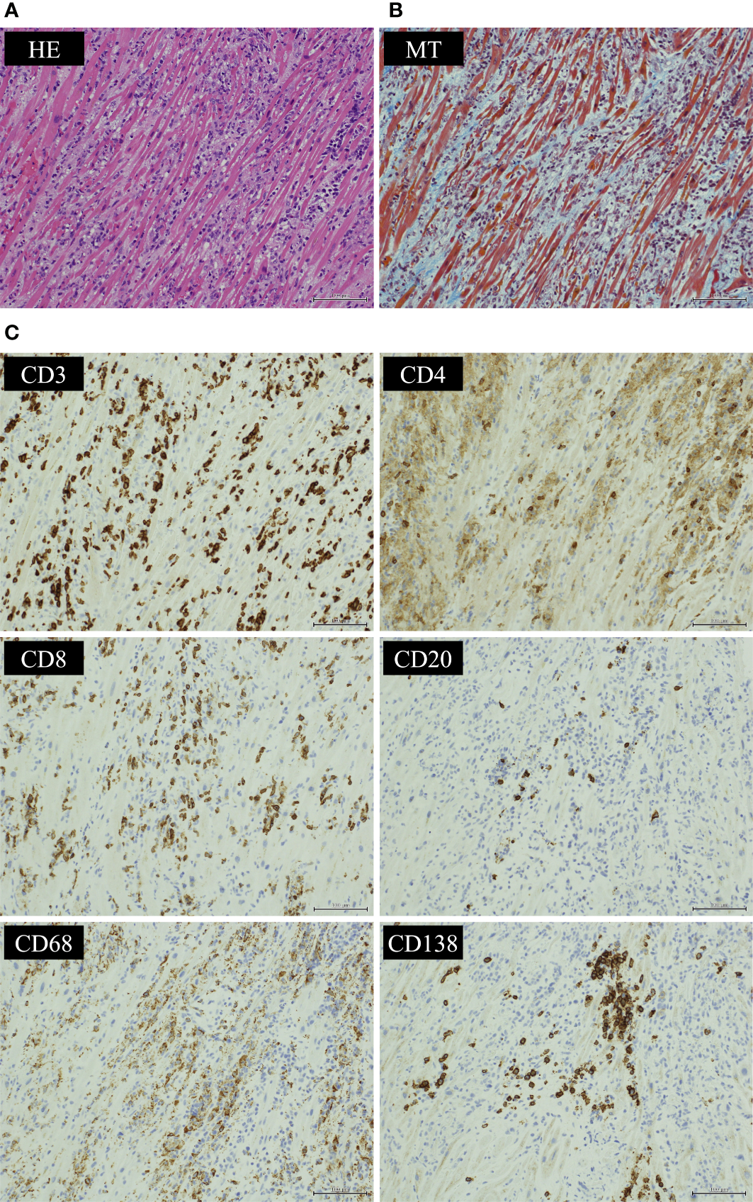
What to see. A: lymphocytic infiltration on H&E. B: stromal fibrosis on Masson trichrome. C: immunostains characterize the infiltrate, including CD3-positive and CD8-positive T cells.
What the image cannot establish. This is one reported case after pembrolizumab and recent SARS-CoV-2 infection. The authors could not fully exclude viral contribution. Histology supports myocarditis, while drug causation requires the clinical context.
Figure 4, © 2023 Nishiyama et al., Frontiers in Immunology. Original source · CC BY 4.0. Reproduced without alteration.
M
Mechanical support · pacing · airway — bridges, not destinations
Resuscitation and goals of care proceed togetherBridges
Shock
Activate the shock/MCS team. Select temporary support from hemodynamics, ventricular involvement, oxygenation, reversibility and cancer prognosis. LV unloading during VA-ECMO is individualized.
Pacing
High-grade AV block / symptomatic bradycardia → urgent pacing/EP support. Temporary support may allow recovery. permanent pacing timing is individualized, without a fixed 28-day wait.
Vent · 20/30/40
Urgent airway assessment for bulbar weakness, weak cough, fatigue or deteriorating mechanics. FVC <20 mL/kg, NIF weaker than −30 or MEP <40 cm H₂O are warning signs, not standalone intubation rules. Do not wait for hypercapnia.
SCD prev
EP-guided risk assessment. Consider temporary protection selectively. Sustained VT/VF, scar, ventricular recovery and prognosis guide ICD decisions. Completion of immunosuppression is not a universal prerequisite.
Transplant
MCS as bridge to recovery. Heart-transplant eligibility after malignancy depends on tumor type, stage, recurrence risk, and oncology assessment. no universal 3- or 5-year wait.
ICI
R
Response criteria — when to advance, hold, or step back up
Anchored to troponin trajectoryAdequate response @ 72 h
Troponin ↓ > 50% from peak + rhythm / LV stable. If not met → Step 2.
Begin taper
Sustained trop trending normal · BNP stable · LVEF stable / recovered · no arrhythmias · FVC stable.
Pause / re-intensify
Rising Tn/BNP, recurrent symptoms, or falling FVC/LVEF → urgently assess relapse and other causes. Specialist-guided treatment.
Do not rechallenge
Generally avoid after severe myocarditis. Exceptions need MDT review of recovery and cancer benefit.
⏱
Monitoring intensity — match the trajectory
Serial cardiac and respiratory reassessment| Parameter | Inpatient | Outpatient |
|---|---|---|
| hs-Tn / CK | Serial troponin to establish trend. CK if myositis. Use the same assay and interpret persistent elevation in context. | Early follow-up during taper, with frequency set by response and relapse risk. |
| BNP | When clinical course or congestion makes the result useful. | As indicated by symptoms, volume and treatment response. |
| ECG / telemetry | Continuous. | Weekly early → space as stable. |
| Echo + GLS | At diagnosis and with clinical change. Repeat before discharge or major treatment decisions as indicated. | Repeat to document recovery or investigate symptoms. Individualize the interval. |
| CMR (if feasible) | Prompt CMR when feasible. A normal early study does not exclude ICI myocarditis. | Follow-up CMR when it will change care. Imaging alone does not determine ICD eligibility. |
| FVC · NIF/MIP · MEP | q 4–6h if overlap sx / evolving. Daily if stable on IS. | As clinically indicated. |
↓
Taper schedule — begin after sustained improvement
Response-guided, individualized taper| Phase | Prednisone | Other agents |
|---|---|---|
| After response | After IV pulse and objective improvement: prednisone/prednisolone 1 mg/kg/day, max 80 mg/day (ESC). | Individualize with oncology/neurology for myositis or MG overlap. |
| Each reduction | Follow symptoms, ECG/conduction, LV function, troponin, and respiratory strength. Taper more slowly at low doses. | Monitor adrenal suppression and infection risk. The calendar alone does not determine treatment. |
| Relapse / persistent injury | Pause taper, assess other causes, and discuss escalation with specialists. | MMF, abatacept, ruxolitinib, IVIG, or PLEX selection and withdrawal require an expert plan. |
PX
Prophylaxis & supportive care on prolonged IS
Layer per regimenPJP
PJPTMP-SMX is commonly used when prednisone ≥20 mg/day is expected for ≥4 weeks or combined immunosuppression creates comparable risk. Check renal function, potassium and counts. Alternatives require individual selection, including G6PD testing before dapsone.
HSV / VZV / CMV
HSV / VZV / CMVUse oncology/infectious-disease risk assessment based on serostatus, agents and immune deficit. Pulse steroids alone do not mandate valacyclovir or valganciclovir. CMV prophylaxis or pre-emptive monitoring is regimen-specific.
Fungal
FungalAntifungal prophylaxis is reserved for defined high-risk settings, including expected profound, prolonged neutropenia. MCS alone is not an indication. Review azole interactions and QT effects.
GI
GIAssess ulcer and bleeding risk. Acid suppression is selective, especially with prior ulcer or concurrent antithrombotics, rather than an automatic consequence of steroids.
Bone
Calcium 1000–1200 mg + Vit D 800 IU daily on long-term GCs. Formal bone-health eval if prolonged or pre-existing fracture risk.
Vaccines
No live vaccines on IS. Update inactivated (influenza, pneumococcal, HBV, COVID, recombinant zoster) before / after the IS window.
VTE prophylaxis
VTE preventionAssess thrombosis, bleeding, platelet count and existing therapeutic anticoagulation. Coordinate device anticoagulation with the MCS team rather than adding duplicate prophylaxis.
Goals-of-care
Early palliative-care involvement when oncologic prognosis informs MCS / Tx eligibility. Frame MCS as bridge to recovery, not destination.
Rx
Why these drugs — mechanism · onset · watch-fors
Anchors clinical reasoningAgent
Methylpred
Broad genomic + non-genomic suppression of all leukocyte lineages — fastest available anti-inflammatory.
Onset/t½ hours · 2–3 h · effect 24–36 h
WatchHyperglycemia · HTN · infection · GI bleed · psych · myopathy.
MMF
Inhibits IMPDH → blocks de novo purine synthesis. B/T-lymphocyte-selective. Prevents clonal expansion.
Onset/t½ days–wk · ~17 h (MPA)
WatchCytopenia · GI · opportunistic infection · hold ANC < 1.3.
Abatacept
CTLA-4–Ig blocks CD80/86–CD28 T-cell co-stimulation. Its use in ICI myocarditis is off label and does not literally reverse every ICI target.
Onset/t½ Response timing in ICI myocarditis is not established. Dosing is specialist-led
WatchLatent TB / HBV reactivation · infusion reaction · screen at baseline.
Ruxolitinib
JAK 1/2 inhibitor — dampens IFN-γ / IL-6 signaling driving cytotoxic T-cell effector function.
Onset/t½ hours · ~3 h · PO BID
WatchCytopenia · HSV/VZV reactivation · cytokine rebound on abrupt stop.
IVIG
FcRn saturation, complement modulation, autoantibody neutralization — antibody-mediated MG / antisynthetase overlap.
Onset/t½ days · ~3 wk
WatchRenal injury, volume load, thrombosis, hemolysis and infusion reactions. Review prior immunoglobulin reactions and product-specific precautions.
References
ESC 2022 and ASCO/ESMO irAE guidance inform care. Salem 2023's abatacept/ruxolitinib strategy is observational. It does not establish a universal regimen. Use specialist guidance for infection prevention, respiratory support, and pacing.
PREVENT
2026
Guideline
Guideline
Goals are backLDL-C targets return (not just % reduction). PREVENT-ASCVD replaces Pooled-Cohort risk. Measure Lp(a) once in adulthood.
Lower for longerEarlier therapy, even in younger adults. 10- & 30-yr risk. CAC, apoB & Lp(a) sharpen who to treat.
Cancer patientsContinue lipid-lowering therapy through cancer treatment unless contraindicated. Defer in pregnancy / lactation.
1
Estimate risk — PREVENT-ASCVD (age 30–79, no ASCVD, LDL 70–189)
10- & 30-year riskLOW < 3%
Lifestyle (Life's Essential 8). No routine LLT.
BORDERLINE 3–<5%
LLT reasonable after clinician–patient discussion + risk enhancers.
INTERMEDIATE 5–<10%
LLT recommended. CAC may refine. Moderate-intensity statin.
HIGH ≥ 10%
Statin (often high-intensity). Treat to goal below.
2
Treatment goals — LDL-C & non-HDL-C
Lower LDL = lower riskBorderline / Intermediate
3–<10% · primary prevention
< 100 mg/dL
non-HDL-C < 130. Any CAC → favor statin to this goal.
High
PREVENT-ASCVD ≥10%. Diabetes / CKD have separate risk-based pathways.
< 70 mg/dL
non-HDL-C < 100. High-intensity statin first-line.
Very-High ASCVD
established ASCVD · secondary prev.
< 55 mg/dL
non-HDL-C < 85. Statin + ezetimibe ± PCSK9.
Severe ↑LDL / FH
LDL ≥190 or familial hyperchol.
↓ ≥50%
Goal <100 mg/dL if no added risk. <70 with FH, added risk or CAC. <55 with ASCVD. Add nonstatin therapy as needed.
3
Lipid-lowering therapy — statin first, then add on to goal
Statin remains the foundation| Step | Agent & dose | LDL ↓ | Use when |
|---|---|---|---|
| High-intensity | Atorvastatin (Lipitor) 40–80 mg · Rosuvastatin (Crestor) 20–40 mg | ≥50% | ASCVD · LDL ≥190 · high / very-high risk · diabetes 40–75 + risk. |
| Moderate | Rosuvastatin 5–10 · Atorvastatin 10–20 · Pravastatin (Pravachol) 40–80 · Pitavastatin (Livalo) 1–4 · simvastatin 20–40 | 30–49% | Intermediate risk · statin-associated symptoms on high-intensity. |
| + Ezetimibe | Ezetimibe (Zetia) 10 mg PO daily | +18–25% | 1st add-on when not at goal on max-tolerated statin. |
| + Bempedoic | Bempedoic acid (Nexletol) 180 mg PO daily | +17–18% | Statin-intolerant or needs more. Oral ACL inhibitor. |
| + PCSK9 mAb | Evolocumab (Repatha) 140 mg q2wk · Alirocumab (Praluent) 75–150 mg q2wk SC | +50–60% | Very-high ASCVD or FH not at <55. Large gap to goal. |
Rx
Risk enhancers & advanced testing
Sharpen the borderline patient- Lp(a) measure once in adulthood (universal). Lifestyle won't change it — ↑ Lp(a) raises risk independent of LDL.
- apoB residual-risk marker when CKM syndrome · T2DM · high TG · known CVD already at LDL/non-HDL goal.
- CAC scan men ≥40 / women ≥45 at borderline–intermediate — any CAC → statin, goal <100. Higher score → lower target.
- Enhancers FHx premature ASCVD · LDL ≥160 · metabolic syndrome · CKD · chronic inflammation · S-Asian ethnicity · preeclampsia / early menopause · ABI <0.9.
- Diabetes age 40–75 without ASCVD: at least moderate-intensity statin, usually LDL-C <100. Multiple risk factors can justify a lower goal. Apply separate CKD/HIV recommendations and individualize outside these ages.
- Triglycerides address secondary causes and use statins for ASCVD risk. Icosapent ethyl requires eligible risk and TG criteria. Persistent TG ≥500, especially ≥1000 mg/dL, prompts a pancreatitis-prevention plan.
CO
Cardio-oncology specifics
Treat through cancer · mind DDIs- Continue LLT through active cancer treatment unless contraindicated — do not reflexively stop statins (2026 guideline).
- Pro-atherogenic Rx VEGF/TKI · nilotinib / ponatinib · ADT · everolimus (mTOR) · lorlatinib · mediastinal RT · ICI (accelerated atherosclerosis) — lower the threshold to start a statin.
- CTRCD prevention specialist-selected statin therapy may be considered at HIGH / V·HIGH toxicity risk. Radiation dose alone is not a universal statin indication.
- Drug interactions prefer rosuvastatin · pravastatin · pitavastatin (minimal CYP3A4). avoid simvastatin / lovastatin with strong CYP3A4 inhibitors & many oral TKIs.
- Defer most LLT during conception, pregnancy & lactation.
One-line plan
Run PREVENT-ASCVD → set the LDL goal → start max-tolerated interaction-reviewed statin → add ezetimibe ± PCSK9 to hit goal → check Lp(a) once → reassess interactions and tolerance during cancer therapy.
4
Statin intolerance (SAMS) — keep patients on therapy
True intolerance is uncommon1 · Confirm
Check CK · TSH · vitamin-D. Exclude hypothyroidism & CYP3A4 DDIs (azoles · TKIs). Most "intolerance" is not true myositis.
2 · Rechallenge
Stop 2–4 wk → rechallenge same or lower dose, symptom-guided. Try ≥ 2–3 statins before declaring intolerance.
3 · Switch
Rosuvastatin alternate-day or pravastatin / pitavastatin. Fewer CYP3A interactions does not mean interaction-free. Check transporters, renal function and dose limits.
4 · Non-statin
Still off goal / truly intolerant → ezetimibe → bempedoic acid → PCSK9 mAb to reach target.
5
Monitoring & follow-up
Adherence > titrationLipid panel
Recheck 4–12 wk after start / dose change, then q3–12 mo. Confirm ≥ 50% LDL drop on high-intensity.
Liver
Baseline ALT. Routine repeat testing not required unless symptoms.
Muscle (CK)
Check only if symptomatic. CK ≥ 10× ULN or rhabdomyolysis → stop now.
Glucose
Small ↑ new-onset DM risk — do not stop the statin. Manage glycemia, benefit outweighs.
22%fewer events / 1 mmol/L LDL ↓
Why it pays off — in oncologyStandard ASCVD prevention remains valuable during cancer care. CTT found about 22% relative reduction in major vascular events per 1 mmol/L LDL-C reduction across trial follow-up. This is not a 22% annual reduction or a proven CTRCD-prevention effect.
PREVENT
2025
Guideline
Guideline
Diagnose & targetHTN at ≥130/80. General treatment goal <130/80, individualized for tolerability, orthostasis, frailty and goals of care.
Risk-based with PREVENTPREVENT replaces pooled-cohort risk. Stage 1 → drug if ASCVD · DM · CKD or 10-yr risk ≥7.5%.
Cancer patientsContinue BP therapy through treatment. VEGF / TKI HTN is the commonest cardio-onc HTN — treat and continue.
1
BP categories — same thresholds as 2017
Diagnose ≥ 130/80 · confirm out-of-officeNORMAL
<120 and <80. Reassess yearly.
ELEVATED
120–129 and <80. Lifestyle.
STAGE 1
130–139 or 80–89. Risk-based Rx.
STAGE 2
≥140 or ≥90. Two-drug Rx.
2
When to treat & target — lower for most
Target < 130/80Elevated
120–129 / <80
Lifestyle
No drug unless it progresses. Reassess 3–6 mo.
Stage 1 · lower risk
PREVENT < 7.5%
< 130/80
Lifestyle 3–6 mo, then drug if still ≥130/80.
Stage 1 · higher risk
ASCVD · DM · CKD · ≥7.5%
< 130/80
Start drug now + lifestyle.
Stage 2
≥140 / ≥90
< 130/80
Start 2 agents, ideally single-pill combination.
3
Drug therapy — race-neutral first-line
Thiazide · ACEi/ARB · DHP-CCB| Step | Agents | Use when |
|---|---|---|
| 1st-line | Thiazide-like chlorthalidone 12.5–25 / indapamide 1.25–2.5 · ACEi lisinopril / ARB losartan (example option) · DHP-CCB amlodipine 5–10 | Most adults — any of the four classes. Not ACEi + ARB together. |
| Start 2 | Two classes, ideally a single-pill combination (e.g. ARB + amlodipine) | Stage 2, or BP ≥20/10 above goal. |
| + MRA | Spironolactone 25–50 · eplerenone | Resistant HTN (4th agent). Screen primary aldosteronism first. |
| + Other | β-blocker · hydralazine · others | Compelling indication (CAD · HF · arrhythmia) or still uncontrolled. |
| Cardio-onc | Avoid non-DHP CCB (verapamil / diltiazem) with CYP3A4-metabolized oral TKIs | Use DHP-CCB (amlodipine) + ACEi/ARB instead. |
Lx
Lifestyle — foundational
For every BP category- Sodium <2300 mg/day, ideal <1500 (cut ≥1000/day). Potassium-based salt substitute — not in CKD or K-sparing drugs.
- Diet DASH-style — more fruit / vegetables / whole grains.
- Weight loss if overweight. GLP-1 / weight management where appropriate.
- Activity 90–150 min/wk aerobic + resistance.
- Alcohol ≤2/day (men) · ≤1/day (women). Stress management.
CO
Cardio-oncology specifics
Treat through cancer · mind DDIs- VEGF / TKI HTN monitor home BP daily in the first cycle and after dose increases, then every 2–3 weeks. The goal and treatment plan depend on prognosis, tolerability and the exact agent.
- First-line ACEi/ARB ± DHP-CCB (amlodipine). Avoid non-DHP CCB with CYP3A4 TKIs.
- Abiraterone mineralocorticoid HTN + hypokalemia — prefer eplerenone (spironolactone has anti-androgen / AR-agonist concern in prostate Ca).
- Also drug-induced ibrutinib / BTK (independently ↑ CV events) · mTOR · steroids · calcineurin inhibitors · EPO agents.
- Severe HTN if SBP ≥180 or DBP ≥110 mmHg, hold the causative cancer therapy while urgently assessing and treating BP. Acute organ injury is an emergency. Resume only after control and oncology review.
4
Secondary HTN — when to work up & how
Resistant · onset <30 · abrupt · unprovoked ↓K⁺ · incidentaloma · crisisPrimary aldosteronism
Screen aldosterone-to-renin ratio (↑aldo + suppressed renin). Correct K⁺, don't salt-restrict, stop MRA 4–6 wk first. Confirm saline / Na-load, then subtype CT + AVS. Screen resistant HTN or suggestive features. Screening may also be considered in stage 2 HTN.
Renovascular
Renal duplex · CTA / MRA. Clues: bruit, flash pulmonary edema, Cr ↑ >30% on ACEi/ARB, young woman (FMD).
Pheo / paraganglioma
Plasma-free or 24-h urine metanephrines. Clues: episodic HTN, headache, palpitations, sweats.
Cushing · endocrine
1-mg overnight dexamethasone / late-night salivary cortisol. Also TSH, Ca / PTH.
Renal · OSA
Cr / UA / renal US. STOP-BANG → sleep study. Cheap — do early in every work-up.
5
Monitoring & follow-up
Recheck · home BP · electrolytesRecheck
About 1 month after start/change, and monthly until controlled. Space follow-up once stable.
Home BP
VEGF: daily in cycle 1 and after dose increases, then every 2–3 weeks. Increase checks if BP rises.
Labs
BMP (K⁺ / Cr) at baseline and 1–2 wk after ACEi/ARB/MRA start.
Don't stop
Continue indicated BP treatment. Severe HTN or acute organ injury may require holding the causative cancer drug.
6
Home BP — how, when & time of day
Validated cuff · average ≥ 130/80 = HTNDevice
Validated automated upper-arm cuff, correct size. Not wrist / finger / smartwatch.
Prepare
Empty bladder. 5 min seated rest, back supported, feet flat, arm at heart level. No caffeine / smoke / exercise ×30 min. Don't talk.
When · time of day
Morning (before meds & breakfast) + evening (before bed). 2 readings 1 min apart each.
How long
7 days (min 3) before each visit. discard day 1, average the rest.
Interpret
Average ≥ 130/80 = HTN (office 130 ↔ home 130. Office 140 ↔ home 135/85).
20%fewer CV events / 10 mmHg SBP ↓
Why it pays off — in oncologyTreat hypertension for its established cardiovascular benefits. Choose a tolerable goal, check interacting drugs and monitor for hypotension when a VEGF inhibitor stops. Do not extrapolate a fixed trial risk reduction to every patient with active cancer.
PROTO
T
Hematopoietic stem-cell transplant — cardiac workup
High-dose conditioning · amyloid screenBefore conditioning
Before conditioningHistory and exam, ECG, BP and baseline cardiac imaging. Review prior cardiotoxic exposures and conditioning regimen. Biomarkers and additional testing depend on risk and planned serial use.
During admission
Telemetry + daily weights through conditioning. Watch cyclophosphamide / melphalan hemorrhagic myocarditis / HF (1–3 wk). Strict fluid balance. Correct K⁺ / Mg²⁺.
Post-transplant
After transplantRisk-based follow-up. High-risk survivors need early reassessment and a long-term plan. New dyspnea, edema, chest pain or arrhythmia warrants prompt testing.
Amyloid screen
Suspected AL amyloidosisSerum free light chains plus serum and urine immunofixation, cardiac imaging and hematology review. CMR characterizes involvement but cannot type amyloid. Tissue confirmation/typing may be needed.
Echo & biomarker standardsUse measured LVEF, preferably 3-D when feasible, and comparable GLS acquisition. Use ultrasound enhancement when endocardial definition is inadequate. CMR adjudicates uncertain imaging. BNP or NT-proBNP is acceptable. Keep assay and modality consistent.
D
Common drug–drug interactions to be aware of
Screen before co-prescribing| Interaction | Why it matters | What to do |
|---|---|---|
| Strong CYP3A4 inhibitor + oral TKI / CDK4-6 / AR inhibitor | Strong CYP3A inhibitors can raise exposure to ibrutinib, ribociclib and many TKIs. The effect is drug-specific. Abiraterone does not share this simple rule. | Avoid strong inhibitors. If unavoidable, dose-reduce per label. ECG + electrolytes. Choose an interaction-sparing antimicrobial. |
| Non-DHP CCB (verapamil / diltiazem) + VEGF-TKI / mTOR | CYP3A4 inhibition ↑ anticancer-drug exposure. | Treat HTN with a DHP-CCB (amlodipine). ACEi/ARB first-line. |
| Simvastatin / lovastatin + CYP3A4 inhibitor / TKI | ↑ statin levels → myopathy / rhabdomyolysis. | Consider pravastatin, rosuvastatin or pitavastatin after a full interaction check. Transporter interactions still matter, including darolutamide–rosuvastatin. |
| Warfarin + ibrutinib / capecitabine / TKI | Ibrutinib platelet dysfunction → bleeding. Capecitabine markedly ↑ INR. | Prefer a DOAC with ibrutinib (screen CYP3A4). If warfarin + capecitabine, check INR q3–5 d. |
| QT-additive stacking | Ribociclib · vandetanib · arsenic · nilotinib + ondansetron / azoles / fluoroquinolone / methadone → Torsades. | Minimize stacking. Baseline + serial ECG. Keep K⁺ ≥ 4.0 · Mg²⁺ ≥ 2.0. |
| DOAC + enzalutamide / apalutamide (strong inducers) | Enzalutamide and apalutamide have different transporter effects. Strong CYP induction may reduce apixaban/rivaroxaban exposure. Apalutamide also induces P-gp. | Review CYP3A and P-gp effects for the exact combination. Dabigatran/edoxaban are not automatically safe substitutes. Consider LMWH or another anticoagulant with oncology/pharmacy input. |
| Anthracycline + trastuzumab (concurrent) | Additive cardiotoxicity → higher CTRCD risk. | Sequence when oncologically appropriate. Follow drug-specific echo surveillance. At HIGH / V·HIGH risk, use cardio-onc review for any specialist-selected prevention. |
S
Cancer-survivor evaluation
Annual review · risk-based echo2026 IC-OS/MASCC minimum-care layer for adults treated for adult-onset cancer: after cancer therapy, review symptoms and CV risk factors annually and re-stratify CTR-CVT risk at 5 years. Obtain a 12-month echo after HER2-targeted therapy or, for early-stage cancer treated with curative intent, cumulative doxorubicin ≥250 mg/m² (or equivalent). In asymptomatic survivors, q5y echo may be considered for MODERATE risk (IIb-C). HIGH / V·HIGH risk: years 1, 3, and 5, then q5y (IIa-C). Imaging-frequency evidence is limited. Childhood-cancer survivors follow their separate exposure-based pathway, and new symptoms trigger referral at any time.
| Exposure | Asymptomatic | Symptomatic (chest pain · dyspnea · palpitations · syncope · fatigue) |
|---|---|---|
| Anthracycline | Annual symptoms + CV risk factors. Echo at 12 mo if early-stage/curative intent and cumulative doxorubicin ≥250 mg/m². Thereafter use post-Tx risk tier. | Above + metabolic stress test. CMR ± stress. Consider rest/exercise RHC. Ziopatch / Holter. CTA-Heartflow (intermediate) or coronary angio (high suspicion). |
| Chest RT > 1 y | Annual symptom/risk review. Tailor echo and CAD screening to heart dose, field, elapsed time and risk. Do not wait for year 10 if symptoms develop. | Above + CMR ± stress. BNP / lipids / TSH. CTA-Heartflow or coronary angio (per CAD suspicion). Metabolic stress. Rest/exercise RHC. Ziopatch / Holter. |
| HER2 / trastuzumab | Annual symptoms + CV risk factors. echo at 12 mo post-Tx, then symptoms and post-Tx risk tier guide longer surveillance. | Above + BNP. Repeat echo. CMR if persistent LV dysfunction. resume / continue GDMT if EF not fully recovered. |
| ADT / hormonal | ASCVD risk. BP · lipids · A1c · weight q6–12 mo. DEXA. Lifestyle + statin by risk. | Above + stress test / CTA per CAD suspicion. Optimize metabolic syndrome. Cardiology if MACE. |
Why lifelong: in 5-year childhood-cancer survivors the 30-year cumulative incidence of symptomatic cardiac events (CHF the most common) reached 7.3% after anthracyclines and 12.6% (~1 in 8) after anthracyclines + chest RT — risk climbs exponentially with cumulative dose, so surveillance never fully stops.
Nursing monitoring protocol
VEGF / TKI · BPHome BP daily in cycle 1 and after dose increases, then every 2–3 weeks. Escalate severe BP or symptoms promptly.
Anthracycline / HER2 Ask weight, dyspnea, edema, orthopnea each cycle. Echo per schedule. Call if new HF symptoms.
ICI · myocarditisUse the agreed risk-based ECG/troponin plan. New symptoms, ECG changes or troponin rise require urgent assessment.
QT agents Pre-dose ECG + K⁺ / Mg²⁺. Keep K⁺ ≥4.0, Mg²⁺ ≥2.0. Call if QTc ≥500 or the agent-specific threshold is reached, or for syncope.
WORKUP
Same symptoms, shifted differential
Cancer patients present like everyone else—but therapy reshapes the odds. For every workup ask: which target/pathway, which agent and combinations, what cumulative exposure, and how long since the last cycle? Class effects predict the common toxicity. Agent pharmacology, prior radiation, and host reserve explain the exception. Pair the evaluation with the drug-class card and HFA-ICOS tier. Keep a low threshold for ECG + troponin + echo.
E
Symptom-based work-ups
Think culprit drug firstSYN
Syncope
Rule out arrhythmic & structural- First pass 12-lead ECG (QTc · conduction · ischemia) · orthostatic vitals · glucose · Hgb · electrolytes.
- Reflex / orthostatic volume depletion (vomiting · diarrhea · poor intake) · autonomic neuropathy (vinca · platinum) → rehydrate, med review.
- Arrhythmic — red flags exertional · supine · no prodrome · FHx SCD → admit + telemetry. QT drugs (ribociclib · vandetanib · arsenic). Anthracycline / RT conduction disease.
- Structural severe AS (RT valve) · HCM / amyloid (myeloma) · large PE → echo.
- Escalate echo → ambulatory monitor (Ziopatch / Holter). implantable loop if recurrent unexplained.
SOB
Dyspnea / shortness of breath
Cardiac vs pulmonary vs anemia- First pass SpO₂ · BNP · troponin · ECG · CBC · echo + GLS.
- HF / CTRCD anthracycline · HER2 · VEGF · carfilzomib → echo + BNP. Treat as CTRCD card.
- Pericardial effusion / tamponade (RT · dasatinib · ICI · cyclophosphamide) → echo.
- Vascular PE / VTE (cancer-hypercoagulable · IMiDs · asparaginase) → CTA-PE. PAH (dasatinib) → echo PASP.
- Pulmonary suspected pneumonitis/ILD → CT chest and urgent oncology/pulmonary review. Exclude infection and PE. Holding therapy and corticosteroids depend on the agent and grade. T-DXd ILD has specific permanent-stop rules.
PALP
Palpitations
Capture the rhythm- First pass 12-lead ECG during symptoms · K⁺ / Mg²⁺ · TSH · echo.
- Atrial fibrillation ibrutinib · alkylators / SCT · ICI · melphalan → rate control (BB. Non-DHP CCB mind DDIs). Use CHA₂DS₂-VASc as an input, then add active cancer, thrombocytopenia/coagulopathy, intracranial disease, interactions, procedures, prognosis, and goals. Cancer neither erases the score nor lets it decide alone. Avoid warfarin with ibrutinib.
- Sinus tach / SVT anemia · thyroiditis (TKI / ICI) · pain · hypovolemia · steroids → treat cause. Check TSH.
- Ventricular / QT QT-prolonging drugs → ECG + electrolytes. PVC / NSVT after anthracycline / RT → echo + monitor.
- Capture Ziopatch / Holter. Event monitor if infrequent.
CP
Chest pain
Vasospasm · ischemia · pericardial- First pass 12-lead ECG + troponin (vs baseline) · telemetry if active.
- Vasospasm / ACS 5-FU / capecitabine during or soon after infusion → stop infusion, nitrates + CCB. ICI myocarditis if troponin ↑ → emergency (P.12).
- Fixed CAD mediastinal RT (premature, ostial) · cisplatin / VEGF (accelerated atherosclerosis) → stress imaging / CTA.
- Pericardial RT · cyclophosphamide · ICI · anthracycline → diffuse ST, echo for effusion / tamponade.
- Non-cardiac PE (pleuritic) · RT esophagitis · musculoskeletal.
EDEMA
Edema / volume / weight gain
HF vs drug vs renal vs venous- First pass weight trend · JVP · BNP · echo + GLS · BMP / albumin.
- HF / CTRCD anthracycline · HER2 · VEGF · carfilzomib → echo + BNP, GDMT.
- Drug fluid retention docetaxel · imatinib (periorbital) · abiraterone (mineralocorticoid) · steroids.
- Renal VEGF proteinuria / nephrotic → UPCR. Cisplatin nephrotoxicity.
- Venous unilateral → DVT duplex. Lymphedema (post-op / RT). SVC / IVC obstruction (tumor).
FATIGUE
Fatigue / reduced tolerance
Don't miss early CTRCD- First pass H&P · CBC · TSH · BMP · BNP · echo + GLS. Functional capacity (METs / DASI).
- Early HF / CTRCD subclinical EF / GLS decline (anthracycline · HER2 · VEGF) → echo + GLS, biomarkers.
- Anemia investigate severity and cause. Transfusion and ESA decisions depend on symptoms, treatment intent and oncology guidance. ESAs carry thrombotic risk.
- Endocrine hypothyroidism (TKI / ICI / neck RT) · adrenal insufficiency / hypophysitis (ICI) → TSH, AM cortisol.
- Other deconditioning · cachexia → cardiac rehab after ruling out the above.
When to escalate now
Syncope with exertion or documented arrhythmia · new HF with hypotension · suspected tamponade · sustained VT or QTc ≥ 500 ms · massive PE → admit, telemetry, urgent cardiology. Suspected ICI myocarditis at any presentation → treat as emergency (P.12).
WORKUP
Cancer patients come to surgery often
Resection · access · biopsy — carrying cardiotoxic exposure, hypercoagulability & deconditioning. Co-manage with oncology · anesthesia · surgery, and don't reflexively stop cardiac meds. For every case ask: which drug, what cumulative dose, how long since the last cycle?
1
Who to refer & what to assess
RCRI · METs / DASI · biomarkersREFER
Refer to cardio-onc if
Higher peri-op CV risk- Exposure active / recent cardiotoxic Tx · prior CTRCD · mediastinal RT · HFA-ICOS high / V-high.
- Functional poor capacity (< 4 METs) · new dyspnea / angina / edema.
- Surgery intermediate / high-risk or major oncologic resection.
- Rhythm / device AF · prior arrhythmia · CIED interrogation if pacemaker / ICD.
ASSESS
Pre-op assessment
Function · cardiac · heme- Risk scores RCRI · METs / DASI · surgery-specific risk.
- Cardiac ECG and biomarkers according to surgical/patient risk. Echo for new symptoms, suspected ventricular dysfunction or significant valve disease. Do not order echo or stress testing solely because cancer is present.
- Heme platelets · anticoagulation status. Cancer hypercoagulability & bleeding risk.
- Volume / lytes correct anemia · K⁺ / Mg²⁺ · hydration (cisplatin).
2
Agent-specific peri-op management
Hold windows · wound healing · bleeding| Agent / class | Peri-op action |
|---|---|
| VEGF / anti-VEGF (bevacizumab · TKIs) | Use agent-specific holds. Bevacizumab: ≥28 d before elective surgery. ≥28 d after major surgery and until healed. TKIs have different windows. |
| BTK inhibitors (ibrutinib) | Hold ~3–7 d peri-op — platelet dysfunction / bleeding. |
| Anticoagulant — cancer VTE (DOAC / LMWH) | Time interruption and restart to drug, renal function, procedure, and hemostasis. do not routinely bridge DOACs. Recent VTE needs specialist planning. |
| Antiplatelet / post-PCI DAPT | Continue aspirin if bleeding allows. delay elective surgery per stent-timing rules. |
| Anthracycline / cardiotoxic chemo | Don't schedule major surgery at nadir. Coordinate cycle timing & marrow recovery. |
| ICI | Generally continue. Keep myocarditis vigilance peri-op (troponin if symptoms). |
| Tamoxifen / hormonal | VTE risk — consider holding for high-VTE-risk surgery. Weigh recurrence. |
3
Optimize · timing · post-op
Don't stop secondary preventionOPT
Optimize & time
Before the OR- Individualize continue indicated statin/β-blocker. Manage ASA and RAAS drugs by surgery/HF risk. Hold SGLT2i 3–4 d before elective surgery.
- Do not stop chronic β-blocker / clonidine abruptly (rebound).
- Timing coordinate with oncology — surgery vs chemo nadir. Allow marrow recovery. VEGF washout.
- Prehab exercise / cardiac rehab when time allows. Correct anemia.
POST
Post-op
MINS · VTE · rhythm- MINS troponin surveillance (0 / 24 / 48 h) in high-risk — injury is often silent.
- VTE high — pharmacologic prophylaxis unless contraindicated. extended after major cancer surgery.
- Rhythm watch post-op AF (esp. thoracic). Telemetry in high-risk.
- Resume GDMT early. Restart anticoagulation & cancer therapy per recovery / wound healing.
Defer or escalate
Active ischemia · decompensated HF · severe symptomatic valve disease · uncontrolled arrhythmia · BP ≥ 180/110 → optimize first, defer elective surgery. Suspected ICI myocarditis → emergency (P.12). Emergent surgery → proceed with invasive monitoring & cardiology co-management.
4
Extended VTE prophylaxis — cancer surgery is different
major abdominal / pelvic oncologic surgeryWHO
Candidates for extended prophylaxis
Beyond the standard 7–10 d- Major abdominal / pelvic oncologic surgery with limited mobility, high BMI, or advanced stage.
- High Caprini score (≥7) — most cancer resections qualify.
- Prior VTE or active chemo-related hypercoagulability.
- Duration consider up to 4 weeks after high-risk major abdominal/pelvic cancer surgery, with bleeding and mobility reassessed.
HOW
Agents & practical points
Balance bleeding vs thrombosis- LMWH enoxaparin 40 mg SC daily × 4 wk — best evidence base.
- Oral options apixaban or rivaroxaban may be offered to selected patients after initial LMWH/UFH. ASCO rates this a weak recommendation. PROLAPS II tested rivaroxaban against placebo after the first postoperative week, not non-inferiority to LMWH.
- Bleeding check hold if active bleed / drain output high. Resume once surgical hemostasis stable.
- Selection review bleeding, renal function, drug interactions, absorption and adherence. Do not generalize trial safety to every resection.
5
Peri-op risk scores — quick reference
RCRI · DASI · METs · surgery risk| Tool | Components | Interpretation |
|---|---|---|
| RCRI | High-risk surgery · IHD · HF · CVA/TIA · DM on insulin · Cr >2 mg/dL | Use a validated perioperative calculator with its stated endpoint and population. The same RCRI score does not imply one universal event probability. |
| DASI | 12-item Sx questionnaire → estimated METs (walk 2 blocks, climb stairs, run, etc.) | DASI ≤34 or <4 METs signals poor capacity. Stress testing only in selected elevated-risk cases where it changes management |
| Functional | Can walk 4 mph? Climb 2 flights? Heavy housework? | ≥4 METs = adequate capacity → proceed without testing if no active cardiac condition |
| Surgery risk | Low: endoscopy, cataract. Intermediate: abdominal, ortho, H&N. High: aortic, major vascular | Cancer resection = usually intermediate or high. Combine with RCRI + function |
Cancer therapy may reduce functional capacity through fatigue, anemia and deconditioning. Evaluate reversible causes and use prehabilitation when feasible. Treat anemia by cause and clinical context, without a universal Hb 10 g/dL intervention threshold.
REF
H
Hold / Stop & Resume criteria — by therapy class
Check the exact label · no universal restart threshold| Therapy class | When to HOLD / STOP | Immediate management | When to RESUME (or switch) | Tests before resumption | Post-resumption surveillance |
|---|---|---|---|---|---|
| AnthracyclinesDox · Epi · Idarubicin | Interrupt for moderate/severe asymptomatic CTRCD or clinically important HF. Isolated Tn/GLS change needs assessment. | HF therapy. Exclude other causes. Assess dexrazoxane/liposomal options. | MDT after recovery and benefit-risk review. No universal EF/4-week rule. | TTE/GLS, Tn/NP. Lifetime exposure. | Close monitoring, often each 1–2 cycles after restart. Risk-based survivorship. |
| HER2 therapiesTrastuzumab · ADCs | Interrupt for EF <40% or moderate/severe symptomatic CTRCD. Reconcile exact label. | HF therapy and MDT. Asymptomatic EF 40–49% may continue with protection/monitoring. | ESC: asymptomatic, EF ≥40% (ideally ≥50%). Individual labels differ. | TTE/GLS, Tn/NP. | TTE/biomarkers every 2 cycles initially after restart, then risk-based. |
| VEGF / multi-TKIsSunitinib · Lenvatinib | Severe/uncontrolled HTN, important CTRCD, vascular toxicity or QT per label. | ≥180 systolic or ≥110 diastolic: hold. Treat BP and evaluate organ injury. | BP <160/100 after severe HTN per ESC, plus stricter product recovery rules. | BP log. Renal/electrolytes. ECG/TTE by phenotype. | Home BP and agent/risk-specific ECG, renal, and LV surveillance. |
| Anti-VEGF mAbBevacizumab | Hold severe HTN uncontrolled medically. Discontinue crisis/encephalopathy, severe ATE or grade 4 VTE. | Treat BP/event. Evaluate bleeding, renal injury, proteinuria. | HTN: once medically controlled. No restart after label-defined permanent-stop toxicity. | BP. Urine protein/renal function. Wound status. | BP at least every 2–3 weeks. Serial urine protein assessment. Follow exact label hold thresholds. |
| BCR-ABL TKIsNilotinib · Ponatinib | New ischemic limb/neurologic/coronary symptoms: urgent assessment and label-based hold. | Treat vascular event. Antithrombotics by indication and bleeding risk. | MDT weighs alternative TKI. No universal ABI-normalization restart rule. | Vascular assessment, ECG, lipids/BP. | Agent- and event-specific vascular follow-up. |
| Dasatinib (PAH)CML | Suspected PAH: interrupt/evaluate. RHC confirms hemodynamic phenotype. | PH specialist assessment. Manage RV failure and PAH as indicated. | Permanently discontinue if PAH confirmed. Choose alternate cancer therapy. | Echo, BNP, RHC as indicated. | Follow PH/RV recovery. Do not rechallenge based only on improvement. |
| Immune checkpointPembro · Nivo · Ipi | Suspected myocarditis: hold immediately and urgently assess. | Prompt steroids when likely/unstable. Monitored care. Specialist adjuncts if refractory. | Usually discontinue confirmed disease. Any rechallenge is exceptional MDT judgment. | Clinical/rhythm, Tn, TTE ± CMR. Assess neuromuscular overlap. | Individual plan through taper and after recovery. |
| CAR-T (CRS)Axi-cel · Tisa-cel | CRS is managed after cell infusion. It is not a reversible oral-drug hold. | Grade-based cellular-therapy pathway. Tocilizumab/steroids as indicated. Support perfusion. | Future cancer therapy only after recovery and cellular-therapy team review. | Hemodynamics, oxygenation, rhythm. TTE/biomarkers as indicated. | Product pathway and follow-up of residual injury. |
| CDK4/6Ribociclib | QTcF >480 ms: interrupt. Recurrent >500 ms or serious QT-associated arrhythmia requires permanent discontinuation per label. | Correct electrolytes/DDIs. Serious arrhythmia requires emergency care. | ≤480 ms. Same/lower dose by cancer setting and event history in label. | ECG. K/Mg. Interaction list. | ECG at baseline, ~day 14 C1, and as clinically indicated. |
| ALK / ROS1 TKIsCrizo · Alectinib | Symptomatic bradycardia or significant QT prolongation: apply specific label. | Hold if indicated. Assess nodal drugs, conduction and reversible causes. | Symptoms resolve and agent-specific HR/QT recovery criteria are met. | ECG, pulse, electrolytes. | Agent-specific ECG/pulse monitoring. |
| EGFR TKIOsimertinib | QTc >500 ms on ≥2 ECGs. Permanently stop life-threatening QT arrhythmia or symptomatic CHF. | Hold, correct electrolytes/DDIs. Urgent rhythm/HF care as indicated. | QTc <481 or baseline if baseline ≥481. Resume 40 mg per label. | ECG, electrolytes. LV assessment by risk/symptoms. | ECG/electrolytes in at-risk patients. LV surveillance as indicated. |
| Fluoropyrimidines5-FU · Capecitabine | Chest pain/ACS, ischemic ECG or other acute CV toxicity: stop exposure. | ECG/Tn, ACS evaluation. Nitrates/CCB for vasospasm. | Alternative regimen preferred when feasible. Essential rechallenge needs MDT prophylaxis/monitoring. | Exclude severe CAD. Assess ischemia and LV function. | Monitored re-exposure according to the agreed protocol. |
| High-dose alkylatorsSCT conditioning | Acute HF/instability or suspected myocardial injury: urgent transplant-team evaluation. | Support circulation. Phenotype-directed HF/arrhythmia care. No automatic steroid rule. | Alternative or further therapy after MDT recovery assessment. | TTE/biomarkers. Renal, fluid, infection assessment. | Transplant and cardiac follow-up matched to injury. |
| PlatinumCis · Carbo · Oxali | Acute vascular event, severe renal/electrolyte or hemodynamic toxicity. | ACS/vascular care. Correct electrolytes and manage volume. | Oncology/MDT decision by event, kidney function, alternatives and recovery. | Renal/electrolytes. CV tests driven by event. | Renal/electrolytes during therapy. Long-term vascular risk. |
| BRAF / MEKDab + Tram | LV decline, symptoms or QT toxicity by the individual agent label. | Hold responsible agent(s) as indicated. HF therapy and correction of causes. | Recovery, dose, and permanent-stop thresholds differ by drug. | TTE, ECG, electrolytes as indicated. | Label-directed LV/BP and QT monitoring. |
| BTK inhibitorsIbrutinib | Serious arrhythmia, uncontrolled severe HTN, or bleeding: assess label hold criteria. | Rate/rhythm care. Individual anticoagulation/DDI/platelet review. | MDT may restart, reduce or switch after control. AF alone is not an automatic permanent stop. | ECG, BP. Thrombotic/bleeding risk. | Pulse/BP and bleeding assessment each visit. |
| Proteasome inhibitorsCarfilzomib | Significant HF or CV toxicity: hold per label. BNP rise alone needs context. | Assess congestion, BP, renal function and other causes. Treat HF. | Consider lower-dose restart after recovery and MDT benefit-risk assessment. | TTE/NP as indicated. BP and volume. | BP/volume and risk-directed cardiac testing. |
| Hormonal — prostateAbiraterone · ADT | Severe HTN, hypokalemia, fluid retention or HF: assess and hold as indicated. | Correct K/BP/volume. Verify steroid pairing and interactions. | Label and oncology plan after control. ADT selection individualized. | K, BP, liver tests and volume status. | Abiraterone: BP/K/fluid retention at least monthly. |
| mTOR inhibitorsEverolimus | Significant pneumonitis, infection, metabolic or vascular toxicity per label. | Identify cause. Drug hold/steroids depend on severity and diagnosis. | Recovery and dose-modification rules are product-specific. | Pulmonary evaluation, BP, lipids/glucose, renal and counts. | Periodic metabolic/organ surveillance by label and risk. |
EVIDENCE
E
Recommendation class is not a patient risk score
ESC 2022 · interpretationIRecommended. Expected benefit supports the action in the stated population.
IIaShould be considered. Evidence or opinion favors usefulness.
IIbMay be considered. Usefulness is less well established.
IIINot recommended. No benefit or potential harm in the specified setting.
Evidence levelA: multiple randomized trials/meta-analyses. B: a randomized trial or large non-randomized studies. C: consensus, small studies or registries. A strong recommendation can rest on limited trial data. Neither risk-factor stacking nor clinician confidence changes the published class.
1
Identify which kind of statement you are reading
| Source | What it answers | How to apply it |
|---|---|---|
| Regulatory label | Approved indication, formulation, dose modifications, monitoring and permanent-stop criteria. | Check the exact agent and cancer setting. A class summary cannot replace a product label. |
| Society guideline | A recommendation for a defined clinical population. | Retain its year, population, class and evidence level. Read exceptions and contraindications. |
| Clinical practice statement | Consensus implementation where evidence is evolving. | Distinguish a proposed minimum-care approach from a trial-proven optimum. |
| Randomized trial | A comparison in enrolled patients using specified outcomes. | Check absolute risk, endpoint and follow-up. LVEF preservation does not establish a survival benefit. |
| Observational study | Association, feasibility or a safety signal. | Consider confounding, selection and surveillance bias. Association is not proof that intervention changes outcomes. |
| Example schedule | One way a program may operationalize monitoring. | Adapt to actual drug, risk, symptoms and available care. It is not a universal mandate. |
2
Three decisions where the distinction changes care
HER2 therapy
Selected asymptomatic patients with LVEF 40–49% may continue under a cardio-oncology plan. This does not erase the different hold/resume rules for trastuzumab, T-DM1 and T-DXd.
ICI myocarditis
Urgent treatment follows clinical suspicion and severity. Abatacept/ruxolitinib experience is promising but largely observational. It is not a fixed rescue regimen for every patient.
Primary prevention
Treat established CV indications. Selective high-risk prevention is different from routinely prescribing a multi-drug cardioprotective bundle to every patient.
Primary sourcesESC cardio-oncology 2022, HFA-ICOS baseline assessment 2020, IC-OS/MASCC 2026 and current product labels. The drug cards and hold/resume matrix preserve the clinical context for their use.
EVIDENCE
II
Selective care requires a reason and a reassessment plan
Guideline options · individualized decisionsClass II does not become Class I by accumulation. Multiple risks may strengthen the case for an intervention, but do not change its published recommendation class. Document the expected benefit, competing harms and patient preference.
| Decision | Reason to consider | Guardrail |
|---|---|---|
| ACEi/ARB, beta-blocker or statin before exposure | ESC 2022 offers selective prevention in high/very-high toxicity risk. | Not a routine bundle. Standard hypertension, ASCVD and HF indications remain separate. Review hypotension, renal function and interactions. |
| Dexrazoxane / liposomal anthracycline | Necessary anthracycline treatment with substantial cardiac vulnerability. | Match cancer efficacy, cumulative exposure and regulatory indication. A single 250 mg/m² threshold does not define every eligible or ineligible patient. |
| New isolated GLS or troponin change | A reproducible signal may precede LVEF decline. | Check loading, rhythm, assay, image quality and other causes. Do not use an invented absolute GLS cutoff or automatically stop cancer treatment. |
| CMR / biopsy | Uncertain ventricular function, tissue disease or unresolved suspected myocarditis. | Ask what diagnostic uncertainty the test resolves. Do not delay urgent treatment in an unstable patient. |
| After radiation | Exposure-informed surveillance and conventional CV prevention. | Use field, mean-heart/substructure dose, time and symptoms. No universal radiation dose mandates statin, aspirin or coronary CT. |
| ADT choice | Cardiovascular history can inform agonist versus antagonist discussion. | Trial populations and endpoints differ. Do not promise a cardiovascular advantage for every patient. |
| SGLT2 inhibitor | Established HF, diabetes or CKD indication. | Do not label primary CTRCD prophylaxis an ESC 2022 Class II recommendation. Evaluate the current indication and safety. |
| Rehabilitation / exercise | Functional limitation, deconditioning and eligible cardiac disease. | Prescribe to symptoms and safety. Cancer exposure alone does not establish payer eligibility for cardiac rehabilitation. |
?
Before ordering, answer four questions
1 · What problem is being prevented?
Distinguish HF, an imaging change, a vascular event and treatment interruption. Benefits for one endpoint do not prove benefits for the others.
2 · Does this patient resemble the evidence?
Check age, cancer stage, drug, cumulative dose, baseline function and prior therapy. A lymphoma statin trial is not a universal cancer prevention protocol.
3 · What can the intervention harm?
Assess BP, renal function, electrolytes, thrombocytopenia, interactions and treatment burden. Avoid aspirin solely for TKI exposure without an established indication or a specific specialist plan.
4 · How will the plan be reassessed?
Name the symptom, laboratory or imaging trigger, the clinician responsible and the timing. New toxicity changes the risk assessment.
Keep the treatment goal explicitPreserve effective cancer care while managing cardiovascular risk. A palliative treatment goal does not automatically exclude useful cardiovascular therapy, and curative intent does not make every optional intervention mandatory.
PEARLS
Reframe
the field
the field
BidirectionalCancer therapy → CV disease, and CV disease → cancer (shared biology & risk factors). Not "reverse" — bidirectional.
Beyond a low EFCardiotoxicity is much broader than HFrEF — HFpEF, vascular, metabolic, arrhythmic, pulmonary-vascular.
Permissive cardiotoxicityThe immediate threat is the cancer — don't reflexively stop life-saving therapy for mild LV dysfunction.
1
Bidirectional cardio-oncology
shared biology, not one-way→
Therapy → CV disease
the classic axis- Direct toxicity anthracyclines, HER2, VEGF, ICI, RT — across the CV spectrum.
- Aging survivors >18 M US survivors. Metastatic disease is now chronic → years of cumulative exposure.
←
CV disease → cancer
reverse axis- Association CV disease and cancer share exposures and biological pathways. Observed associations do not establish that one disease directly causes the other in an individual patient.
- HF and cancer cohort estimates vary with age, smoking, comorbidity and surveillance. Interpret relative risk alongside the population and absolute incidence.
- Mechanism inflammation · clonal hematopoiesis · neurohormonal activation · shared risk factors.
2
Cardiotoxicity is broader than a low EF
phenotype by drugHFpEF
HFpEFTreatment-related HF can occur with preserved EF. Symptomatic CTRCD includes a confirmed HF syndrome. Investigate filling pressure, valves, pericardium and alternative causes.
Vascular
accelerated atherosclerosis (RT, TKI), vasospasm / ACS (5-FU, capecitabine), PAD.
Metabolic
ADT → ↑ fat mass, diabetes, HTN, metabolic syndrome, fatigue.
Hypertension
VEGF / VEGF-TKI — on-target, within the first week (~↑10 mm Hg).
Arrhythmia
BTK inhibitors → AF and VT. QT prolongation with several agents.
PH · valve · pericardium
dasatinib → PAH. Radiation → valve, pericardial & conduction disease.
3
Anthracyclines & permissive cardiotoxicity
dose · mechanism · don't stop reflexivelyA
Anthracyclines
still in wide use- Dose-dependent risk rises sharply > 250 mg/m², but there is no truly safe dose.
- Mechanism topoisomerase-2β, ROS, iron handling, titin → systolic and diastolic (HFpEF) injury.
- Prognosis cardiotoxicity can complicate cancer care. An association with worse survival does not prove that prevention restores a fixed amount of survival.
↔
Permissive cardiotoxicity
HER2 & beyond- HER2 EF decline common (3–33%, highest after anthracycline), usually reversible.
- Don't reflexively stop life-saving therapy for mild LV dysfunction → start neurohormonal Rx, monitor closely, continue cancer therapy.
- Small prospective studies SAFE-HEaRt: 27/30 evaluable patients completed planned HER2 therapy. SCHOLAR: 18/20 completed trastuzumab, but 2/20 developed clinically important HF. Both used specialist follow-up and HF therapy.
4
Why the field matters
a growing, aging, exposed populationSurvivors
Cancer survivors may live for decades after treatment. Exposure history and competing risks shape long-term CV care.
Competing mortality
CV disease is a leading cause of non-cancer death in survivors.
Metastatic = chronic
prolonged survival on serial cardiotoxic regimens → cumulative risk.
Risk varies
by cancer & therapy — AF with targeted agents, IHD in lung cancer, HF with anthracyclines.
Association is not treatment effectAAML0531 linked cardiotoxicity with worse pediatric AML outcomes. It did not randomize cardioprotection or show that avoiding cardiotoxicity recovers a 20% survival benefit.
5
Clonal hematopoiesis — the biological bridge
CHIP / CCUS · shared biology · emerging targetCH
What it is
somatic mutations in blood- CHIP somatic mutations (DNMT3A, TET2, ASXL1, TP53, JAK2) in hematopoietic cells at VAF ≥2% without cytopenias or hematologic malignancy.
- Prevalence 10–20% of adults >70 y. Rises with age, smoking, prior chemo / RT.
- CV link CHIP is associated with vascular risk, with important variation by gene and clone size. These associations do not yet define a routine cardio-oncology screening or treatment pathway.
CO
Cardio-onc relevance
why it matters here- Cancer Tx → CHIP cytotoxic chemo (alkylators, platinums) and RT select for pre-existing clones (esp. TP53, PPM1D) — cancer treatment accelerates CHIP.
- Progression risk depends on mutation, clone size, cytopenias and other features. Avoid applying a single annual transformation rate to every clone.
- Double hit CHIP patients who receive anthracyclines may face compounded CV + hematologic risk — an emerging reason to integrate hematology and cardiology surveillance.
6
Key terminology — precision in language
what the terms actually meanCTRCD
CTRCDSymptomatic: a confirmed HF syndrome. Asymptomatic: severe LVEF <40%, moderate LVEF 40–49% with the specified EF/GLS/biomarker changes, or mild LVEF ≥50% with new relative GLS reduction >15% and/or biomarker rise. Use the complete severity definition.
Reversibility
RecoveryDescribe symptoms, LVEF, GLS and biomarkers over time. EF normalization does not guarantee complete myocardial recovery. Avoid an unsupported universal 5-point/12-month irreversibility rule.
Cardioprotection
Primary = before exposure. CV drugs are not a routine bundle, though specialist-selected neurohormonal/statin therapy may be considered at HIGH / V·HIGH risk. Secondary follows detected injury.
PEARLS
1
Radiation heart disease
no threshold doseGy
The dose-response
Darby, NEJM 2013- +7.4% / Gy major coronary events per Gy mean heart dose, no threshold. Begins <5 yr, persists >20 yr.
- Modern era mean heart dose is far lower (~1–2 Gy vs ~4.9 historically)—but historical mantle-field risk does not transport directly to modern conformal therapy.
- Read the plan record field and era, mean-heart dose, and relevant substructure dose. Late disease follows beam geometry and tissue dose—not a generic “chest radiation” label. LV dose informs HF risk and pulmonary-vein dose may inform AF risk.
Rx
What it causes
screen + modify risk- Spectrum CAD, HFpEF, valve disease, pericardial disease, conduction disease.
- Manage control conventional risk factors. Statins follow ASCVD/risk indications. Select CAD screening by exposure and clinical risk, rather than treating all prior radiation identically.
- Late, additive often a "second hit" decades later — keep it on the survivorship problem list.
2
Monitoring — what actually predicts
change > baselineAnthracycline echo
baseline. At 12 mo post-Tx if early-stage/curative intent and cumulative doxorubicin ≥250 mg/m². Then risk-based (limited evidence).
GLS
the change (not baseline) predicts subsequent EF decline. Clinical actionability still evolving.
Biomarkers
not a universal baseline bundle. Consider hs-Tn/BNP at HIGH / V·HIGH risk, then interpret trends in clinical context.
CMR
EF adjudication (gold standard), ICI myocarditis dx, post-RT fibrosis / tissue characterization.
Serial imaging is a measurement contract. Keep modality, acquisition, loading, rhythm, vendor/software, and analysis constant. Prefer 3-D EF when feasible, and ask whether change exceeds repeatability. GLS change is prognostic, but SUCCOUR did not prove that every GLS-guided strategy improves outcomes.
“Late toxicity” is usually a second hit. Anthracycline EF decline is often detectable early. Later presentations commonly acquire a trigger such as HTN, AF, or coronary disease.
“Late toxicity” is usually a second hit. Anthracycline EF decline is often detectable early. Later presentations commonly acquire a trigger such as HTN, AF, or coronary disease.
3
Prevention — risk-guided, then go hard on risk factors
intensive RF modification is the leverCp
Cardioprotection
not one-size-fits-all- Not routine ACEi/ARB, β-blocker, statin, or SGLT2i solely for primary CTR-CVT prophylaxis. Treat standard CV indications. Specialist-selected neurohormonal/statin therapy may be considered at HIGH / V·HIGH risk.
- Enalapril Cardinale 2006: LVEF decline 43% control vs 0% enalapril (P <0.001) in troponin-positive patients after high-dose chemotherapy (114 randomized).
- HER2 cardioprot. Guglin 2019: in the anthracycline-pretreated subgroup, cardiotoxicity-free survival longer on carvedilol (P = 0.009) and lisinopril (P = 0.015) vs placebo (overall cohort NS).
- STOP-CA lymphoma patients: LVEF decline ≥10 points to <55% at 12 months occurred in 13/150 (9%) on atorvastatin and 33/150 (22%) on placebo. HF events did not differ significantly. Generalizability is limited.
- PRADA II sacubitril/valsartan did not significantly improve the primary LVEF endpoint in early breast cancer. Secondary imaging/biomarker signals require separate interpretation.
RF
Intensive risk-factor control
the biggest win- Blood pressure use evidence-based treatment with individual tolerability. Do not assume general-population intensive-BP trial results apply unchanged during active cancer.
- VEGF-TKI HTN rises in the first week → treat & continue the drug.
- Opportunistic CAC use calcium on oncology CT to inform ASCVD discussion and treatment. Integrate burden, age, competing illness and patient preference.
4
Drug-specific prevention
tailor to the agentADT (prostate)
metabolic syndrome — treat BP, lipids, glucose. Statin. CV risk burden is high & undertreated.
VEGF / TKI
VEGF / TKIHome BP daily in cycle 1 and after dose increases, then every 2–3 weeks. Severe HTN needs a treatment hold and coordinated care.
Anthracycline
baseline echo. Minimize cumulative dose. Use cardio-onc review for any specialist-selected prevention at HIGH / V·HIGH risk—no routine CV-drug bundle.
Fluoropyrimidine
5-FU / capecitabine → coronary vasospasm / ACS — counsel, recognize, treat.
5
Equity & the cardiologist's role
don't write the patient off- Disparities assess medication cost, transport, access to BP devices and reliable follow-up. Do not attribute one fixed proportion of racial disparities to a single pathway without a specific study and population.
- Environment air pollution (PM2.5, ozone) associates with systolic dysfunction. Neighborhood walkability with lower HTN / diabetes / obesity.
- The role a cancer diagnosis is not a reason to under-treat CV disease — manage it intensively so the patient can complete the life-saving cancer therapy & avoid an interrupting CV event.
6
Exercise & cardiac rehabilitation in cancer
underused, evidence-based, safeRx
Why exercise matters
during & after treatment- Exercise improves fitness, function and fatigue in selected cancer populations. Prevention of clinical anthracycline HF remains unproven.
- Mortality physical activity is associated with better survival in observational studies. Magnitude varies by cancer and study design. Do not promise a universal mortality reduction.
- Function combats cancer-related fatigue (the #1 patient complaint), deconditioning, sarcopenia from ADT / steroids.
- Mental health reduces depression & anxiety. Improves QoL scores across cancer types.
How
Prescribing exercise
structured & supervised- Goal 150 min/wk moderate-intensity aerobic + 2 d/wk resistance training (ACS / ACSM guideline).
- Rehabilitation refer to oncology exercise or cardiac rehabilitation when clinically appropriate. Standard cardiac diagnoses and payer rules determine covered cardiac rehabilitation.
- Prehab 2–4 wk structured exercise before major surgery ↓ LOS & complications.
- Safety exercise is safe during chemo/RT with appropriate screening. avoid high-intensity if active myocarditis, unstable arrhythmia, or symptomatic HF.
Shared treatment goalSupport effective cancer treatment through timely CV care. The oncologic consequence of a hold depends on regimen, setting, duration and alternatives. Some treatment interruptions are necessary to prevent serious harm.
REF
D
Disease-group cardio-onc workup matrix
Expanded reference · cross-ref P.3–10 drug cardsHow to use this pageMatch the disease group to the actual agents, dose, stage and treatment intent. These prompts do not replace current labels or the individualized surveillance plan. New symptoms take priority over a routine interval.
| Disease | Cardiotoxic regimens | CV signals | Assessment prompts | Clinical pearls | Escalate to cardio-onc |
|---|---|---|---|---|---|
| BreastHER2+ · HR+ · TNBC |
|
|
|
|
|
| GI / PancreaticCRC · Gastric · HCC |
|
|
|
|
|
| GUProstate · RCC · Testicular |
|
|
|
|
|
| Thoracic / H&NNSCLC · SCLC · H&N SCC |
|
|
|
|
|
| GynecologicOvarian · Cervical · Endometrial |
|
|
|
|
|
| Mal HemLeukemia · Lymphoma · Myeloma |
|
|
|
|
|
| Melanoma / SarcomaICI-heavy · anthracycline |
|
|
|
|
|
| Neuro-OncGBM · CNS mets |
|
|
|
|
|
| Benign Hem / Sickle CellSCD · Thalassemia |
|
|
|
|
|
Baseline assessmentBefore potentially cardiotoxic therapy, document CV history and conventional risk factors. Use a validated therapy-specific HFA-ICOS proforma where applicable. The disease label alone does not determine risk or surveillance.
Rx
R
Representative cancer therapies — identify the CV exposure
For the cardiologist — what your patient is receivingWhy this page existsUse these examples to recognize cardiovascular exposures, not to select an oncology regimen. Stage, biomarkers, prior therapy, approvals and treatment intent determine sequencing. This table is not exhaustive and does not rank current first-line options.
| Cancer | Common treatment examples | Other settings / later therapies | Key CV-toxic agents (→ drug card page) |
|---|---|---|---|
| SOLID TUMORS | |||
| Breast, HER2+ |
|
|
|
| Breast, HR+/HER2− |
|
|
|
| Breast, TNBC |
|
|
|
| Gynecologic / Ovarian |
|
|
|
| Colorectal |
|
|
|
| Pancreatic |
|
|
|
| Gastric / Esoph |
|
|
|
| RCC |
|
|
|
| ProstateSeparate mCSPC from mCRPC |
|
|
|
| NSCLC |
|
|
|
| SCLC |
|
|
|
| Head & Neck |
|
|
|
| Melanoma |
|
|
|
| HEMATOLOGIC MALIGNANCIES | |||
| DLBCL |
|
|
|
| CML |
|
|
|
| Mult Myeloma |
|
|
|
| AML / APL |
|
|
|
| CLL / SLL |
|
|
|
| Hodgkin |
|
|
|
| SPECIAL POPULATIONS | |||
| Sarcoma |
|
|
|
| Testicular |
|
|
|
Page numbers after each agent (→ P.3, P.5, etc.) point to the drug-class card with full toxicity profile, HFA-ICOS scoring, and management. CV = significant cardiotoxicity. LO = lower risk but monitor.
Rx
A
Common regimen acronyms — what your patient is actually getting
Shorthand decoded · CV = cardiotoxic
Why this page exists
When a patient's chart says "on R-CHOP" or "completed KEYNOTE-522," a cardiologist needs to know which drugs are inside that acronym — and which ones are cardiotoxic. The bolded letter in each component maps to the acronym. Cross-reference with P.3–10 drug cards for management.
| Regimen | Components (generic) | Used in | CV-toxic agent(s) |
|---|---|---|---|
| R-CHOP | Rituximab · Cyclophosphamide · Hydroxydaunorubicin (CV doxorubicin) · Oncovin (vincristine) · Prednisone | DLBCL · NHL | CV Doxorubicin → P.3 |
| ddAC-T | Dose-dense CV Adriamycin (doxorubicin) + Cyclophosphamide q2wk → Taxane (paclitaxel q2wk) | Breast (neo/adj) | CV Doxorubicin → P.3 |
| KEYNOTE-522 | CV Pembrolizumab + carboplatin/paclitaxel, then anthracycline + cyclophosphamide, followed by adjuvant pembrolizumab after surgery. Original trial AC/EC was every 3 weeks. Dose-dense variants are modified regimens. | TNBC (neoadj) | CV Pembro → P.4. Doxo → P.3 |
| FOLFOX | CV Fluorouracil (5-FU) + Leucovorin (FOL) + Oxaliplatin | CRC · Gastric | CV 5-FU → P.6 (vasospasm) |
| FOLFIRI | CV Fluorouracil + Leucovorin + Irinotecan | CRC, in several treatment settings | CV 5-FU → P.6 |
| FOLFIRINOX | CV 5-FU + leucovorin + irinotecan + oxaliplatin | Pancreatic | CV 5-FU → P.6 |
| CapeOx | CV Capecitabine (oral 5-FU) + Oxaliplatin | CRC · Gastric | CV Capecitabine → P.6 |
| BEP | Bleomycin + Etoposide + cisPlatin | Testicular (curative) | CV Cisplatin → P.9 |
| ABVD | CV Adriamycin (doxorubicin) + Bleomycin + Vinblastine + Dacarbazine | Hodgkin lymphoma | CV Doxorubicin → P.3 |
| TCHP | Taxotere (docetaxel) + Carboplatin + CV Herceptin (trastuzumab) + Perjeta (pertuzumab) | Breast, HER2+ | CV Trastuzumab → P.5 |
| VRd | Velcade (bortezomib) + Revlimid (lenalidomide) + dexamethasone | Mult myeloma (1L) | LO Lenalidomide (VTE) |
| KRd | CV Kyprolis (carfilzomib) + Revlimid + dexamethasone | Multiple myeloma, selected treatment settings | CV Carfilzomib → P.7 |
| R-ICE | Rituximab + Ifosfamide + Carboplatin + Etoposide | DLBCL salvage → SCT | CV Ifosfamide → P.4 |
| BEAM | BCNU (carmustine) + Etoposide + Ara-C (cytarabine) + Melphalan | Auto-SCT conditioning | CV High-dose alkylators → P.4 |
| 7+3 | Cytarabine × 7 d + daunorubicin × 3 d | AML induction | CV Daunorubicin → P.3 |