Assess perfusion and rhythm together. Tachyarrhythmia with hypotension, ischemic chest pain, acutely altered mentation, shock, or acute heart failure that is caused by the rate → immediate synchronized cardioversion, except sustained polymorphic VT, which needs an unsynchronized shock. Do not shock compensatory sinus tachycardia. Obtain a 12-lead when feasible without delaying treatment: classify by QRS width × regularity.
▤
Tachycardia decision algorithm
stable? → width × regularityRegular + narrow <120 ms
For SVT: modified Valsalva → adenosine. Sinus tachycardia needs treatment of its cause. Flutter may be unmasked without terminating.
Regular + wide ≥120 ms
Manage uncertain WCT as VT. Consider an antiarrhythmic infusion or synchronized cardioversion. Adenosine only if stable, regular and monomorphic.
Irregular + narrow
AF, variable-block flutter or MAT. Choose rate/rhythm therapy by ventricular function and mechanism. Anticoagulation applies to AF/flutter risk, not MAT alone.
Irregular + wide: separate two emergencies
Pre-excited AF: no AV-nodal blockers, including IV amiodarone. If stable, procainamide/ibutilide with expert support. If unstable, synchronized cardioversion.
Sustained polymorphic VT: immediate unsynchronized shock. If long QT, treat recurrent torsades with magnesium and cause correction.
A
Synchronized cardioversion — energy & technique
sync the R-wave| Rhythm | Initial biphasic | Notes |
|---|---|---|
| Narrow regular (SVT) | Device guidance | Use the manufacturer's recommended energy. If unknown, use the maximum energy setting (AHA 2025). Sedate when feasible. SEDATION P.5 → |
| Atrial flutter | 200 J | A 200-J starting biphasic shock may be reasonable (AHA 2025). Escalate for failure. |
| Atrial fibrillation | ≥200 J | Start at ≥200 J, escalate if unsuccessful, and reconfirm SYNC before every shock. |
| Monomorphic VT (w/ pulse) | Device guidance | Synchronized. Follow device energy guidance, or maximum if unknown. Reconfirm SYNC for each shock. |
| Sustained polymorphic VT / pulseless VT or VF | DEFIB | Unsynchronized — synchronization is unreliable in polymorphic VT. Use defibrillation energy. |
B
Adenosine — how to give it
diagnostic & therapeutic- Dose 6 mg rapid IV + 20 mL flush, arm raised → 12 mg ×1 (use the current device/drug protocol for further dosing).
- Why transient AV block terminates AVNRT/AVRT and unmasks flutter/atrial activity (diagnostic) in other SVT.
- Enhanced effect Central delivery or heart transplant may need a much smaller initial dose (AHA notes 1 mg may suffice). Dipyridamole/carbamazepine also potentiate the effect. Use monitored, individualized dosing.
- Avoid in wide-irregular / pre-excited AF (may favor accessory-pathway conduction and trigger VF) & asthma/active bronchospasm. Warn about brief flushing/chest pressure/asystole.
C
Read the strip — ECG clue → diagnosis → move
pattern recognition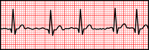

| ECG morphology | Likely rhythm | First move |
|---|---|---|
| Regular narrow, no visible P, 150–250 | AVNRT / AVRT | Vagal → adenosine · terminates re-entry |
| Sawtooth flutter waves · ventricular ~150 (2:1) | Atrial flutter | Adenosine unmasks · rate-control / DCCV |
| Irregularly irregular, no organized P | Atrial fibrillation | Rate control + anticoagulate (CHA₂DS₂-VASc) |
| ≥3 P-wave morphologies, irregular | MAT | Treat lung disease · ↓ β-agonist · avoid DCCV |
| Regular wide + AV dissociation / fusion-capture | VT | Amiodarone / procainamide · DCCV if unstable |
| Irregular wide, very fast, varying QRS width | Pre-excited AF | Procainamide / DCCV — no AV-nodal blockers |
| P before every QRS, rate tracks demand | Sinus tachycardia | Treat the cause (pain, fever, hypovolemia, PE) |
Once stable and classified, match therapy to mechanism. The two traps that kill: treating VT as SVT, and giving an AV-nodal blocker in pre-excited AF. Either can precipitate collapse. Match therapy to the rhythm and ventricular function.
A
Regular SVT — AVNRT · AVRT · atrial tach
break the re-entry circuitStep 1 · vagal Modified Valsalva for stable regular SVT. REVERT: sinus rhythm at 1 minute in 93/214 (43%) vs 37/214 (17%) with standard Valsalva. Avoid unsupervised carotid massage.
Step 2 · adenosine 6 → 12 mg rapid IV push — terminates node-dependent SVT (AVNRT/AVRT). Diagnostic in atrial flutter/AT.
Step 3 · rate / rhythm agents IV β-blocker or non-DHP CCB (diltiazem/verapamil) if adenosine fails & QRS narrow & not pre-excited.
Any instability → synchronized cardioversion. Recurrent/symptomatic SVT → EP referral for catheter ablation (success and risk depend on the mechanism and target).
B
Atrial fibrillation / flutter — rate vs rhythm + stroke
three parallel decisions| Decision | Approach |
|---|---|
| Rate control | If stable, β-blocker or diltiazem/verapamil only with EF >40%. Avoid IV non-DHP CCB in significant LV systolic dysfunction. Digoxin may help when these are ineffective/contraindicated. IV amiodarone is an option in selected critically ill/decompensated HF patients, with cardioversion and stroke risk considered. Resting <100–110 bpm is symptom-guided in AF without HF. |
| Rhythm control | Electrical cardioversion or selected antiarrhythmic therapy for symptoms or clinical benefit. Drug choice depends on EF, QT, structural disease and anticoagulation. Avoid ibutilide with depressed EF or prolonged QT. Early rhythm control is a strategy, not a mandate for an acute drug. |
| Cardioversion timing | ≥48 h / unknown → therapeutic anticoagulation ×3 wk or imaging to exclude intracardiac thrombus first. <48 h is not automatically low risk: if not anticoagulated and CHA₂DS₂-VASc ≥2, consider precardioversion imaging. Continue OAC ≥4 wk after. Long-term by stroke risk. See dedicated AF guide P.6. |
CHA₂DS₂-VASc — stroke risk
CHF +1 · HTN +1
A₂ age ≥75 +2 · DM +1
S₂ prior stroke/TIA +2
V vascular dz +1
A age 65–74 +1
Sc female +1
Anticoagulate ≥2 (♂) / ≥3 (♀). Reasonable at 1 (♂) / 2 (♀)
C
Wide-complex: VT vs SVT-with-aberrancy
when in doubt, it's VTFindings that favor VT
- AV dissociation, capture or fusion beats strongly support VT.
- Very wide: RBBB >140 / LBBB >160 ms. NW ("northwest") axis.
- Precordial concordance (all V1–V6 same polarity). R-to-S >100 ms.
- Known structural heart disease / prior MI → assume VT.
Favors SVT + aberrancy
- Typical RBBB / LBBB morphology or QRS identical to a prior tracing supports aberrancy but does not exclude VT.
- Prior MI, ventricular scar and reduced EF increase the prior probability of VT. ECG algorithms have imperfect validation.
- If uncertain, treat as VT — do not give verapamil/adenosine empirically to undiagnosed WCT (except a classic regular-monomorphic trial).
D
Pre-excited AF (WPW)
the one that gets the wrong drugWide, irregular, often very fast (>200) with varying QRS width. Avoid adenosine, β-blockers, diltiazem/verapamil, digoxin, and IV amiodarone. Preferential accessory-pathway conduction can precipitate VF. Give procainamide or ibutilide. If unstable (or any doubt) → synchronized cardioversion. Definitive: accessory-pathway ablation.
E
Electrical storm & sustained VT
≥3 treated episodes in 24 h, ≥5 min apart| Lever | Action |
|---|---|
| Sympatholysis | β-blockade + IV amiodarone when tolerated in structural-heart-disease storm. A 60-patient ICD trial favored oral propranolol over oral metoprolol, both with IV amiodarone. Esmolol allows rapid titration. No universal single best therapy. |
| Antiarrhythmic | Amiodarone IV for structural-disease VT (150 mg over 10 min, then 1 mg/min for 6 h per AHA). Lidocaine may help ischemic VT. For tolerated WCT, avoid procainamide with CHF or prolonged QT. |
| Sedation | Analgesia and sedation reduce distress from repeated shocks. Deep sedation/intubation is an escalation for refractory storm, with hemodynamic support. |
| Fix the substrate | Urgent angiography for STEMI or ongoing ischemia. Correct K/Mg/Ca deficits, hypoxia, acidosis, HF and drug triggers. Revascularization alone does not remove scar-mediated VT. |
| ICD | Interrogate to separate appropriate from inappropriate shocks. A magnet can suspend all tachy therapies in many ICDs, not selectively inappropriate shocks. Use external pads/monitoring and model-specific guidance until reprogrammed. |
| Escalate | Stellate ganglion block, urgent catheter ablation, and MCS (ECMO/Impella) as a bridge in refractory storm. |
| Torsades | For long-QT torsades: IV magnesium, correct K⁺ and withdraw QT drugs. Pacing/isoproterenol may help recurrent acquired, pause-dependent TdP with expert input. Isoproterenol is not generic therapy for congenital LQTS or ischemic polymorphic VT. |
In acute bradycardia with hemodynamic compromise, atropine is reasonable while reversible causes are treated. If response is inadequate—or unstable high-degree AV block makes delay unsafe—move promptly to transcutaneous pacing and/or epinephrine or dopamine while preparing transvenous pacing. Avoid atropine after heart transplant because paradoxical AV block or sinus arrest can occur.
A
Symptomatic bradycardia — ACLS ladder
drug → drip → paceStep 1 · atropine 1 mg IV q3–5 min, max 3 mg for hemodynamically significant bradycardia · reassess immediately.
Step 2 · adrenergic support and/or pace If atropine is ineffective: dopamine 5–20 µg/kg/min, epinephrine 2–10 µg/min, and/or transcutaneous pacing while preparing definitive support.
Step 3 · transvenous pacing For persistent unstable bradycardia or symptomatic high-grade block refractory to medical therapy. Temporary pacing is a bridge. Routine pacing for established asystolic cardiac arrest is not recommended. Assess permanent pacing after reversible causes are addressed.
B
AV block — read the ladder
where it blocks = how dangerous| Pattern | Interpretation and next question |
|---|---|
| 1° AV block | PR >200 ms with every P conducted. Usually no pacing if asymptomatic. Marked PR prolongation can cause symptoms. |
| Mobitz I | Progressive PR prolongation before a dropped QRS. Often nodal, but symptoms and evidence of infranodal disease matter. |
| Mobitz II / high-grade | Unexpected nonconducted P waves with unchanged PR in conducted beats suggests His–Purkinje disease. Acquired nonreversible Mobitz II, high-grade or complete block generally requires permanent pacing even without symptoms. |
| 2:1 / complete block | 2:1 cannot be classified as Mobitz I or II from PR alone. Complete block has independent atrial/ventricular rhythms. An escape can be narrow or wide. Stabilize before localization. |
C
Transcutaneous (TCP) vs transvenous (TVP) pacing
bridge vs definitive| Transcutaneous (pads) | Transvenous (RV lead) | |
|---|---|---|
| Role | Immediate bridge · every code cart has it | Definitive temporary · for sustained high-grade block |
| Setup | Place pads per device instructions. Set a rate supporting perfusion, raise current until capture and add a device-specific margin (ZOLL M2: usually ~10%). | Experienced operator with ultrasound-guided access and echo/fluoroscopic guidance. Choose access and lead type with the definitive-device plan. |
| Capture | Confirm mechanical capture — palpate femoral pulse / art-line / Doppler (electrical ≠ perfusion) | Confirm electrical and mechanical capture. For Medtronic 5392, output ≥2–3× stimulation threshold. Follow the actual generator manual. |
| Comfort | Painful — sedate & give analgesia. SEDATION P.5 → | Well tolerated once seated |
Rate
60–80 bpm
common starting range, individualize to perfusion
Output
TVP 2–3× threshold
5392 example, not a universal generator setting
Mode
demand (VVI)
asynchronous pacing risks competition and R-on-T
Sequence playing
D
Pacing pitfalls & complications
capture · sense · perforate- Failure to capture spike, no QRS → ↑ output, reposition lead, correct hyperK / acidosis / ischemia, check connections.
- Failure to sense (undersensing): lower mV increases sensitivity. Oversensing may require higher mV. First check leads, cables, artifact and mode. Preserve reliable QRS sensing with monitoring/backup.
- Complications RV perforation → tamponade, induced VT, lead dislodgement, infection, pneumo/access injury.
- Pre-empt Pre-existing LBBB during RHC/PA-cath can develop complete block (RBBB injury) — have pacing ready.
One-liner: tachy → stable vs unstable → if unstable, choose synchronized cardioversion or unsynchronized shock by rhythm. If stable, width × regularity picks the drug — never give an AV-nodal blocker to pre-excited AF, and treat any uncertain wide tachy as VT. Hemodynamically significant brady → treat reversible causes + atropine. If ineffective, use adrenergic support and/or pacing without delay and confirm mechanical capture.