Scope: a public clinical guide for durable continuous-flow LVAD physiology and common presentations. It does not replace the current device instructions, the patient-specific emergency plan, or early involvement of the implanting LVAD team.
1
The circulation in one line
follow the bloodRight heartDelivers preload through the lungs.
Left ventricleReceives pulmonary venous return.
Inflow cannulaDraws blood from the LV apex.
Rotary pumpAdds continuous forward energy.
Outflow graftReturns blood to the aorta.
2
The first 60 seconds
patient + pump + power| Question | What to do | Why it matters |
|---|---|---|
| Who is this patient? | Identify device, implanting team, baseline speed/flow/power/PI, anticoagulant, and usual BP method. | One “normal” value does not transport across devices or patients. |
| Is the pump powered? | Look, listen, feel for the pump hum. Inspect controller, driveline, and both power connections without disconnecting a functioning source. | A pump hum supports rotor activity. It does not establish adequate blood flow or systemic perfusion. A power or cable problem can still mimic patient deterioration. |
| Is there perfusion? | Assess mentation, skin, capillary refill, Doppler pressure, breathing, urine, lactate, and echo when needed. | A continuous-flow patient may have no palpable pulse even when well. |
| What changed? | Compare alarms and trends with baseline. Seek bleeding, volume loss, RV failure, arrhythmia, tamponade, hypertension, or obstruction. | An alarm names a device state—not its cause. |
PEARL
Low flow is a syndrome. Do not answer it reflexively with fluid or speed. First determine whether blood cannot reach the pump, cannot cross the pump, cannot leave the graft, or is recirculating instead of reaching the body.
1
Pump generations: gains and tradeoffs
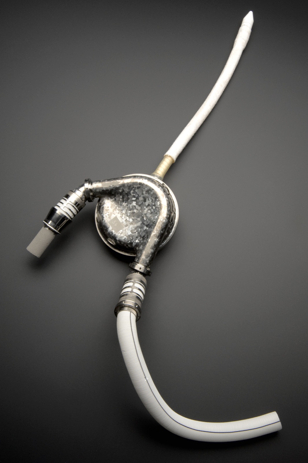
pulsatile → axial → centrifugalREMATCH made durable support a therapy. The large pulsatile HeartMate VE improved survival versus medical therapy but carried major infection and device-failure burdens. HeartMate II then made continuous axial flow practical. HVAD moved a compact centrifugal pump with hybrid passive-magnetic/hydrodynamic suspension into the pericardium. HeartMate 3 moved to full magnetic levitation.

2
The three pumps still encountered in practice
architecture has consequences| Pump | Architecture and status | What changes at the bedside |
|---|---|---|
| HeartMate II | Mechanical-bearing axial pump positioned below the diaphragm in an abdominal/preperitoneal pocket. No longer implanted, although legacy recipients remain. | No programmed artificial pulse. Power rise plus hemolysis is a classic intrapump-thrombosis pattern, suggestive rather than diagnostic. Thrombosis and exchange were more common than with HM3. |
| HeartWare HVAD | Intrapericardial centrifugal pump with passive-magnetic/hydrodynamic suspension. Medtronic stopped sale and distribution on June 3, 2021 and recommended no new implants. | FDA cited excess neurologic events, mortality, and pumps that could delay or fail to restart. Existing recipients need HVAD-specific support. Exchange is individualized, not automatic. |
| HeartMate 3 | Intrapericardial centrifugal pump with a fully magnetically levitated rotor. Current routine adult U.S. Durable-LVAD platform. | Programmed speed modulation produces an artificial pulse 30 times/min, asynchronous with the native heartbeat. Thrombosis, stroke, bleeding, and exchange are lower than with HMII, not absent. |
3
The investigational landscape is widening
pipeline, not practiceBrioVAD
A fully magnetically levitated centrifugal pump in INNOVATE, a randomized comparison with HM3. The September 9, 2026 registry update lists recruiting, with no results posted. It remains investigational.EVAHEART 2
A centrifugal pump with a water-purged hydrodynamic noncontact bearing in the COMPETENCE comparison with HM3. The March 27, 2026 registry update lists recruitment suspended for device modification, with no results posted. The record does not identify the triggering problem.EFICAS — total heart replacement. This prospective study at 10 French centers enrolled 55 adults with refractory biventricular HF who were unsuitable for LVAD and listed or eligible for transplant. With the Aeson bioprosthetic total artificial heart, 40/55 (72.7%, 95% CI 57.1–85.1) met the 180-day composite of survival free of disabling stroke and device malfunction requiring reoperation or urgent transplant, or elective transplantation. At 180 days, 96% were free of disabling stroke and 76% had been discharged. The comparison was against a prespecified 38% historical performance threshold. It had no randomized or contemporaneous control, so these results cannot establish superiority to another device.
DO NOT PORT
RPM ranges, alarms, waveform or PI semantics, flow-power relationships, controller exchanges, and ramp expectations are device specific. Wireless energy transfer, physiologic automatic speed control, and further hemocompatibility gains remain investigational.
1
The system is more than the pump
blood + power + data2
Localize the failing pathway
the symptom may be shared| Pathway | Components | Bedside consequence |
|---|---|---|
| Blood | LV apex → inflow cannula → pump → outflow graft → ascending aorta | Underfilling, cannula malposition, pump resistance, graft obstruction, and aortic regurgitant recirculation can all reduce useful systemic delivery. |
| Power | Driveline → controller → two external power connections | A damaged lead, loose connection, depleted source, or controller problem can threaten pump support. Never disconnect both sources from a functioning system. |
| Information | Controller estimates, alarms, event history, and logs | The display describes a device state. It must be reconciled with perfusion, loading, rhythm, imaging, and the patient’s baseline. |
| Skin–hardware interface | Percutaneous driveline and exit site | The driveline is both the power conduit and a portal for trauma and infection. Traction prevention is physiology-preserving care. |
ORIENT
When the patient deteriorates, draw the circuit mentally: Can blood reach the pump? Can the rotor move it? Can it leave the graft? Is it reaching the body rather than recirculating?
HeartMate 3 mental model: the controller estimates flow from rotor speed and power. The estimate changes with preload, afterload, blood viscosity, and obstruction. Confirm that the programmed hematocrit reflects the current laboratory value. Displayed flow is not an independent measurement of systemic delivery.
A
The four numbers
trend, do not worship| Display | What it represents | Common interpretation error |
|---|---|---|
| Speed | Programmed rotor revolutions per minute. The prescribed operating point. | Assuming more speed always means more useful output. |
| Flow | Calculated estimate derived largely from speed and power. | Calling it cardiac output or using it alone to clear perfusion. |
| Power | Energy required to maintain the programmed speed. | Ignoring a rise from intrapump resistance or a fall with severe underfilling. |
| Pulsatility index | Power-derived measure of pulsatility associated with native cardiac-cycle flow variation. | Calling every PI event “suction”. Abrupt loading or rhythm changes can also trigger it. |
B
What changes the pump
preload + afterloadLess preload
Bleeding, dehydration, RV failure, tamponade, tension physiology, arrhythmia, or inflow malposition can reduce flow and provoke suction.More afterload
Hypertension or outflow obstruction can reduce forward flow at the same speed.More intrapump resistance
Thrombus can alter power, flow estimation, hemolysis, sound, and unloading.Recirculation
Important aortic regurgitation can yield reassuring pump flow while net systemic output falls.C
Outcome context
device-specific comparisonIn MOMENTUM 3, fully magnetically levitated HeartMate 3 support had better five-year event-free survival than the axial-flow HeartMate II comparator, with fewer strokes, bleeding events, and pump thromboses. This is a device-versus-device result—not an LVAD-versus-medical-therapy estimate.
Check the component, alarm and current advisory. Abbott’s 2025–2026 notices cover selected power units, power cords and backup batteries. The October 2025 backup-battery fault notice advises keeping external power connected and obtaining clinical guidance, rather than attempting an immediate controller exchange. The May 2026 date-code notice calls for affected batteries to be identified, taken out of use, replaced and returned through the LVAD team.
SAFETY
Never disconnect both power sources. HeartMate 3 is MR Unsafe. Use the exact implanted-system instructions for procedures and controller exchange, with trained-team support.
1
Pattern map
controller + patient + imaging| Pattern | Think first | Discriminator |
|---|---|---|
| Low flow + low/normal power | Underfilling, RV failure, arrhythmia, tamponade, inflow/outflow obstruction, high afterload | CVP/JVP, LV size, septum, RV, effusion, BP, cannulae, CTA when hardware disease is suspected |
| Unexpected high power ± high displayed flow | Intrapump resistance/thrombus. Power-driven estimation can make displayed flow rise while useful output falls | Hemolysis trend, urine, log timing, loss of LV unloading, and CT to exclude inflow/outflow disease |
| Repeated PI events | Rapid loading or rhythm change. Possible suction, not proof of suction | Episode timing, rhythm, symptoms, LV size, inflow position, volume and RV phenotype |
| Normal/high displayed flow + shock | Sepsis/vasodilation, aortic regurgitant loop, estimation error, competing physiology | Perfusion, lactate, echo, invasive output when necessary |
Tamponade may first look like an LVAD low-flow problem. In unexplained low flow, obtain focused echo early and look specifically for pericardial effusion or organized clot with a small or compressed LV or RV. Integrate chamber size, septal position, RV function, and the device trend. A reassuring controller display does not exclude tamponade.
2
Preload dependent does not mean “give fluid”
the RV may be full and failingTrue underfill: bleeding or volume loss, low venous pressure, small LV, suction, fluid responsiveness. Stop the loss and restore appropriate filling.
Congested underdelivery: high venous pressure, dilated/dysfunctional RV, TR, septal shift. More volume can worsen LV filling and pump flow.
3
Persistent unexplained low flow
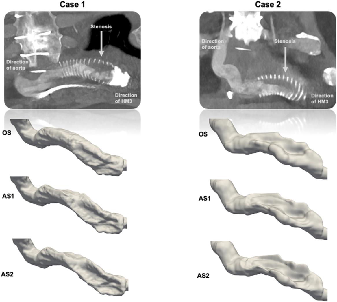
remember the graftLate or persistent low-flow alarms can reflect extrinsic outflow-graft obstruction from material accumulating between the graft and its surrounding components. After common loading and BP causes are assessed, CTA is central to defining outflow-graft compression, twisting, kinking, or stenosis.
MOVE
Stabilize perfusion, preserve power, obtain ECG and focused echo early, send hemolysis/end-organ studies when malfunction is possible, and involve the LVAD team while the phenotype is being localized.
2025 AHA update: in an unresponsive durable-LVAD patient with impaired perfusion, perform chest compressions. It may be reasonable to begin immediately while another rescuer simultaneously assesses device-related reversible causes (Class 2b, expert opinion).
A
Parallel resuscitation
do not serially troubleshoot1 · PatientResponsiveness, breathing, skin, capillary refill, Doppler pressure, rhythm, ultrasound when immediately available.
2 · DevicePower sources, controller screen, driveline connection, pump hum, visible damage. Bring backup equipment.
3 · CauseBleeding, hypovolemia, RV failure, tamponade, tension pneumothorax, arrhythmia, obstruction, thrombosis, sepsis.
B
Pump stoppage
time and certainty matter| Situation | Principle | Boundary |
|---|---|---|
| Brief witnessed power interruption | Correct the power/connection problem promptly using the implanted-device plan. | Do not disconnect a working lead while investigating the other. |
| Unknown or prolonged downtime | Begin patient resuscitation and obtain immediate expert device guidance. | There is no evidence-defined universal elapsed time that settles restart safety. Thrombosis/embolization risk and device state must be assessed. |
| Controller fault or exchange | Use the patient’s backup controller only with current instructions and trained support when feasible. | Generic memorized exchange choreography can stop a functioning pump. |
C
VT/VF may look deceptively tolerated
the RV still matters- The LVAD may preserve consciousness transiently, but ventricular arrhythmia removes effective RV contraction, reduces LV filling, and can progress to low flow and end-organ injury.
- Treat hemodynamic compromise and the rhythm. Also seek suction, electrolytes, ischemia, medication effects, and RV failure.
- For external defibrillation/cardioversion, leave the functioning pump connected. Open-heart defibrillation follows different manufacturer precautions.
RULE
Do not spend the arrest interval proving that the device is the problem. Restore circulation while device causes are checked in parallel.
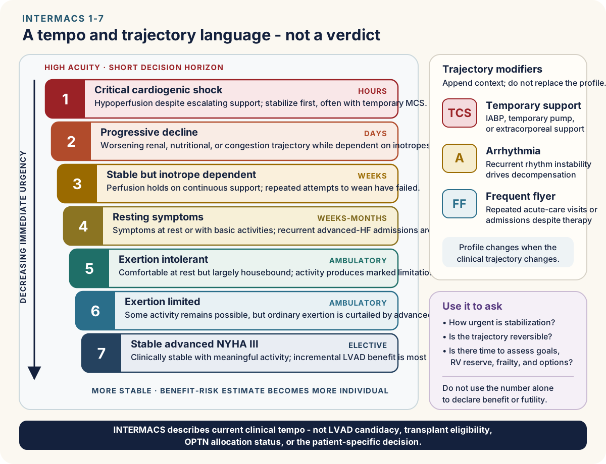
| Profile | Clinical anchor | Conceptual clock |
|---|---|---|
| 1 · Critical shock | Hypotension and rising lactate or organ injury despite escalating support. Stabilize first, often with temporary MCS. | Hours |
| 2 · Progressive decline | Worsening nutrition, kidney/liver function, congestion, or perfusion despite inotropes. | Days |
| 3 · Stable on inotropes | Inotrope dependent or repeatedly unable to wean without recurrent low output. | Weeks |
| 4 · Resting symptoms | Symptoms at rest or with activities of daily living, often with recurrent admissions. | Weeks–months |
| 5 · Exertion intolerant | Comfortable at rest but effectively housebound by symptoms with minimal activity. | Months |
| 6 · Exertion limited | Can leave home, but meaningful activity remains substantially limited—the “walking wounded.” | Months |
| 7 · Advanced class III | Clinically stable but markedly limited. Elective benefit is increasingly preference- and trajectory-sensitive. | Elective horizon |
Modifiers add context without changing the profile: TCS temporary circulatory support, A clinically important arrhythmia, and FF frequent HF admissions. Profile and modifiers describe illness. They do not confer candidacy or OPTN status.
BTT · bridge to transplant
Transplant remains the intended destination.BTC · bridge to candidacy
Support creates time to reassess a potentially reversible barrier.BTR · bridge to recovery
Recovery is plausible and tested deliberately.DT · long-term therapy
Durable support is the intended destination. Strategy can evolve. It is not a promise.ROADMAP
Ambulatory, non-inotrope-dependent patients are the uncertainty boundary. ROADMAP found more survival with functional improvement and better quality of life after LVAD, but also more adverse events. Its prospective nonrandomized design cannot remove selection bias or establish a universal threshold.
1
What the implant is meant to accomplish
purpose can evolve| Frame | Clinical question | Teaching point |
|---|---|---|
| Bridge to transplant | Can durable support restore stability while transplantation remains intended? | Candidacy and allocation status require separate reassessment. |
| Long-term therapy | Will survival, function, and quality of life improve enough to justify device burden? | Goals, caregiver capacity, frailty, cognition, adherence, and competing illness matter. |
| Bridge to candidacy/decision | Could organ dysfunction, pulmonary pressure, nutrition, or uncertainty improve with support? | Define the barrier, reassessment, and fallback before implant. |
| Bridge to recovery | Is the cardiomyopathy plausibly reversible? | Recovery is screened deliberately. It is not inferred from one better EF. |
2
The RV is the peri-implant pivot
the LVAD cannot replace RV delivery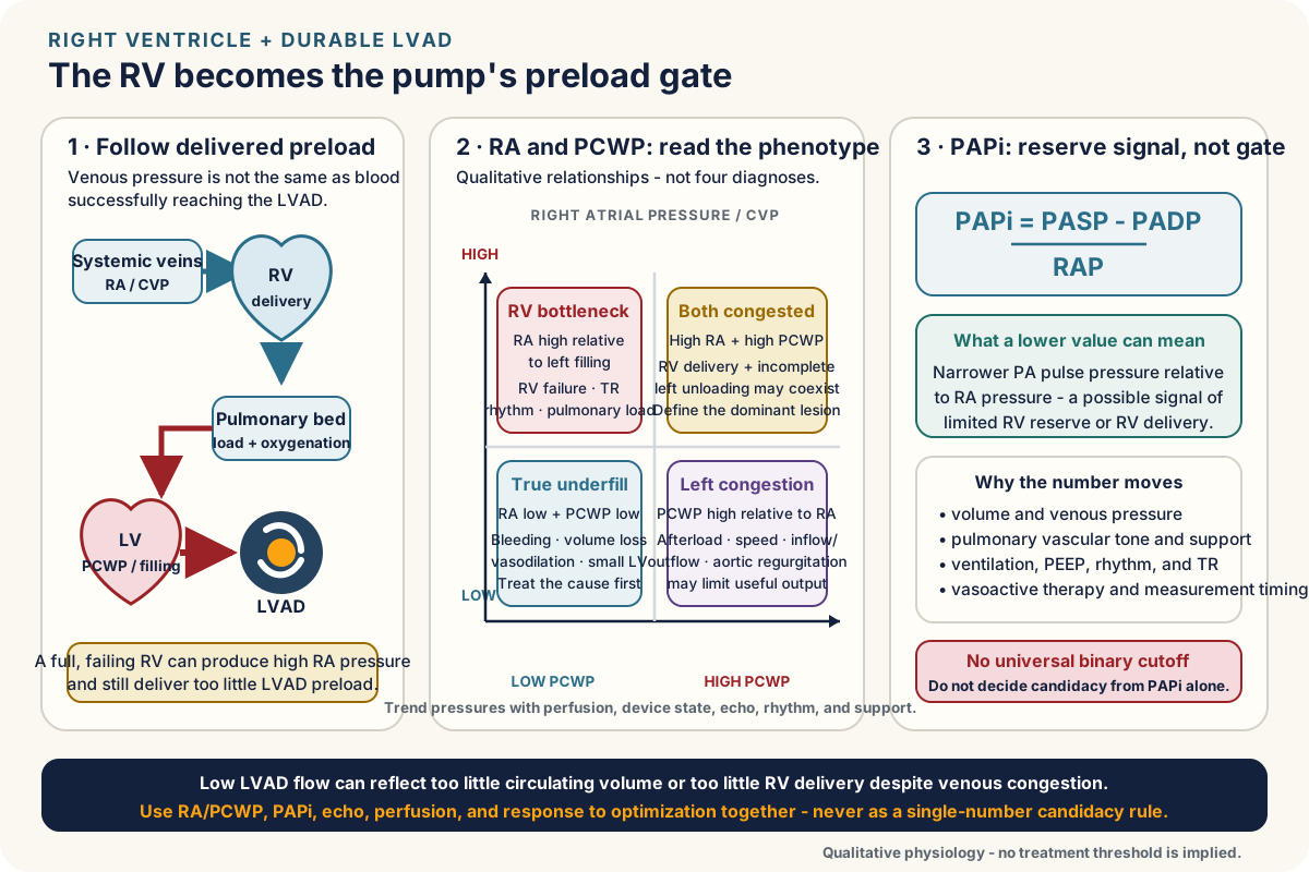
Read the loading state
PAPi and RA/PCWP change with volume, vasoactive support, ventilation, severe TR, rhythm, and measurement timing. A single resting number may describe the moment more than the reserve.Test reserve
Integrate RV structure/function, TR, renal-hepatic congestion, pressor or ventilatory dependence, and response to diuresis or hemodynamic optimization. Dynamic response can add more than an isolated threshold.3
Geometry becomes physiology
implant choices persist- Inflow direction, apical position, outflow course, graft angle, and relief of twisting affect unloading, suction, thrombosis, obstruction, and stroke risk.
- After bypass, low flow may reflect RV failure, underfill, tamponade, cannula position, or excessive support. “Turn it up” is not a diagnosis.
TIMING
Discuss durable support before irreversible end-organ decline and severe deconditioning narrow the benefit window—but only after goals, alternatives, device burden, and the exit strategy are explicit.
1
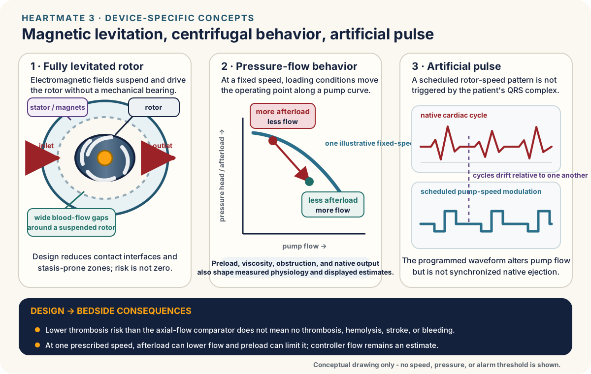
Design feature → clinical consequence
name the platform| HeartMate 3 feature | What it changes | What it does not prove |
|---|---|---|
| Fully magnetically levitated rotor | No mechanical or hydrodynamic bearing contact. Wider blood-flow gaps reduce stasis and shear hot spots. | Hemocompatibility events are lower, not zero. |
| Centrifugal pressure–flow behavior | At a fixed operating point, higher afterload reduces flow. Inadequate RV delivery limits preload. | More speed cannot repair bleeding, RV failure, tamponade, graft obstruction, or recirculation. |
| Artificial pulse | Programmed speed modulation changes pump flow about 30 times/min and is asynchronous with the native heartbeat. | It does not represent native ventricular ejection or validate systemic perfusion. |
2
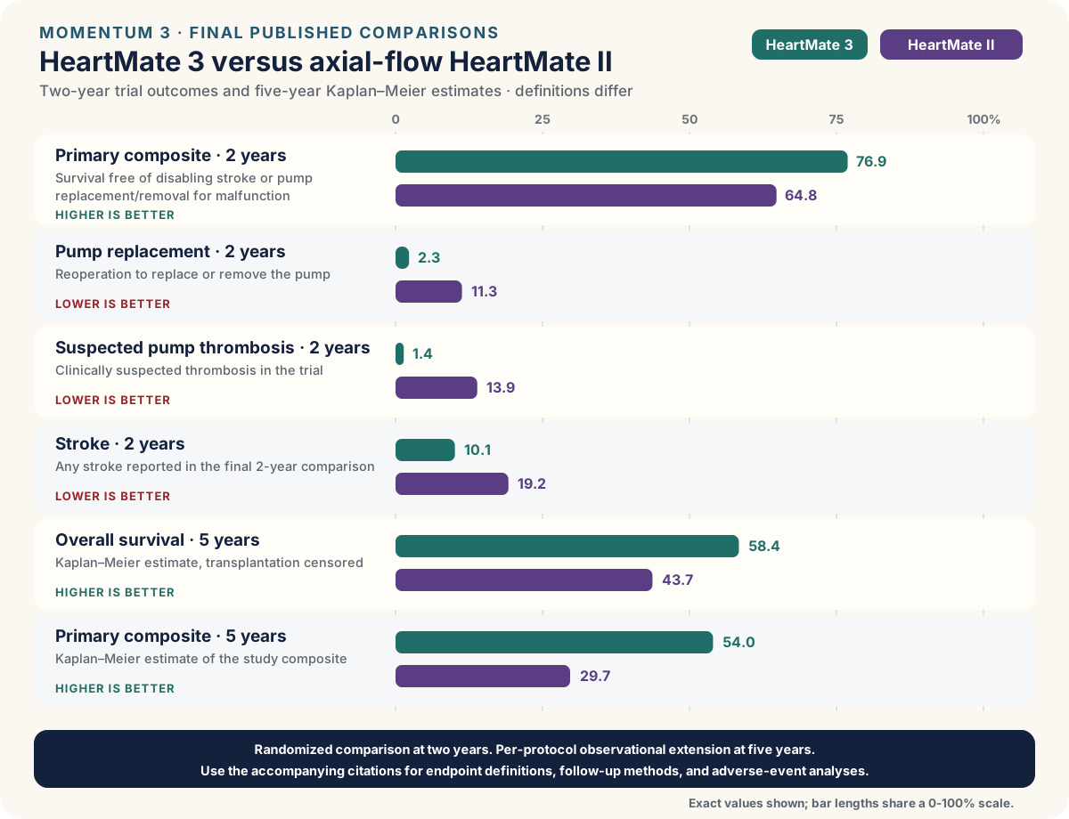
MOMENTUM 3: use the final comparison
HM3 vs HMII| Outcome | HeartMate 3 | HeartMate II |
|---|---|---|
| 2-year survival free of disabling stroke or reoperation to replace/remove a malfunctioning pump | 76.9% | 64.8% |
| 2-year pump replacement | 2.3% | 11.3% |
| 2-year suspected pump thrombosis | 1.4% | 13.9% |
| 2-year stroke | 10.1% | 19.2% |
| 5-year survival (Kaplan–Meier estimate) | 58.4% | 43.7% |
| 5-year composite event-free survival (Kaplan–Meier estimate) | 54.0% | 29.7% |
BOUNDARY
The two-year trial randomized 1,028 patients. Five-year results are per-protocol Kaplan–Meier estimates from extended observational follow-up, with transplantation censored. They are not simple percentages of all randomized patients still alive on their original pump. These results do not make old-pump alarm patterns, ramp rules, or antithrombotic assumptions portable to HeartMate 3—and it does not eliminate the need to localize a new event.
1
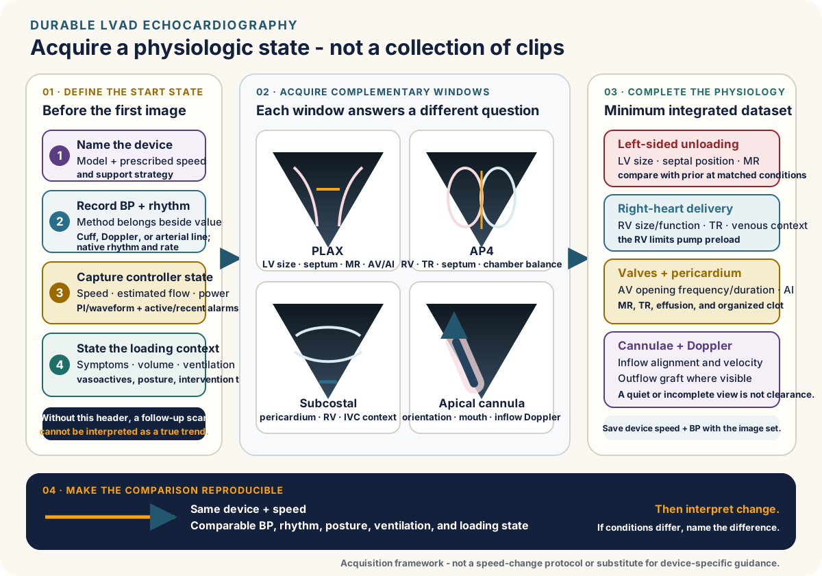
Before the first view
freeze the contextDevice state
Record exact platform, prescribed and current speed, displayed flow, power, PI when applicable, active alarms, and recent controller trend.Patient state
Record BP method and value, rhythm, symptoms, volume/loading state, oxygen or ventilatory support, and vasoactive or inotropic therapy. State the clinical question and retrieve the prior baseline.2
Acquire the anatomy and the flow
standard + device-specific views| Views | Acquire and report | Question answered |
|---|---|---|
| PLAX + short axis | LV size, septal position, wall motion, MR, and RV geometry across more than one plane. | Is unloading inadequate, excessive, or asymmetric? |
| Apical | RV size/function, TR, estimated pressure, and chamber relationships. Use off-axis views when hardware requires them. | Can the RV deliver preload to the LVAD? |
| Aortic valve | Opening frequency and duration over multiple cycles. Characterize AI with LVAD-specific Doppler and recognize that conventional grading may underestimate it. | Is the root being washed and is flow recirculating? |
| Subcostal / pericardium | IVC and venous congestion. Search deliberately for effusion or organized postoperative collection compressing either ventricle. | Underfill, RV congestion, or tamponade physiology? |
| Inflow cannula | Position and direction toward the mitral inflow. Color and spectral Doppler for turbulence or abnormal velocity pattern. | Malposition, obstruction, or suction? |
| Outflow graft | Color and spectral Doppler wherever seen. Document the sampled segment and image quality. | Abnormal flow supports a lesion. A quiet or unseen segment does not exclude one. |
3
Turn findings into a phenotype
focused troubleshootingSmall LV / leftward septum
True underfill, RV delivery failure, suction, or excessive unloading.Dilated LV / persistent MR
High afterload, insufficient unloading, inflow–pump–outflow dysfunction, or important AI.Dilated RV / TR / venous congestion
The pump is preload dependent but the failing RV may be overfilled. Reflex fluid can worsen delivery.Displayed flow and patient disagree
Seek recirculating AI, controller-estimation error, shunt, vasodilation, or competing shock.Routine study: image at the prescribed setting and do not casually adjust speed. Supervised ramp: establish explicit indication, safety gates, standardized loading/BP, staged changes, and prespecified endpoints with the LVAD team.
REPORT
State device and speed, BP method/value, rhythm, controller values, loading and support state, image limitations, and comparison with baseline. A conclusion without the operating conditions is not reproducible.
A ramp is not one test. Optimization, suspected malfunction, and myocardial-recovery or turn-down studies have different starting conditions, safety gates, and endpoints. Do not transport one protocol into another.
1
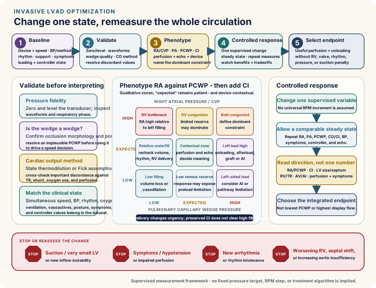
Standardize before interpreting
indication + conditions + baselineWhen it helps
Persistent symptoms or congestion, repeated admissions, discordant echo/controller/perfusion data, uncertain RV reserve, suspected inadequate unloading or AI, and supervised optimization or recovery assessment.Preconditions
Identify device and current speed. Document BP and method, volume and rhythm, vasoactive/ventilatory state, anticoagulation and procedure plan. Zero and level the transducer correctly.Baseline hemodynamics
Record RA, RV, PA, and PCWP. Validate a questionable wedge with waveform and oxygen saturation rather than accepting the first number. Obtain arterial and mixed venous oxygen saturation. Calculate PVR and SVR.Output and simultaneous data
Measure CO/CI by thermodilution and/or direct or estimated Fick. Severe TR can distort thermodilution. Shunt, supplemental oxygen, sampling, and estimated oxygen consumption can distort Fick. Record echo, BP, and controller values at the same operating point.2
Name the baseline phenotype
pressures tell a story| RA | PCWP | Working phenotype | Questions to pursue |
|---|---|---|---|
| High | High | Biventricular congestion | Volume, RV failure/TR, inadequate unloading, afterload, AI, or mixed disease |
| High | Normal / low | RV delivery bottleneck | RV contractility, rhythm, pulmonary load, septum, TR, excessive unloading |
| Normal / low | High | Inadequate LV unloading or recirculation | Afterload, speed context, inflow/pump/outflow dysfunction, MR, AI |
| Low | Low | Underfill | Diuresis, bleeding, vasodilation, low intake, suction, or excessive unloading |
3
Change one operating point at a time
program-specific, never memorized RPMControlled sequence
Use supervised, device- and program-specific speed steps with adequate equilibration. At each stage repeat RA/PA/PCWP, measured CO/CI, BP, symptoms, controller values, and focused echo.Desired direction
Seek lower filling pressures and adequate measured output while avoiding suction, progressive leftward septal shift, RV worsening, arrhythmia, hypotension, worse AI, or a persistently closed aortic valve with root stasis.Context-only targets: RA/CVP <12 mm Hg, PCWP <18 mm Hg, and CI >2.2 L/min/m² are often-used trial or optimization goals—not universal thresholds. Symptoms, perfusion, device, RV/valve phenotype, and the patient-specific plan remain decisive.
STOP / INTERPRET
Defer elective optimization when procedural or hemodynamic safety is not established. Stop for suction, symptomatic hypotension, clinically important arrhythmia, progressive leftward septal shift or RV failure, or worsening AI. The classic Columbia LVEDD slope was derived in axial-flow HeartMate II and is load sensitive. Failure to unload can identify important dysfunction but does not locate it. Successful unloading does not exclude early thrombus. Do not transport a fixed slope to HeartMate 3.

1
Name the hardware, then ask the modality’s question
recognition ≠ diagnosis| Modality | Best question | Blind spot |
|---|---|---|
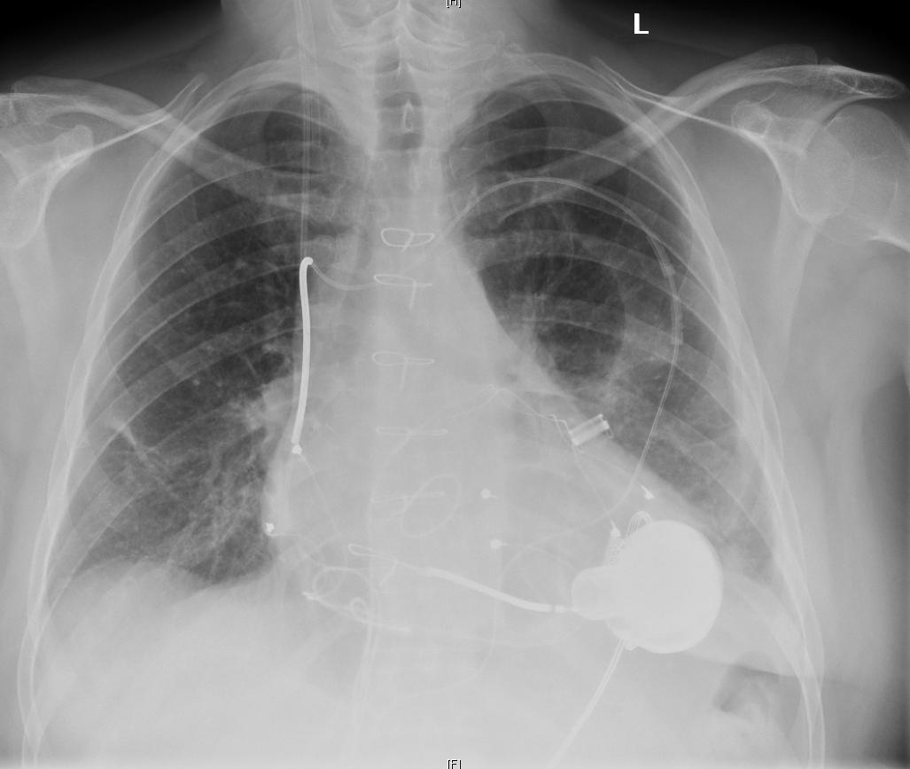
| Chest radiograph | Where are pump, inflow, visible outflow course, driveline, and other hardware? Is there an obvious interval positional change or pulmonary process? | Cannot establish pump flow, thrombus, subtle graft lumen disease, or adequate systemic perfusion. |
| Echocardiography | Is the LV unloaded? What are septal position, RV function, valves, effusion, inflow direction/velocity, and visible outflow Doppler? | A nondiagnostic or quiet outflow signal does not exclude obstruction. |
| CT angiography | Is the inflow malpositioned or the outflow graft kinked, twisted, compressed, stenosed, or otherwise anatomically compromised? | Defines graft anatomy, but not net delivery. Metal artifact limits the pump interior, so CT cannot reliably exclude intrapump thrombus. |
2
Device recognition changes the next move
legacy recipients remain- Confirm the device with the controller, implant card, operative record, or implanting team. Do not infer platform or perform controller actions from silhouette alone.
- HVAD is no longer implanted in the United States, but supported recipients remain. Its restart concerns, controller behavior, and exchange decisions are not HeartMate 3 instructions.
- For HeartMate 3, interpret imaging beside controller trends and the current implanted-system labeling. Normal geometry does not validate the flow estimate.
CTA
Persistent or unexplained low flow after loading, blood pressure, rhythm, and RV causes are assessed should trigger an anatomy question. CTA is central when inflow or outflow hardware disease remains plausible.
1
Blood pressure: first ask if there is a pulse
method changes meaning| Flow phenotype | Measurement | Consensus teaching range |
|---|---|---|
| Meaningfully pulsatile | Validated cuff can estimate systolic/diastolic pressure and MAP | Cuff-derived MAP is commonly targeted around 75–90 mm Hg. |
| Minimally/nonpulsatile | Doppler opening pressure | Opening pressure around 80–100 mm Hg. Do not automatically relabel it “MAP.” |
These consensus ranges apply to stable, non-critically ill patients. Doppler opening pressure often approximates MAP with little pulsatility but approaches systolic pressure as pulsatility increases. In shock or when measurements disagree, use an arterial line when feasible. Avoid overtreatment based on an unverified pressure.
2
Antithrombotics: name the device
old pump evidence does not transport- Vitamin K antagonist remains standard. INR 2.0–3.0 is the usual starting range for contemporary durable continuous-flow support, then individualized for device and events.
- For HeartMate 3, ARIES-HM3 randomized 628 patients to aspirin 100 mg/day or placebo with VKA (INR 2.0–3.0). In the 589-patient primary analysis population, aspirin avoidance was noninferior for 12-month survival free of major nonsurgical hemocompatibility events and reduced nonsurgical bleeding (RR 0.66, 95% CI 0.51–0.85). The FDA incorporated ARIES into labeling in 2024.
- Aspirin avoidance does not mean anticoagulation avoidance. Do not extend it to HMII or HVAD. A separate current antiplatelet indication, such as recent PCI, needs an individualized plan. Direct oral anticoagulants remain investigational/off-label for LVAD support and are not routine VKA substitutes.
3
Longitudinal care
device care is whole-person careVolume and RV
Daily trend, diuretics, renal/hepatic congestion, arrhythmia, pulmonary pressure, and speed interact.HF therapy
Reassess guideline-directed therapy when BP, kidney function, potassium, RV physiology, and recovery goals permit.Prevention
Vaccines, dental care, nutrition, exercise/rehabilitation, cancer screening, bone and mental health remain active care.Surveillance
Symptoms, exam, BP method, controller trends, INR/TTR, hemolysis, end organs, echo, driveline, and adverse events.WHY
The best antithrombotic regimen cannot compensate for uncontrolled pressure, poor time in therapeutic range, occult infection, or a failing right ventricle.
A
Three mechanical lesions
same alarm, different treatment| Lesion | Clues | Best next discriminators |
|---|---|---|
| Inflow obstruction | Low flow, poor unloading, suction/position dependence, abnormal inflow Doppler | Echo across positions and loading. CT for anatomy when needed |
| Intrapump thrombus | Hemolysis trajectory, pigmenturia, power change, loss of unloading, log abnormalities | Hemolysis panel, device logs, echo/ramp, CTA to exclude graft disease |
| Outflow obstruction | Low flow with poor unloading. External compression, twist, kink, anastomotic or intraluminal disease | CTA is central. Outflow Doppler may rise or fall depending on whether the sample meets the lesion. A quiet signal is not reassuring. |
B
Hemolysis is a pattern
compare with baseline- Trend LDH with plasma-free hemoglobin, haptoglobin, bilirubin, hemoglobin, urine, renal function, power, symptoms, and anticoagulation.
- LDH has non-device causes. Low haptoglobin alone is common during continuous-flow support and is not diagnostic. Graft obstruction can occur without marked hemolysis. Normal power or one reassuring LDH value does not clear the circuit.
- HeartMate 3 markedly reduced pump thrombosis versus HeartMate II, but “rare” is not “never.”
C
Treatment follows lesion and stability
specialist decisionStabilize
Perfusion, organ injury, arrhythmia, BP, anticoagulation history, and bleeding risk.Define
Logs, labs, echo/ramp, CT. Involve surgery/intervention before anatomy is lost in a generic “thrombosis” label.Treat
Anticoagulation, intervention, graft revision, pump exchange, or transplantation depend on lesion, device, contraindications, and trajectory.Do not reflexively intensify
Current consensus does not support automatic high INR, high-dose aspirin, added clopidogrel, or platelet-function testing after every suspected event.PEARL
A new pulse is not automatically recovery. In a previously nonpulsatile patient, returning pulsatility can reflect improved native function or lost unloading. Pair it with LV size, aortic-valve opening, power, logs, and hemolysis.
1
Why GI bleeding recurs
flow + vessel + medicationContinuous flowNarrows pulse pressure.
ShearProduces acquired von Willebrand dysfunction.
AngiodysplasiaFragile mucosal vascular lesions develop.
AnticoagulationNecessary therapy amplifies bleeding.
Re-exposureRestart decisions reset risk.
2
Acute bleeding sequence
resuscitate + keep watching the pumpSeverityAirway/hemodynamics, bleeding source, hemoglobin trend, INR, platelets, renal/hepatic function.
CircuitPower/flow trend, LDH and hemolysis, device type, prior thrombosis, aortic regurgitation.
DecisionHold/reverse/resume therapy according to bleed severity and thrombosis risk with GI, LVAD, and procedural input.
For HeartMate 3 patients on VKA, aspirin omission is now a central bleeding-reduction strategy when no unusual thrombotic indication overrides it.
3
Stroke: move fast, identify type
ischemic ≠ hemorrhagic| Immediate problem | What matters | Boundary |
|---|---|---|
| Focal deficit / encephalopathy | Last known well, CT/CTA, BP, INR, platelets, device function, infection, embolic source | Absence of pulse does not invalidate neurologic assessment or delay imaging. |
| Ischemic stroke | Large-vessel anatomy and thrombectomy candidacy. Anticoagulation and hardware complicate lysis decisions | Use a stroke/LVAD team. Do not apply a generic reperfusion recipe. |
| Intracranial hemorrhage | Rapid BP and anticoagulation reversal strategy, neurosurgical anatomy, serial imaging | Continue pump and hemolysis surveillance while thrombosis prophylaxis is interrupted. |
PREVENT
Use the correct BP method, keep VKA time in range, remove unnecessary antiplatelet therapy for eligible HeartMate 3 patients, treat infection, and investigate recurrent alarms rather than normalizing them.
A
RV failure: full veins, empty pump
delivery failure- Rising JVP/CVP, edema/ascites, renal-hepatic dysfunction, low flow, dilated RV, TR, septal shift, low PAPi.
- Triggers: volume excess, hypoxia/acidosis, pulmonary vascular load, arrhythmia, ischemia, infection, speed, valve disease.
Why fluid can hurt: the failing RV dilates, TR worsens, the septum shifts left, venous pressure rises, and LVAD preload can fall further. “Preload dependent” means optimally filled—not maximally filled.
B
Arrhythmia: tolerated is not benign
restore RV contribution| Rhythm | LVAD interaction | Clinical move |
|---|---|---|
| AF/flutter | Loss of atrial contribution and rapid rate can worsen RV filling/output. | Assess perfusion, congestion, rate/rhythm strategy, anticoagulation, and triggers. |
| VT/VF | Some LVAD flow may persist, but RV delivery and end-organ perfusion decline. | Treat clinically important arrhythmia. Correct suction, electrolytes, ischemia, and RV failure. |
| Bradycardia/device interaction | Low native output or AV dyssynchrony may reduce RV-to-LV delivery. | Interrogate implanted rhythm devices and assess the whole circulation. |
C
Aortic regurgitation creates recirculation
pump flow ≠ net outputPumpMoves blood forward.
AortaReceives pump flow.
Incompetent valveLeaks toward the LV.
LVRefills without systemic delivery.
Pump againMoves recirculating blood again.
- Clues: worsening congestion, poor exercise tolerance, low pulse pressure, persistent aortic-valve closure, abnormal continuous-flow AI Doppler, and unexpectedly reassuring displayed flow.
- Management integrates BP, volume, speed, RV state, transplant candidacy, and surgical/transcatheter options. Conventional AI grading can underestimate LVAD-specific regurgitation.
PEARL
When displayed flow and the patient disagree, trust neither in isolation. Follow the blood from right heart to systemic bed—and ask whether any of it is going in circles.
1
Classify before choosing treatment
category + complexity| 2024 category | Clinical pattern | Implication |
|---|---|---|
| Percutaneous lead infection | Driveline-site pain, erythema, induration or drainage. Uncomplicated disease has negative blood cultures, no systemic illness or collection, and improves with antibiotics. | Systemic illness, bacteremia, abscess/tract extension, resistant or fungal organisms, or internal-component involvement support complicated infection. |
| Device-specific bloodstream infection / device endocarditis | Bloodstream infection linked to hardware or persistent same-organism bacteremia. Device endocarditis adds evidence of intravascular device infection or septic embolic disease. | These are MCS-specific categories in the 2024 definitions. Persistent bacteremia warrants TEE and assessment for device and metastatic infection. |
| External surface of an implanted component | Infected tissue or fluid around pump, cannula or graft, sometimes extending into the mediastinum. | Define the extent and plan source control. A quiet exit site does not exclude this. |
| Non–MCS-specific infection | Pneumonia, urinary infection, non-device bloodstream infection or another independent source. | Still assess effects on perfusion, anticoagulation and risk of secondary device seeding. |
Use the 2024 categories rather than the older MCS-specific / MCS-related / non-MCS triad. Complicated percutaneous infection is not determined by depth alone.
2
Work up the question you actually have
microbiology + anatomyMicrobiology
Culture drainage or deep tissue and obtain peripheral blood cultures in suspected driveline infection, ideally before antibiotics when this will not delay urgent treatment. A dry surface swab is weak evidence.Ultrasound / CT
Ultrasound can define superficial collections. CT assesses deeper extension, pocket, graft, and other anatomy.PET/CT
Selected when occult extension or internal-component disease remains uncertain. Inflammation and postoperative timing affect specificity.TEE and metastatic search
Persistent bacteremia or endovascular concern requires valve/device assessment and a search for secondary foci.3
Treatment has three layers
drug + source + destination- Antimicrobial therapy: severity, organism, susceptibility, depth, organ function, interactions, and retained material determine drug and duration.
- Source control: drain a collection. Debride infected tissue. Assess driveline relocation, component exchange, transplantation, or other surgical strategy for deep disease.
- Suppression: may control selected retained-hardware infection when cure is not achievable. It suppresses risk. It does not sterilize infected hardware.
- Prevention: secure the driveline, minimize traction/trauma, use consistent exit-site care, and teach early reporting of drainage, pain, separation, fever, or alarms.
PEARL
At a collection, source control is treatment. At retained infected hardware, a quiet wound on suppressive antibiotics is controlled disease—not proven cure.
1
Recovery: look deliberately
rare, real, selected- Every recipient should have at least one post-implant recovery assessment. Potentially reversible nonischemic phenotypes deserve longitudinal surveillance.
- Follow structure, EF, valve disease, RV function, symptoms, biomarkers, exercise capacity, and hemodynamics while optimizing tolerated HF therapy.
- A formal low-speed or turn-down study requires the LVAD team, anticoagulation safeguards, echo, and often invasive hemodynamics. Successful explant/decommissioning depends on durable LV and RV reserve. RESTAGE-HF enrolled 40 selected, predominantly young nonischemic patients on HeartMate II. Its recovery rate and low-speed protocol do not generalize to all recipients or to HeartMate 3.
2
Every procedure is a coordination problem
device + anticoagulation + venue| Before | During | After |
|---|---|---|
| Identify device and baseline. Involve LVAD/anesthesia/procedural teams. Define power, monitoring, BP method, anticoagulation, antibiotics when indicated, and rescue capability. | Secure power and driveline. Monitor perfusion with a method that works for the flow phenotype. Anticipate electrocautery, positioning, volume, RV, and bleeding effects. | Reconnect usual equipment safely. Reassess controller values, perfusion, bleeding, neurologic status, anticoagulation restart, and driveline integrity. |
3
Daily safety: teach the why
patient + caregiverPower
Keep a functioning source connected, maintain charged backup power/controller equipment, and rehearse the device-specific emergency plan.Driveline
Prevent traction, keep the exit site protected as instructed, and report new drainage, pain, separation, or trauma early.Water, travel, activity
Follow implanted-system restrictions. Plan power, supplies, destination support, medications, and emergency information before travel.Call early
Persistent alarms, syncope, dyspnea, bleeding, fever, neurologic symptoms, dark urine, new edema, or major equipment damage are not “watch and wait” findings.Revisit goals before a crisis. Discuss acceptable outcomes, caregiver burden, an advance directive and a designated surrogate during evaluation and follow-up. Include palliative care alongside active device treatment. Any later decision to withdraw support needs a planned multidisciplinary conversation and symptom-control plan.
4
Six durable bedside pearls
carry these forward| 1 | No pulse is not arrest. | 2 | Displayed flow is estimated—not systemic output. |
| 3 | Most PI events are not suction. | 4 | Preload dependent does not mean “give fluid.” |
| 5 | A ramp study is controlled physiology. | 6 | Suppression controls. Cure requires source control when achievable. |
VERIFY
Confirm the exact implanted device, current manufacturer labeling, patient-specific settings and antithrombotic plan, and the implanting team’s emergency instructions before acting.