How to read an echo Understand how the machine formed the image, name the mechanism, prove severity with concordant independent domains, state the hemodynamic consequence, and explain discordance. Never grade a valve from one attractive color frame.
1
The 60-second read
order mattersAcquire
Rhythm · HR · BP · loading
Multiple windows; optimize PW/CW/color; document image quality.
Rhythm · HR · BP · loading
Multiple windows; optimize PW/CW/color; document image quality.
Anatomy
What is structurally wrong?
Leaflet/cusp, annulus, chord, papillary muscle, root, RV/LV.
What is structurally wrong?
Leaflet/cusp, annulus, chord, papillary muscle, root, RV/LV.
Hemodynamics
Stenosis or regurgitation?
Velocity/gradient/area or VC/PISA/volumetrics/flow reversal.
Stenosis or regurgitation?
Velocity/gradient/area or VC/PISA/volumetrics/flow reversal.
Consequences
Does the heart agree?
Chamber size/function, pressure, flow, chronicity; report uncertainty.
Does the heart agree?
Chamber size/function, pressure, flow, chronicity; report uncertainty.
2
Interpretation hierarchy — strongest evidence first
integrate, do not average| Domain | Regurgitation | Stenosis | What breaks it |
|---|---|---|---|
| Specific anatomy | Flail, rupture, perforation, large coaptation gap | Restricted opening, commissural fusion, heavy calcification | Shadowing; wrong plane; TTE resolution |
| Direct lesion | VC/3D VCA; flow convergence; defect | Planimetry; highest valid Vmax/mean gradient | Noncircular orifice; flow dependence; alignment |
| Quantitative | EROA · RVol · RF; volumetric/CMR | Continuity AVA/MVA; DVI; projected AVA | Squared diameter error; mixed lesions; low flow |
| Consequences | LV/LA or RV/RA remodeling; venous reversal | Hypertrophy, low flow, pressure burden | Acute lesion; competing cardiomyopathy/PH |
3
Current-source map
as of 08 Aug 2026Diastology
ASE 2025 supersedes 2016 in the US. Diagnose DD separately from estimating mean LAP and grade.Native valves
ASE 2017 measurement tables + EACVI 2022 four-grade regurgitation + ESC 2025 current disease context.Prostheses / right heart
ASE 2024 prosthetic function; ASE 2025 right heart; TVARC/ACC for expanded TR.Call now · do not let a tidy number delay action
Don’t miss
Acute MRFlail/rupture, shock, hyperdynamic small LV; jet may be eccentric, short, or deceptively small.
Acute ARDissection, endocarditis, premature MV closure; normal LV size does not reassure.
TamponadeRight-sided collapse plus respiratory flow variation, plethoric IVC, and clinical pressure.
Prosthetic failureNew obstruction or regurgitation with shock, fever, embolus, or unexpected baseline change.
RV catastropheAcute pressure load, severe TR, McConnell-pattern differential, or failing RV with a low-gradient jet.
Guide mantra Severe-looking morphology with mild numbers? Suspect low flow, acute pressure equalization, eccentricity, poor alignment, or timing. Severe numbers with normal chambers? Ask acute lesion, high flow, small body size, measurement error, or mixed disease.
Pulse → wait → listen A piezoelectric crystal converts voltage into ultrasound and returning pressure into voltage. Echo position comes from round-trip time; brightness comes from return amplitude. Everything else is optimization or artifact.
A
The five equations behind the screen
units matter| Relationship | Meaning | Clinical consequence |
|---|---|---|
| c = fλ | Soft-tissue speed is assumed 1540 m/s. | Higher frequency → shorter wavelength → better resolution but more attenuation. |
| depth = ct/2 | Divide by 2 for out-and-back travel. | Wrong assumed speed causes range error; deeper views reduce frame rate. |
| PRFmax = c/(2d) | One pulse must return before the next leaves. | Greater depth lowers pulse-repetition frequency and the Doppler Nyquist limit. |
| Z = ρc | Acoustic impedance = density × speed. | Large impedance boundary → strong reflection; gel removes the air boundary. |
| dB = 10 log(P₂/P₁) | Logarithmic intensity comparison. | Gain amplifies received signal; it does not create penetration or information. |
B
Resolution: ask “which direction?”
there is no single resolutionAxial
Along the beam; about half the spatial pulse length. Improve with short pulses, high frequency, broad bandwidth.Lateral
Side-by-side; governed by beam width. Best at the focal zone; worsens away from focus.Temporal
Frames per second. Improve by less depth, narrow sector, fewer focal zones, lower line density.C
Artifact translator
name the violated assumptionReverberation repeated strong reflectors; mirror duplicates across a strong boundary; side/grating lobes place off-axis energy on-axis; shadow lies beyond high attenuation.
Enhancement lies behind low attenuation; refraction bends/duplicates; range ambiguity assigns a late echo to the next pulse; speckle is interference texture.
See the assumptions
Beam geometry · schematicAxial resolution follows spatial pulse length; lateral resolution follows beam width and is best at focus.Move focus before adding gain.
Depth tax · schematicThe system must wait for the deepest pulse to return. More depth means lower PRF, lower frame rate, and earlier PW aliasing.Crop depth before blaming Doppler.
Attenuation · schematicStrong attenuation leaves a dark shadow; weak attenuation makes distal tissue falsely bright. Ask what sits in the beam path.Artifacts obey geometry.
Worked example At 15 cm, maximum round-trip PRF ≈ 1540/(2×0.15) = 5133 Hz. Going to 30 cm halves PRF and therefore halves the ideal Nyquist limit before other system constraints.
Tip & trick If the target is too dark, first lower frequency or use harmonics appropriately; turning up gain brightens noise too. Keep output power as low as reasonably achievable and monitor thermal/mechanical indices when exposure is prolonged or contrast is used.
A
2D controls: fix geometry before cosmetics
depth → sector → focus → gainFrequency
↑ resolution, ↓ penetration. Use the highest frequency that reaches the far wall.
↑ resolution, ↓ penetration. Use the highest frequency that reaches the far wall.
Depth
Just beyond the structure. Too deep wastes pixels and frame rate.
Just beyond the structure. Too deep wastes pixels and frame rate.
Sector width
Narrow around the target to gain frame rate and line density.
Narrow around the target to gain frame rate and line density.
Focus
Place at/just below the structure. Extra focal zones cost temporal resolution.
Place at/just below the structure. Extra focal zones cost temporal resolution.
Overall gain
Amplifies every return. Reduce until chambers are black without erasing tissue.
Amplifies every return. Reduce until chambers are black without erasing tissue.
TGC
Time-gain compensation changes near/far brightness by depth; flatten the myocardium.
Time-gain compensation changes near/far brightness by depth; flatten the myocardium.
Dynamic range
Wide = many grays/soft image; narrow = high contrast/crisp but can erase subtle tissue.
Wide = many grays/soft image; narrow = high contrast/crisp but can erase subtle tissue.
Harmonics
Improve endocardial definition/reduce near-field clutter; may thicken structures and obscure wires.
Improve endocardial definition/reduce near-field clutter; may thicken structures and obscure wires.
Persistence
Frame averaging reduces noise but blurs rapidly moving valves and contrast.
Frame averaging reduces noise but blurs rapidly moving valves and contrast.
B
Color and spectral Doppler controls
optimize for the question| Control | Turn it up | Turn it down | Common misuse |
|---|---|---|---|
| Color scale / PRF | Less aliasing; high-velocity survey | More sensitivity to slow flow; PISA 20–40 cm/s | Changing scale changes apparent jet area. |
| Color gain | More signal, then color bleeds through walls | Cleaner borders, then real flow disappears | Set just below random speckle. |
| Wall filter | Rejects tissue/clutter | Preserves low-velocity venous flow | High filter erases pulmonary/venous signal. |
| Sample volume | More signal, worse range/velocity purity | Precise location, fewer counts | Large LVOT gate mixes acceleration. |
| Spectral gain | Fills envelope and noise | Defines modal edge but may erase signal | Trace dense edge, not blooming fringe. |
| Sweep speed | Spreads beats for timing/duration | Shows respiratory trend | Use 100 mm/s for diastology; 25–50 for respiration. |
C
The 20-second rescue sequence
when an image looks bad1 · Reposition
Patient, probe, window, breath. A knob cannot fix the wrong window.
Patient, probe, window, breath. A knob cannot fix the wrong window.
2 · Crop
Reduce depth and sector; place one focus at the target.
Reduce depth and sector; place one focus at the target.
3 · Balance
Frequency/harmonics, gain/TGC, dynamic range.
Frequency/harmonics, gain/TGC, dynamic range.
4 · Mode
Then tune color/PW/CW for velocity, timing, or localization.
Then tune color/PW/CW for velocity, timing, or localization.
Image rescue · live readout
DEPTH 16 → 12 cm · target fills sector
FOCUS at mitral tips · one focal zone
GAIN down until LV cavity is black
COLOR BOX shallow + narrow · scale set for task
Order: patient/probe → geometry → grayscale → Doppler
DEPTH 16 → 12 cm · target fills sector
FOCUS at mitral tips · one focal zone
GAIN down until LV cavity is black
COLOR BOX shallow + narrow · scale set for task
Order: patient/probe → geometry → grayscale → Doppler
Depthcrop first
Focustarget level
Gain / TGCblack blood, intact wall
Scale / filtermatch velocity
Color jet suddenly “worse”
Scale fell from 70 to 35 cm/s and gain rose. Do not upgrade severity: restore standardized settings and quantify.TR CW envelope disappears
Reposition for parallel CW from multiple windows; lower the velocity scale and optimize spectral gain; use contrast if the edge remains incomplete. Sample-gate and wall-filter fixes belong to hepatic-vein PW, not CW TR.Factoid Color “red toward, blue away” is relative to the transducer, not artery versus vein or oxygenated versus deoxygenated blood. Variance maps add turbulence information but are vendor-specific.
Tip & trick A wide color box is a frame-rate tax. Make it shallow and narrow, then steer only enough to align the beam; excessive steering degrades penetration and lateral resolution.
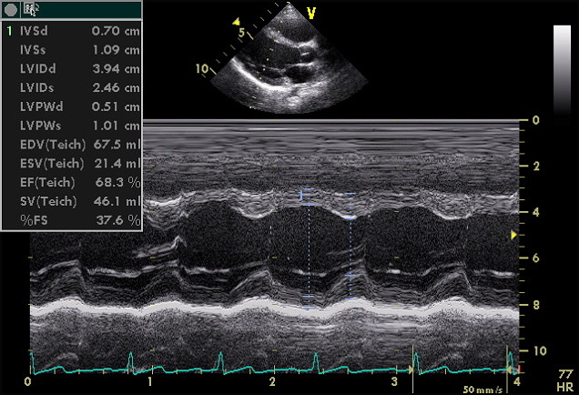
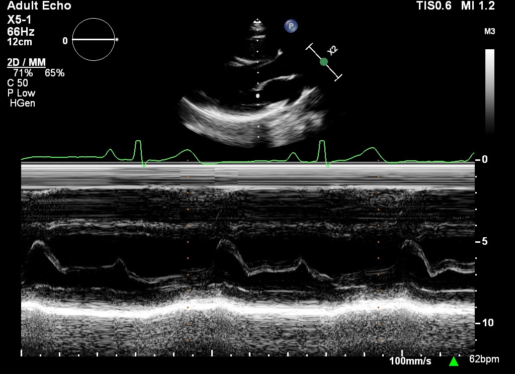
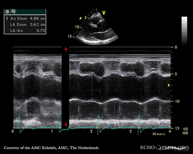
M-mode = motion over time The horizontal axis is time; vertical position is depth from the transducer. One beam line is sampled extremely fast, so timing is superb—but a misplaced line produces a beautifully precise wrong answer.


A
How to place the line
2D guides M-mode| Target | Cursor | Read | Trap |
|---|---|---|---|
| LV dimensions | PLAX, perpendicular to LV long axis at chordal tips | IVS/LVID/PW at end diastole; LVIDs at minimum cavity | Oblique line exaggerates diameter; 2D/volumes preferred for distorted geometry. |
| Mitral valve | Through leaflet tips | D–E–F–A–C motion; E-point septal separation (EPSS) | Wrong chordal plane imitates restricted motion. |
| Aortic root / LA | Through AV cusps, perpendicular to root | Box-like cusp opening, systolic LA posterior-wall motion | Off-axis underestimates cusp separation and root size. |
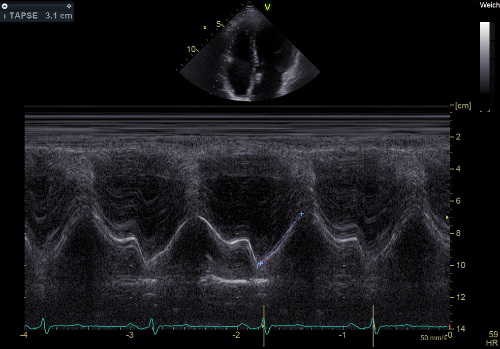
| TAPSE | Apical 4C, through lateral tricuspid annulus | End-diastole to peak-systole excursion | Angle and global translation; not isolated RV contractility. |
AcquireUse 2D to find the target; shrink depth/sector. For dimensions, place the cursor perpendicular to the structure/LV long axis; for excursion, align it with motion.
OrientTime runs left→right; depth increases downward. Confirm anatomy above the trace and ECG timing below it.
MeasureUse leading-edge conventions and the correct cardiac phase; average representative beats when rhythm varies.
ReconcileIf M-mode and 2D disagree, reacquire the line. Obliquity is more likely than exotic physiology.
Worked measurement LVIDd 5.6 cm, LVIDs 3.7 cm → fractional shortening = (5.6−3.7)/5.6 ×100 = 34%. Do not convert this to EF in regional disease, remodeling, pacing, or abnormal LV geometry.
LV M-mode was obtained in PLAX at the chordal level with a cursor perpendicular to the LV long axis: LVIDd __ cm, LVIDs __ cm, IVSd __ cm, PWd __ cm, fractional shortening __%. [State obliquity/geometry limitation.]
Direct vs reconstructed Direct M-mode repeatedly samples one beam line at native high temporal resolution. Anatomic/reconstructed M-mode reslices stored 2D cine and inherits its frame rate: it improves cursor placement but creates no new temporal information. Both can be precise yet off-axis or nonrepresentative.


A
Pattern → mechanism → confirmation
M-mode is a clue, not a verdict| M-mode clue | Suggests | Confirm / caveat |
|---|---|---|
| EPSS increased | Reduced LV stroke volume/LVEF or dilated LV | Measure E-point to septum; AR, MS, tachycardia, septal motion confound. Use biplane/3D EF. |
| Flat E–F slope + reduced excursion | Mitral stenosis | Doming/fusion and planimetry; low output also flattens slope. |
| Late-diastolic “B bump” | High LVEDP / delayed MV closure | Nonspecific; PR prolongation and rhythm matter. |
| Fine diastolic AML flutter | Aortic regurgitant jet strikes anterior MV leaflet | Confirm AR direction/severity; may be absent despite severe AR. |
| Coarse systolic flutter | High-velocity systolic turbulence | May accompany MR/LVOTO; localize with color and CW. |
| SAM–septal contact | Dynamic LVOT obstruction | Measure CW gradient and MR; loading/contractility are dynamic. |
| Early diastolic RV collapse | Tamponade physiology | Duration matters; integrate RA collapse, respiratory Doppler, IVC, BP. |
| TAPSE ≤1.7 cm | Abnormal RV longitudinal shortening | Angle/load/surgery dependent; integrate FAC, S′, strain. |
M-mode recognition atlas · all traces schematic
EPSS increasedE point remains far from septum: low forward stroke volume, reduced EF, or LV dilation. AR/MS and abnormal septal motion confound.Confirm with volumes, not eyeballing.
Late-diastolic B-bumpA shoulder on the A–C closure slope, after atrial contraction, can accompany high LVEDP. Rhythm and PR interval matter.Suggestive, never diagnostic alone.
SAMAnterior leaflet approaches or contacts septum during systole; duration of contact tracks obstruction better than a still frame.Confirm dagger CW + posterior MR.
Mitral flutter in ARAortic regurgitant flow may strike the anterior leaflet and produce rapid diastolic oscillation.Its absence never excludes severe AR.
Pulmonic midsystolic notchAbnormal RV–PA impedance/PH can interrupt systolic cusp opening.Integrate RVOT AT, TR velocity, and RV load.
Paradoxical septal motionPost-op state, LBBB/pacing, RV volume/pressure load, and constriction can reverse normal septal timing.Name the timing and clinical substrate.
Unknown trace
Thin high-frequency diastolic oscillation on the anterior mitral leaflet. First answer: AR-directed flutter; then prove AR mechanism and severity with color/CW/descending-aortic flow.Board trap
A 1.6-cm TAPSE after cardiac surgery does not by itself equal severe RV failure. Translation and altered contraction pattern demand FAC, S′, strain, size, and clinical context.Factoid The classic mitral “M” and posterior “W” are mirror-like only when leaflet motion and cursor placement are favorable. Calcification, prostheses, AF, tachycardia, and off-axis sampling can erase the alphabet.
Worked example TAPSE moves from 2.7 cm at end diastole to 1.1 cm at peak systole: excursion = 1.6 cm, below the ASE 2025 normal boundary. Report it as one abnormal RV index—not “RVEF 30%.”
Tip & trick When M-mode seems to contradict 2D, place the cursor again under 2D guidance. First suspect obliquity, translation, tethering, or the wrong anatomic level.
Doppler measures motion along the beam Δf = 2f₀v cosθ / c. At 0° the velocity is correct; at 20° it is ≈6% low; at 60° it is half. A spectral display is velocity versus time—not anatomy.
A
Choose the mode by the question
localize or measure the peak?| Mode | Knows depth? | Aliases? | Use | Failure mode |
|---|---|---|---|---|
| Pulsed-wave (PW) | Yes: sample gate | Yes, above Nyquist = PRF/2 | MV/PV/LVOT/venous flow; localize acceleration | Cannot record very high velocity; range ambiguity at high PRF. |
| Continuous-wave (CW) | No: entire beam | No clinically relevant aliasing | AS, MR, TR, high gradients | Range ambiguity: cannot tell where along beam signal arose. |
| Color | Yes, many PW samples | Yes; displays mean velocity/direction | Survey jets, convergence, timing, shunts | Jet size depends on gain, scale, depth, frame rate, wall impingement. |
| Tissue Doppler imaging (TDI) | Yes | Low-velocity scale | Annular e′/a′/s′, timing, mechanics | Angle, tethering, translation, surgery/MAC alter values. |
Pulsed-wave · localize “where”
Place a small gate in laminar flow. Walk it toward acceleration; raise PRF or move to CW when the signal wraps.Continuous-wave · measure “how fast”
Trace the dense modal edge and use the window that gives the highest valid velocity; avoid confusing MR, LVOTO, and AS.B
Aliasing: fix it deliberately
do not turn every knobPW spectral
Raise scale/PRF; shift baseline; lower frequency; reduce depth; use high-PRF or CW.
Raise scale/PRF; shift baseline; lower frequency; reduce depth; use high-PRF or CW.
Color survey
Raise scale, narrow box, reduce depth; aliasing itself helps show turbulent convergence.
Raise scale, narrow box, reduce depth; aliasing itself helps show turbulent convergence.
PISA
Intentionally shift baseline toward jet to Va ≈20–40 cm/s; record exact Va.
Intentionally shift baseline toward jet to Va ≈20–40 cm/s; record exact Va.
Never
Do not software angle-correct CW; obtain a more parallel window/PEDOF.
Do not software angle-correct CW; obtain a more parallel window/PEDOF.
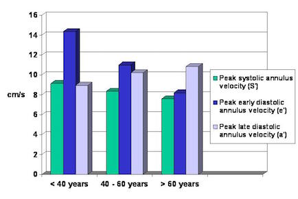

Worked angle error True jet 4.0 m/s at 30°: measured V = 4×cos30° = 3.46 m/s. Gradient falls from 64 to 48 mmHg because velocity is squared.
Tip & trick PW sample volume answers “where?”; CW answers “how fast?” Use PW to walk from LV cavity into LVOT, then CW from every window for the highest valid aortic velocity.
Factoid Spectral broadening can be real turbulence or excessive gain/large sample volume. A filled-in spectrum is not automatically severe disease.
Why proximal isovelocity surface area (PISA) works As blood approaches a small regurgitant orifice, equal-velocity shells crowd together. If the first aliasing shell is hemispheric, its area is 2πr²; area × aliasing velocity gives instantaneous flow.
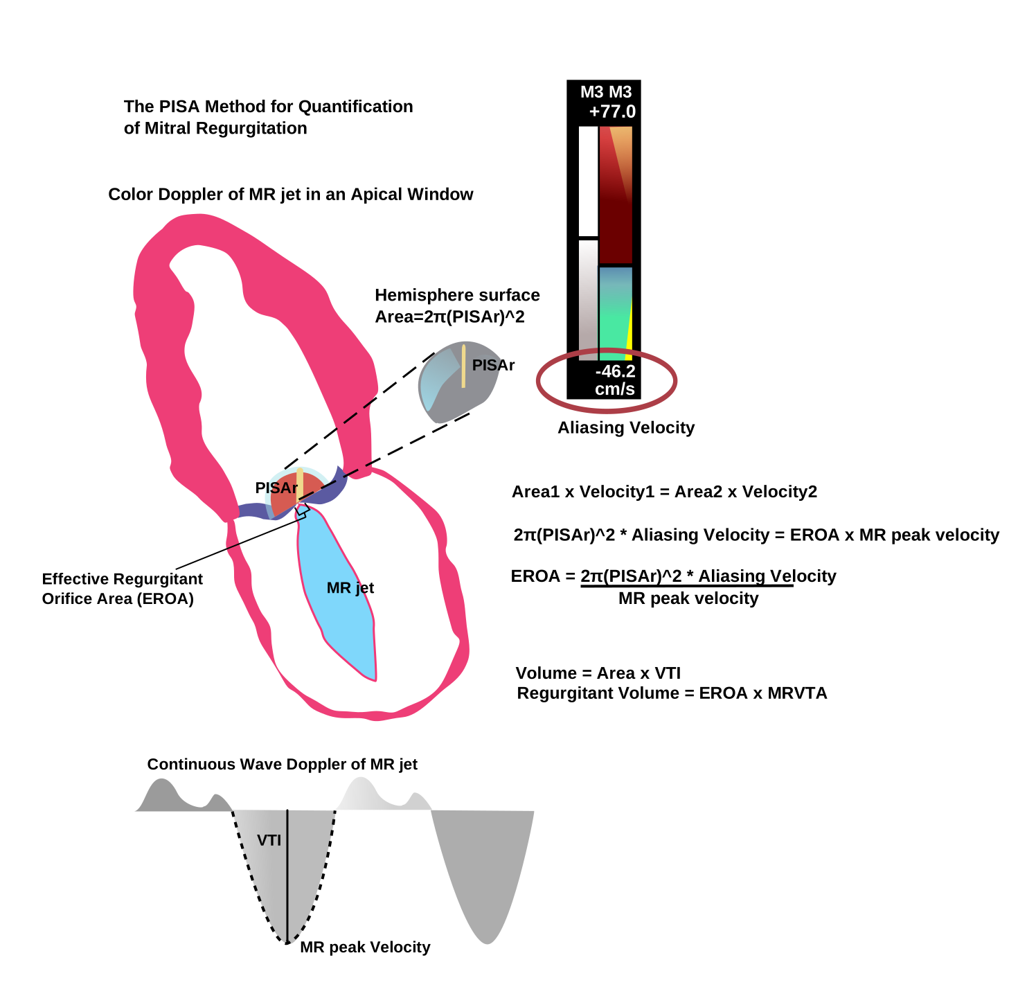

Three-step derivation
Q = 2πr² × Va
EROA = Q / Vregurg,peak
RVol = EROA × VTIregurg
Use one velocity unit throughout. cm² × cm/s = cm³/s = mL/s.
Worked PISA r = 1.0 cm, Va = 40 cm/s → Q = 2π(1.0²)(40) = 251 mL/s. MR Vmax 500 cm/s → EROA = 251/500 = 0.50 cm² (50 mm²). MR VTI 120 cm → RVol = 0.50×120 = 60 mL.
Tip & trick Zoom; set color scale; shift baseline toward the regurgitant jet; freeze the frame aligned to peak CW velocity; measure from the orifice to the first aliasing boundary—not to the outer color edge.
A
What breaks the hemisphere
geometry and time| Problem | Direction of error | Better move |
|---|---|---|
| Radius placement | Error is squared: +20% r → +44% flow | Optimize zoom/frame; repeat; reject a fuzzy shell. |
| Constrained angle α | Hemisphere overestimates if convergence is wall-constrained | Angle-correct Q by α/180° when geometry is clear. |
| Crescentic secondary MR/TR | Single-plane circular PISA often underestimates | Biplane VC, 3D VCA, volumetric echo or CMR. |
| Late/dynamic MR | Peak EROA overstates whole-beat burden | Prove duration on color/CW; integrate instantaneous flow or use volumetrics. |
| Multiple jets | Shells merge; one radius misses total orifice | 3D VCA or volumetric RVol; do not add arbitrary radii. |
| Poor CW alignment | Low denominator falsely raises calculated EROA | Reacquire parallel CW before dividing. |
B
Independent volumetric check
flow must balanceMR
RVol = LV total SV − LVOT forward SVRF = RVol / LV total SV ×100%
If important AR coexists, this subtraction still isolates MR because AR is contained in LV total SV and LVOT systolic output.
AR
RVol = LVOT SV − mitral inflow SVInvalid when important MR changes transmitral inflow. CMR: ventricular SV − phase-contrast forward SV.
Factoid Effective regurgitant orifice area is a hemodynamic effective area, not a literal anatomic hole. Three-dimensional vena-contracta area is closer to direct planimetry but remains dynamic and resolution-dependent.
A
Chamber consequence is part of the grade
but acute disease may be small| Chamber | Measure correctly | Clinical interpretation |
|---|---|---|
| LV | Biplane Simpson from nonforeshortened A4C/A2C; contrast if ≥2 contiguous segments poorly seen; 3D when feasible. | Volume overload enlarges LV in chronic MR/AR; concentric remodeling and low SVi support paradoxical LFLG AS. |
| LA | Biplane end-systolic volume, exclude LAA/PVs; index to BSA. >34 mL/m² is enlarged. | Chronic pressure/volume memory—not real-time LAP. AF, athlete, anemia, MV disease confound. |
| RV | RV-focused A4C; basal diameter, FAC, TAPSE, S′, free-wall strain; integrate, do not rely on TAPSE alone. | Afterload mismatch can reduce RV function; severe TR makes load-dependent indices deceptively preserved. |
| RA / IVC | RA end-systolic area/volume; IVC diameter/collapse plus secondary RAP signs. | RA/IVC enlargement supports chronic TR/high RAP, but not lesion-specific. |
Serial LVEF/GLS is a measurement contract. Keep modality, acquisition protocol, loading state, rhythm, vendor/software, and analysis convention constant; prefer 3-D volumes when feasible. Do not average a technically invalid study into a trend—state comparability, uncertainty, and whether change exceeds laboratory repeatability.
B
Right-heart anchors
ASE 2025TAPSE
≤1.7 cmAbnormal longitudinal shortening; angle/load dependent.
S′
≤9.5 cm/sAbnormal basal longitudinal velocity; fusion/tethering matter.
FAC
≤35%Abnormal; includes longitudinal + radial change, area-tracing dependent.
RV FWLS
−20% or less negativeAbnormal supportive threshold; vendor/load dependent.
C
PASP and coupling
know when Bernoulli fails- PASP 4(TR Vmax)² + RAP; use a complete envelope from multiple windows.
- Contrast agitated saline/UEA can complete a weak signal; avoid blooming the peak.
- Do not infer PASP with RVOT/PS obstruction; report RV–RA gradient instead.
- Severe TR early RA–RV equalization can yield a low Vmax and underestimate PASP.
- TAPSE/PASP is an RV–PA coupling surrogate; both numerator and denominator can fail in severe TR.
- PH cause echo estimates probability; RHC classifies pre- vs post-capillary disease.
| IVC pattern | RAP estimate |
|---|---|
| ≤2.1 cm + >50% collapse | 3 mmHg (0–5) |
| >2.1 cm + <50% collapse | 15 mmHg (10–20) |
| Discordant size/collapse | 8 mmHg (5–10); use secondary signs |
Worked PASP TR Vmax 3.2 m/s and RAP 8 → 4(3.2²)+8=49 mmHg. If TAPSE is 1.4 cm, TAPSE/PASP = 0.29 mm/mmHg; this suggests impaired RV–PA coupling but requires method- and population-specific interpretation.
Factoid Positive-pressure ventilation, athletic physiology, pregnancy and poor inspiratory effort can uncouple IVC size/collapse from invasive RAP. State the ventilation context.
Acute valve catastrophe Normal chamber size never excludes acute severe MR/AR/TR. Acute pressure equalization can shorten and soften the Doppler signal precisely when the lesion is most dangerous.
ASE 2025 separates two questions This page asks whether diastolic dysfunction exists. Page 10 separately estimates mean LAP and assigns Grade 1–3. Do not import TR velocity into this presence algorithm.
Optional age-specific relaxation limits
e′ 20–39: septal <7 · lateral <10 · avg <9e′ 40–65: <6 · <8 · <7; >65: <6 · <7 · <6.5 cm/s
E/A lower limit: <0.88 (20–39) · <0.69 (40–60) · <0.50 (60–80)
Exact-operator trap
Presence Fig 2 uses avg E/e′ >14. The pressure Fig 3 uses avg E/e′ ≥14. Preserve the operator. LVMI >95 women / >115 men g/m² is supportive, not one of the four count chips.Worked patient · carry the same numbers to page 10
Acquire68-year-old with hypertension and exertional dyspnea; sinus 72 bpm, BP 146/82, LVEF 60%. Septal/lateral e′ 5/6 cm/s; average E/e′ 16; E/A 1.3; LARS 17%; LAVI 42 mL/m²; PASP 39 mmHg.
Determine presenceAverage e′ is reduced. E/e′, LARS, and LAVI meet pressure/remodeling markers. Diastolic dysfunction is present.
Do not stopThis page does not grade. Move the same patient to page 10 and separately ask whether mean LAP is elevated.
Why the split matters A patient can have impaired relaxation with normal mean LAP (Grade 1), or preserved annular e′ with enough pressure/remodeling evidence to establish DD. “Normal EF” does not answer either question.
General algorithm eligibility Sinus rhythm; any EF. Do not use this pathway for severe primary MR, any MS, moderate/severe MAC, MV repair/replacement/TEER, AF, transplant, noncardiac PH, constriction, or LVAD. Use the dedicated page 14 route.
Mixed pattern includes: reduced e′ only with E/A >0.8; isolated high E/e′; isolated high TR/PASP; or any two abnormal. If measured, reliable adjudicators are negative, LAP is likely normal. Missing or unreliable adjudicators require supplemental evidence. Symptomatic patients proceed to exercise testing. Grade 2 is any E/A <2 once elevated LAP is established—a key change from 2016.
Core testsFrom page 9: e′ reduced, average E/e′ 16, and PASP 39 mmHg. All three core tests are abnormal.
PressureAll three abnormal → elevated mean LAP; no adjudicator is needed.
GradeE/A 1.3 is <2, therefore Grade 2 DD—not Grade 1 and not “pseudonormal only.”
ReportGrade 2 diastolic dysfunction with elevated mean LAP; sinus 72 bpm, BP 146/82. Evidence: low e′, average E/e′ 16, PASP 39, E/A 1.3; LARS 17% and LAVI 42 supportive.
Grade is not a waveform label Establish mean LAP with the page 10 multiparametric algorithm; only then use E/A to separate Grade 2 from 3. Pseudonormal filling can look “normal.”
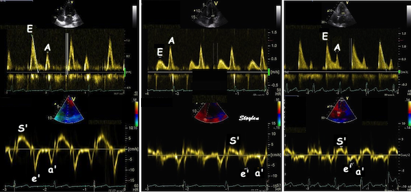

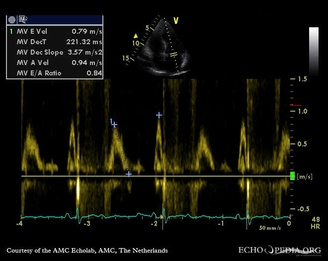

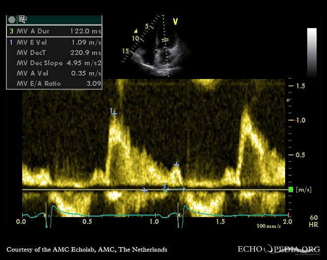

Grade 1 ≠ no disease
Impaired relaxation with normal mean LAP. If dyspneic, pressure may rise only with exercise.Grade 2
Once elevated LAP is established, any E/A <2. The old 0.8–<2 shortcut is obsolete in ASE 2025.Grade 3
E/A ≥2 with elevated LAP. A Valsalva-induced shift toward Grade 1 implies reversibility; persistence suggests fixed restriction.A
Mean LAP is not LVEDP
two pressures, different timestampsMean LAP / mean PCWP
Left atrial pressure or its wedge-pressure surrogate averaged across the cardiac cycle. It is the pulmonary venous back-pressure relevant to congestion and the target of ASE grading.LVEDP
Left-ventricular end-diastolic pressure, measured just before systole. It can rise before mean LAP; a prolonged pulmonary-vein Ar relative to mitral A supports this late-diastolic mismatch.B
LARS — what it means and how it is derived
left atrial reservoir strain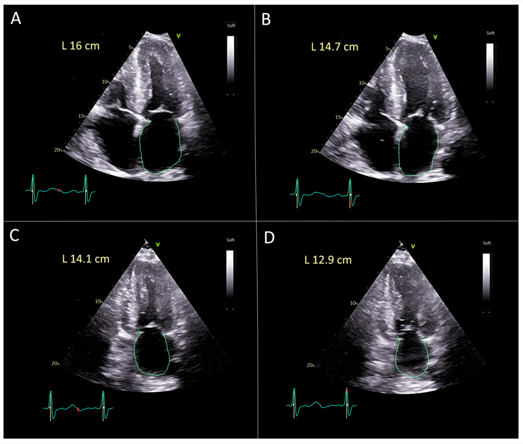
Acquire
A4C + A2C, dedicated nonforeshortened LA, 50–70 fps, stable R–R gating, 3–5 similar cycles. Trace LA endocardium; exclude pulmonary veins and appendage; use validated LA speckle-tracking software.Concept
strain = (Lpeak reservoir − Lreference)/Lreference ×100%With end-diastolic/R-wave zero reference, peak positive strain during ventricular systole is LARS. Vendor/gating conventions matter.
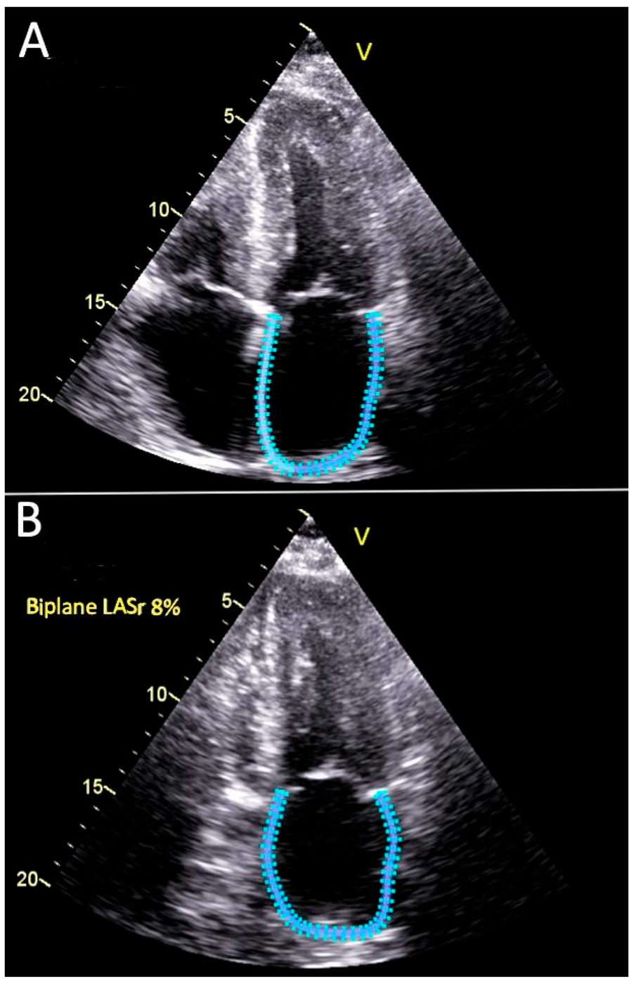
Illustrative calculation Reference LA wall length 14.7 cm; reservoir peak 16.0 cm → (16.0−14.7)/14.7 ×100 = 8.8%. Clinical LARS is software-derived, often biplane; this manual length method only explains the physics.
C
IVRT, mitral L-wave, and pulmonary-vein Ar−A
see the timingMitral L-wave
ELA
L = mid-diastolic transmitral flow between E and A. ≥50 cm/s supports high pressure/advanced relaxation abnormality, but bradycardia and MR/MS confound.
Pulmonary vein Ar−A
SDAr
Measure PV Ar duration and transmitral A duration at 100 mm/s. Ar−A >30 ms supports elevated LVEDP; align both to the ECG.
IVRT
AV closeMV openIVRT
Isovolumic relaxation time = aortic valve closure to mitral valve opening. Record both flows in one apical CW/PW sweep. ≤70 ms supports high LAP; HR, conduction and AR confound.
Tip & trick MV PW gate 1–3 mm at leaflet tips; PV gate 3–5 mm placed 5–10 mm inside a right pulmonary vein. Use 100 mm/s for durations, 25–50 mm/s for respiratory trends. LARS ≤18% is a specific supportive threshold—not the lower limit of normal and never a stand-alone rule-out.
Use when symptoms and rest disagree Unexplained exertional dyspnea with resting Grade 1 or indeterminate pressure. Use exercise—supine bicycle or treadmill—not dobutamine.
A
Invasive reference when stakes remain high
pressure–flow relationshipRest PCWP
>15 mmHgRest LVEDP
>16 mmHgExercise
PCWP ≥25or LVEDP ≥23
Slope
PCWP/CO >2mmHg/L/min
2026 safety update In a large invasively proven ambulatory HFpEF cohort, ASE 2025 rest/stress categories had important false negatives. A normal/Grade 1 rest study—or negative stress echo—must not exclude HFpEF when clinical probability remains high; use HFpEF-specific scoring and/or invasive exercise hemodynamics.
RestRecord MV E/A, septal+lateral e′, TR envelope, BP, HR, rhythm, symptoms.
ExerciseUse symptom-limited supine bike or treadmill; preserve a narrow acquisition list.
Peak / recoveryAcquire average E/e′ and TR Vmax at peak or within 1–2 minutes; record workload.
IntegrateBoth high supports exercise elevation; discordance is inconclusive, not negative.
Worked stress case A different patient has unexplained exertional dyspnea, preserved EF, and resting Grade 1 DD with normal mean LAP. At 75 W, peak average E/e′ = 15 and TR Vmax = 3.3 m/s. Both are positive: exercise filling pressure is abnormal and supports HFpEF. If only TR rose, the result would be inconclusive. The page-9/10 Grade-2 patient is already positive at rest and does not need diagnostic stress grading.
Nuance: E/e′ >14 with TR >2.8 but <3.2 is “likely” HFpEF; isolated exercise TR >3.2 with E/e′ <14 can reflect normal flow-related pressure rise. Normal natriuretic peptide—especially with obesity—also does not exclude HFpEF.
Stop before page 10 Mitral disease/repair, AF, PH, transplant, constriction and LVAD change the relationship between Doppler and pressure. Use the phenotype-specific evidence below.
| Phenotype | Pressure route / high-yield thresholds | Pitfall |
|---|---|---|
| AF | Tier 1: E ≥100 cm/s; septal E/e′ >11; TR >2.8 or PASP >35; DT ≤160 ms. ≥3 high → ↑LAP; 0–1 → normal; exactly 2 → LARS <18, PV S/D <1, BMI >30: none normal, 1/unavailable indeterminate, ≥2 elevated. | Average representative cycles. No A wave. LARS is only one secondary item. |
| PH | E/A ≤0.8 + E ≤50 → normal; E/A ≥2 + low e′ → high. Intermediate: LARS >18 normal, ≤18 high; if absent, lateral E/e′ <8 normal, >13 high. | Use lateral e′; septal flattening confounds septal TDI. Echo does not replace RHC classification. |
| MAC | E/A <0.8 → normal; >1.8 → high; 0.8–1.8: IVRT ≥80 normal, <80 high. | Standard E/e′ and PHT can fail; separate degenerative MS. |
| MS / significant MR | MS: IVRT <60; mitral A >1.5 m/s; IVRT/(TE−e′) <4.2. MR: Ar−A ≥30; IVRT <60; IVRT/(TE−e′) <5.6; avg E/e′ >14 mainly when EF reduced. | High E and altered PV flow are lesion physiology, not generic LAP votes. |
| HCM / restrictive CM | HCM: avg E/e′ >14; Ar−A ≥30; TR >2.8; LAVI >34. RCM: avg E/e′ >14; DT <140; E/A >2.5; IVRT <50. | Amyloid “5-5-5”: e′, s′, a′ all <5 cm/s in advanced disease. |
| Transplant | Avg E/e′ <7 normal; >14 high. Between: E/SRIVR ≤200 normal, >200 high; if unavailable use TR ≤ or >2.8. | E/A ≥2 can be normal early after transplant. |
| LVAD | Integrate E/A >2, RAP >10, PASP >40, avg E/e′ >14 or septal ≥15, LAVI >33, septal position. | Speed, unloading, RV function and AI alter every signal. |
| LBBB / pacing / tachy | Favor lateral E/e′ with caution. Tachy: IVRT ≤70, PV systolic fraction ≤40; avg E/e′ >14 specific but insensitive. | Septal e′ and fusion mislead; a post-PVC pause may separate E/A. |
AF · exactly two Tier-1 positives
E 110 cm/s and septal E/e′ 12 are high; TR 2.6 and DT 180 are not. Adjudicate with LARS, PV S/D, and BMI: ≥2 secondary positives → elevated LAP; one or unavailable → indeterminate.HCM · do not trust one E/e′
Average E/e′ 16 with Ar−A 35 ms, TR 3.0 m/s, and LAVI 48 is concordant. A single high septal E/e′ in paced/LBBB physiology is not.R
Reporting line that earns points
say what you knowDiastolic function: [normal / Grade 1 / Grade 2 / Grade 3 / not gradeable]. Mean left atrial pressure: [normal / elevated / indeterminate]. Evidence: MV E/A __; septal/lateral e′ __/__ cm/s; average E/e′ __; PASP __ mmHg (or TR Vmax __ m/s); adjudicator __. Rhythm __; HR __; BP __. Limitation/special-population route: __.
2025 labels Grade 1 = normal mean LAP · Grade 2 = mild–moderate elevation · Grade 3 = marked elevation. If grade is indeterminate but LAP is supportable, report the pressure conclusion anyway.
The paradox Constriction preserves or exaggerates longitudinal relaxation while dissociating intrathoracic from intracardiac pressure. Restriction impairs myocardial relaxation. Look for the pattern, not a single respiratory percentage.
A
Mayo echo sequence
high-yield discriminator set| Finding | Favors constriction | Important nuance |
|---|---|---|
| Septal motion | Respiration-related septal shift / bounce | Post-op state, LBBB and RV overload can mimic abnormal septal motion. |
| Medial e′ | >8 cm/s strongly favors; 6–8 mixed; <6 favors restriction | Calcification at medial annulus can lower e′ despite constriction. |
| Hepatic veins | Prominent expiratory diastolic reversal | Severe TR causes systolic reversal instead. |
| Annulus signs | Reversus: medial e′ > lateral e′. Paradoxus: E/e′ may vary inversely with filling pressure. | Neither is diagnostic alone; verify the full constriction pattern. |
Case A · constriction pattern
Septal shift with inspiration, medial e′ 10 cm/s, lateral e′ 6 cm/s, mitral inflow variation 31%, and expiratory hepatic diastolic reversal/forward 1.0. Integrative conclusion: physiology strongly favors constriction.Case B · myocardial restriction
No respiratory septal shift; medial e′ 4 cm/s, E/A 2.7, DT 125 ms, IVRT 45 ms, biatrial enlargement and apical-sparing strain. Integrative conclusion: restrictive myocardial physiology.Respiratory septal shift [present/absent]; medial/lateral e′ __/__ cm/s; mitral/tricuspid inflow variation __/__%; hepatic expiratory diastolic reversal/forward __. Integrated physiology [favors constriction / favors restriction / indeterminate]; [CMR/CT/catheterization] recommended because __.
False-negative respiration Markedly elevated LAP, dehydration, irregular breathing or mixed constriction/restriction can blunt variation. Repeat after preload adjustment or use CT/CMR and simultaneous LV/RV hemodynamics when the phenotype matters.
AcquireRecord BP/rhythm. Measure LVOT inner-edge in mid-systole; place PW before acceleration. Beat-match LVOT/AV VTIs and use apical, right-parasternal, and suprasternal PEDOF.
GradeUse highest valid Vmax/mean, then AVA/AVAi and DVI. Severe: Vmax ≥4.0, mean ≥40, AVA ≤1.0, AVAi ≤0.60, or DVI ≤0.25. Calculate SVi; low flow ≤35 mL/m².
ReconcileDiscordant? Recheck LVOT diameter/gate, CW alignment, beat match, BP, body size, and flow. Consider AR, dynamic LVOTO, pressure recovery, and high flow.
Report / escalateState highest window, Vmax/mean, AVA/AVAi, DVI, SVi, BP/rhythm, and grade. AVA ≤1.0 with mean <40 → label discordance and enter the low-flow pathway.
AS begins with flow Vmax and mean gradient are the primary hemodynamic measures; continuity AVA and DVI resolve flow dependence. Use the highest valid CW signal—not the apical default.
A
Native AS severity table
ASE grading| Grade | Vmax | Mean gradient | AVA | Indexed AVA | DVI |
|---|---|---|---|---|---|
| Sclerosis | ≤2.5 m/s | — | — | — | — |
| Mild | 2.6–2.9 | <20 mmHg | >1.5 cm² | >0.85 cm²/m² | >0.50 |
| Moderate | 3.0–3.9 | 20–39 | 1.0–1.5 | 0.60–0.85 | 0.25–0.50 |
| Severe | ≥4.0 | ≥40 | <1.0 | <0.60 | <0.25 |
| Very severe | >5.0 m/s | ≥60 mmHg | — | — | — |
ASE table operators avoid overlap at the exact boundary; current clinical severe-AS criteria commonly use AVA ≤1.0 cm², AVAi ≤0.60 cm²/m² and DVI ≤0.25. Integrate exact-boundary discordance. ESC 2025 uses a 25–40 mmHg mean-gradient range in its normal-flow “moderate AS” management definition; ASE’s echo grading convention remains 20–39.
B
Continuity equation audit
most discordance is technical or flow-relatedLVOT diameter
Inner edge to inner edge, mid-systole, just below cusps. Error is squared. Elliptical LVOT makes 2D area too small.LVOT VTI
PW gate in laminar flow before acceleration. Match heart rate/beat to AV CW; exclude subvalvular acceleration.AV VTI
Apical + right parasternal + suprasternal; PEDOF. Trace modal edge. Do not mistake eccentric MR for AS.C
Flow-independent helpers and traps
context, not shortcuts- DVI ≤0.25 supports severe AS and avoids diameter error, but PW position still matters.
- Low flow SVi ≤35 mL/m². Compute SVi whenever AVA and gradient disagree.
- Blood pressure repeat after SBP control when feasible; afterload can suppress flow/gradient.
- Indexing AVAi may overcall severe AS in obesity; integrate absolute area and flow.
- Dynamic LVOTO late-peaking dagger; localize with PW before applying Bernoulli.
- Pressure recovery can explain Doppler–cath mismatch, especially a small ascending aorta.
- Mixed AS/AR AR raises Vmax/gradient; AVA less flow dependent; report combined burden.
- High flow anemia, thyrotoxicosis, AV fistula can create high gradient with AVA >1.
Pattern requiring skepticism “Paradoxical low-flow severe AS” with Vmax <3 m/s or mean gradient <20 deserves an especially aggressive search for LVOT, tracing, BP, and body-size error before the label sticks.
AcquireVerify BP/rhythm, LVOT diameter and PW gate, beat matching, multiwindow PEDOF, DVI, SVi, and LVEF before naming a phenotype.
GradeClassical LFLG: SVi ≤35 + EF <50%. Paradoxical: SVi ≤35 + EF ≥50%. Normal-flow low-gradient: SVi >35 + EF ≥50%; integrate morphology and CT.
ReconcileClassical LFLG → low-dose DSE; projected AVA at 250 mL/s if flow increases enough to estimate the AVA–flow slope. Use CT calcium if unresolved. Lack of gradient rise without flow augmentation does not prove pseudo-severe.
Report / escalateName phenotype, verification performed, and DSE/CT result. CT supports likely severe at >2000 men/>1200 women and very likely at >3000/>1600; note low-calcium exceptions.
Flow reserve
≥20% SV increase on DSE. Its absence does not exclude true severe AS or benefit; use projected AVA/CT.Low-calcium exceptions
Bicuspid, amyloid, rheumatic, radiation/inflammatory or predominantly fibrotic stenosis may be severe below thresholds.High-gradient AVA >1
Often high flow; if no reversible cause, high-gradient physiology generally carries severe burden—recheck LVOT and mixed AR.Projected AVA Standardize to flow 250 mL/s when flow augmentation permits a reliable AVA–flow slope. Do not extrapolate from essentially unchanged flow. Never call pseudo-severe merely because the gradient failed to reach 40 in a patient without flow augmentation.
AcquireDefine cusp number/motion/coaptation and measure annulus, sinuses, STJ, and ascending aorta. Obtain VC/jet-width ratio, CW PHT, descending-aortic flow, EROA/RVol/RF, and LV response.
GradeSevere anchors: VC >6 mm, jet/LVOT ≥65%, EROA ≥30 mm², RVol ≥60 mL, RF ≥50%, or holodiastolic descending reversal with EDV ≥20 cm/s.
ReconcileEccentric jets defeat width ratios; PHT changes with LVEDP/compliance/vasodilation; reversal has non-AR causes. A normal LV argues against chronicity—not acute severe AR.
Report / escalateState leaflet versus root mechanism, four-grade severity, quantitative support, LV consequence, and aortic dimensions. Acute/dissection/endocarditis → urgent TEE pathway; chronic discordance → CMR.
A
Mechanism: primary valve vs secondary root
describe both cusp and aortaPrimary leaflet disease
Bicuspid/unicuspid; prolapse or flail; endocarditis/perforation; rheumatic restriction; degeneration; trauma. Describe cusp number, thickening, motion and coaptation.Secondary / functional AR
Aortic root or ascending dilation, sinotubular-junction effacement, dissection, annuloaortic ectasia; cusps may be normal but fail to coapt. Measure annulus, sinuses, STJ and ascending aorta.Leaflet failure · schematicProlapse, flail, perforation, or restriction prevents cusp coaptation and often creates an eccentric jet.
Root/STJ failure · schematicDilated root or sinotubular junction separates otherwise normal cusps. Report every aortic level.
Flow reversal · schematicPlace PW in the proximal descending aorta. Persistent diastolic flow below baseline with EDV ≥20 cm/s is a severe anchor.
B
Four-grade AR table
integrated echo| Parameter | Mild 1+ | Mild–mod 2+ | Mod–severe 3+ | Severe 4+ |
|---|---|---|---|---|
| VC width | <3 mm | 3–6 | 3–6 | >6 mm |
| Central jet / LVOT width | <25% | 25–45% | 46–64% | ≥65% |
| Jet CSA / LVOT CSA | <5% | 5–20% | 21–59% | ≥60% |
| Pressure half-time | >500 ms | 200–500 | 200–500 | <200 ms |
| EROA | <10 mm² | 10–19 | 20–29 | ≥30 mm² |
| RVol | <30 mL | 30–44 | 45–59 | ≥60 mL |
| RF | <30% | 30–39% | 40–49% | ≥50% echo; CMR >40% ESC 2025 |
| Descending aortic flow | Brief early reversal | Intermediate | Holodiastolic; EDV 10–<20 | Holodiastolic; EDV ≥20 cm/s |
C
AR traps and acute severe pattern
pressure equalization can hide catastrophe- Jet length is unreliable. Width ratios apply only to central jets; eccentric jets underestimate.
- PHT shortens with high LVEDP/poor compliance and vasodilators; may be long in compensated chronic severe AR.
- Flow reversal varies with age/aortic compliance and may reflect PDA, AV fistula, dissection or ruptured sinus.
- Acute severe AR normal LV, short/low-velocity eccentric jet, premature MV closure, diastolic MR, rapid equalization.
- Chronic severe AR should usually enlarge LV; normal LV argues against chronicity, not acute severity.
- Escalate urgent TEE for suspected dissection/endocarditis or discordant acute physiology; CMR for chronic RF/volumes.
AcquireDefine rheumatic fusion versus MAC tunnel. Use 3D-guided planimetry at the smallest leaflet-tip orifice; obtain CW mean gradient, PHT, and PASP with HR/rhythm. In AF average ≥5 beats.
GradeClinically severe rheumatic MS: MVA ≤1.5 cm². Support: PHT ≥150 ms, mean ≥10 mmHg at HR 60–80, and PASP ≥50; gradient remains flow/HR dependent.
ReconcilePHT fails with altered compliance, LVH, AR/MR, fibrosis, or recent commissurotomy. High flow/MR raises gradient; low flow lowers it. MAC-MS lacks a validated full grade table.
Report / escalateReport mechanism, MVA+method, mean gradient at stated HR/rhythm, PHT/PASP, MR, and anatomy. Symptom discordance → exercise; uncertain MAC/intervention anatomy → TEE/CT.
Modern threshold alert Current ASE/ACC/ESC terminology defines MVA ≤1.5 cm² as severe/clinically severe rheumatic MS. The old “severe only below 1.0” convention is legacy; <1.0 may be called very severe.
A
Rheumatic MS grading
gradient assumes HR 60–80| Grade | MVA | PHT | Mean gradient | PASP |
|---|---|---|---|---|
| Mild / progressive | >2.5 cm² | <100 ms | <5 mmHg | <30 mmHg |
| Moderate / progressive | 1.6–2.5 | 100–149 | 5–9 | 30–49 |
| Severe | ≤1.5 cm² | ≥150 ms | ≥10 mmHg | ≥50 mmHg |
B
Measure by hierarchy
anatomy → area → hemodynamics3D-guided planimetry
Best anatomic area at smallest leaflet-tip orifice. Rheumatic: commissural fusion, tip thickening/calcification, doming AML, restricted PML, subvalvular disease.PHT
MVA = 220/PHTFails with altered LA/LV compliance, LVH, AR, MR, LA fibrosis, and immediately after balloon commissurotomy.
Mean gradient
Flow/HR dependent. Report rhythm and HR; in AF average ≥5 representative beats. High flow/MR raises gradient; low flow lowers it.C
Stress, continuity and degenerative MS
when symptoms do not fit| Issue | Practical answer |
|---|---|
| Exercise | Prefer exercise. Mean gradient >15 mmHg or PASP >60 supports hemodynamically important MS; dobutamine gradient ≥18 if exercise impossible. |
| Continuity area | Invalid with significant MR or AR because MV and LVOT forward flows differ. Isolated AR may permit RVOT flow substitution. |
| MAC-related MS | No universally validated mild/moderate/severe table. Nonplanar tunnel and shadowing limit planimetry; PHT is especially unreliable. Integrate morphology, trustworthy area, gradient at stated HR, symptoms, LA/PASP/RV and MR; use TEE/CT for intervention. |
| Balloon suitability | Unfavorable: Wilkins >8, severe commissural calcification, >mild MR, LA thrombus, severe TR or absent commissural fusion. |
Mechanism first Rheumatic MS is a commissural-fusion disease; degenerative MAC stenosis is a calcific tunnel without fusion. Balloon commissurotomy makes physiologic sense for the former, generally not the latter.
AcquireStandardize color and show origin/direction; then obtain morphology/direct lesion, VC/PISA or volumetrics, CW shape/density, venous flow, and chamber response.
GradeUse ≥3 domains and valve-specific thresholds; weight specific anatomy/direct signs above supportive findings. Native AR/MR/PR use trace–mild–moderate–severe; TR may add massive/torrential.
ReconcileSmall jets can reflect Coanda, low driving pressure, or acute equalization; large jets can reflect high pressure/gain or low scale. Chambers show chronicity—not always grade.
Report / escalateReport mechanism + defined grade + measurements + consequence + limitation. Name the cause of discordance and resolving modality; acute rupture/dissection/trauma → urgent pathway.
Use at least three domains Specific anatomy/direct jet, quantitative severity, and chamber/venous consequences. If discordant, name why and obtain the resolving modality—do not create a false “average grade.”
A
Cross-valve severe anchors
not interchangeable across valves| Valve | VC | EROA | RVol | RF | High-specificity corroborator |
|---|---|---|---|---|---|
| AR | >6 mm | ≥30 mm² | ≥60 mL | ≥50% echo | Holodiastolic descending reversal, EDV ≥20; central jet ≥65% LVOT |
| Primary MR | ≥7 mm; ≥8 biplane | ≥40 mm² | ≥60 mL | ≥50% | Flail/large defect; PV systolic reversal; MV/LVOT VTI >1.4 |
| Secondary MR | integrative | ≥30 mm² may be severe | ≥45 mL may be severe | ≥50% | Elliptical orifice/low forward flow; grade after GDMT/CRT and stable loading |
| TR | ≥7 single; >9 biplane | ≥40 mm² | ≥45 mL | ≥50% | Hepatic systolic reversal; dense triangular CW; 3D VCA ≥75 mm² |
| PR | No validated absolute | No echo partition | CMR ≥60 mL | >40% CMR | Branch-PA reversal; jet/annulus >65–70%; PHT <100 supports ≥moderate |
B
Color jet area: diagnose, rarely grade
momentum image, not flow meterLooks too small
Eccentric Coanda jet; low driving pressure; acute pressure equalization; huge receiving chamber; high Nyquist or low gain settings.Looks too large
Hypertension/high driving velocity; high gain; low scale; small receiving chamber; broad low-velocity entrainment.What to do
Standardize color; show jet origin/direction; then move to VC, PISA/volumetrics, CW density/shape, venous flow and chamber response.C
Acute vs chronic severity
chambers tell time, not always gradeChronic
- MR/AR → LV and usually LA enlargement; TR/PR → RV/RA enlargement.
- Remodeling supports hemodynamic significance and duration, but competing CM can enlarge chambers.
Acute
- Normal chamber size, high pressure, short/soft or eccentric jet, rapid equalization.
- Think rupture, endocarditis, dissection, trauma; urgent TEE/surgical pathway outranks perfect quantification.
Reporting language Native AR/MR/PR: trace, mild, moderate, severe. TR may add massive and torrential within severe. Always report mechanism + severity + supporting measurements; avoid “trivial/minimal” as a substitute for a defined grade.
AcquireAssess leaflet motion, scallop, jet direction, annulus, coaptation, LV/LA geometry, and jet count; use 3D en-face TEE to confirm segment or commissure.
Grade mechanismClassify Carpentier I, II, IIIa, or IIIb and phenotype as primary, ventricular-secondary, atrial-secondary, or mixed. Motion class does not grade severity.
ReconcileP2 usually jets anteriorly and A2 posteriorly, but commissural/multiple jets need 3D. Multiple drivers may coexist—do not force one label.
Report / escalateState phenotype, Carpentier type, A/P segment or commissure, jet number/direction, annular/coaptation geometry, flail gap/width, calcification, and every mixed driver.
MR is a mechanism diagnosis before it is a number State leaflet motion (Carpentier), lesion location/scallop, jet direction, annular/LV/LA geometry and whether the valve is primary, ventricular secondary, atrial secondary, or mixed.
A
Carpentier motion → likely mechanism
jet direction helps localize| Type | Leaflet motion | Common causes | Typical jet clue |
|---|---|---|---|
| I | Normal | Annular dilation; leaflet perforation/cleft | Central with annular dilation; perforation jet arises through leaflet |
| II | Excessive | Prolapse, flail, chordal/papillary rupture | Jet generally directed away from prolapsing/flail leaflet |
| IIIa | Restricted systole + diastole | Rheumatic disease, radiation, inflammatory restriction | Restricted tips; often MR + MS |
| IIIb | Restricted in systole | Ischemic/nonischemic LV remodeling; papillary displacement/tethering | Often posteriorly directed with posterior tethering; may be central |
Geometry before numbers · schematic MR phenotypes
Primary / excessive motionLeaflet crosses the annular plane; flail tip points toward the LA. Jet usually travels away from the diseased leaflet.
Ventricular secondaryRemodeled LV displaces papillary muscles, increases tenting, and reduces closing force; leaflets are usually normal.
Atrial secondaryLA/annular dilation dominates with preserved EF and little LV tethering; posterior annular displacement can add atriogenic restriction.
B
Three MR phenotypes
primary vs two secondary formsPrimary / degenerative
Leaflet/chord/papillary pathology creates MR. MVP/flail, fibroelastic deficiency, Barlow, endocarditis, rheumatic restriction, cleft, radiation. Repairability hinges on scallop, calcification and tissue quality.Ventricular secondary MR
MR is “born in the ventricle”: LV remodeling → papillary displacement → tenting/tethering exceeds closing force. Leaflets are usually structurally normal. Ischemic regional or nonischemic global.Atrial secondary MR
LA/annular dilation from AF/HFpEF with preserved EF and little LV tethering. ESC phenotype: LVEF ≥50%, no regional dysfunction, no/mild LV enlargement, annular AP diameter >35 mm, LAVI >34.C
TEE surgical map
standard LA view: aorta 12 · LAA 9Describe
Anterior A1–A3 · posterior P1–P3 · commissure · cleft · flail gap/width · calcification · annular dimensions · coaptation length/depth · multiple jets.Orient & verify
Standard LA/surgical view: aorta at 12 o’clock, LAA near 9; P1→P3 runs lateral-to-medial. P2 prolapse usually jets anteriorly; A2 posteriorly. Renderings rotate/crop—confirm landmarks and orthogonal 2D/MPR/color. Prove flail by systolic free-edge eversion/free motion on cine; a static 3D still cannot prove flail.Mixed mechanism A patient can have degenerative P2 prolapse plus LV dilation/tethering, or atrial annular dilation plus MAC. Report each driver; a single Carpentier label is not a complete mechanism statement.
AcquireProve lesion and systolic duration; map scallop, jets, flail gap/width, and calcification. Obtain VC/PISA or whole-beat volumes, PV flow, LV/LA, PASP/TR/RV.
GradeHolosystolic flail fits standard VC/PISA/RVol thresholds. Late MVP requires duration and whole-beat RVol/CMR; multiple jets favor 3D VCA or volumetrics.
ReconcileAcute rupture/endocarditis may show a small eccentric low-velocity jet, normal chambers, and hyperdynamic EF. Coanda and peak-frame PISA mislead; do not add single-plane VC widths.
Report / escalateReport Carpentier/scallop, timing, jets, repair anatomy, LV/LA/PASP. Severe MR with EF ≤60% or LVESD ≥40 mm = LV dysfunction. Acute disaster → urgent TEE.
Primary MR: the valve is the disease Prove structural lesion, establish systolic duration, quantify by independent methods, then document LV/LA/PASP response and repair anatomy.
A
High-specificity severe morphology
anatomy can outrank a weak jetDisrupted apparatus
Flail leaflet, ruptured chord/papillary muscle, large perforation, major coaptation defect.Severe restriction
Marked retraction/fibrosis with broad noncoaptation; mixed MR/MS common.Acute disaster
Papillary rupture/endocarditis: normal chambers, hyperdynamic EF, low MR velocity, small eccentric jet and giant V wave.B
MVP is a timing exam
peak EROA ≠ whole-beat burden| Pattern | What the image shows | Quantitation response |
|---|---|---|
| Holosystolic flail | Persistent noncoaptation; dense complete CW; often PV reversal | PISA/VC/RVol thresholds apply best; confirm biplane/volumetric. |
| Late-systolic MVP | MR begins after mid-systole; peak may be dramatic | Single-frame PISA and VC can overgrade. Use duration, volumetric RVol or CMR. |
| Multiple scallops/jets | Barlow or clefts; distributed orifices | 3D VCA/volumetric method; do not add single-plane VC widths. |
| Eccentric Coanda | Wall-hugging jet looks small in LA | Jet area underestimates; seek VC, PISA origin, PVs and volume burden. |
C
Chronic primary MR consequence set
EF is deceptive- LV volume-loaded EF is augmented; “normal” 55% may represent contractile dysfunction in severe MR. Track dimensions/volumes and GLS.
- LA enlargement supports chronicity and burden; AF may be cause/consequence.
- PASP/TR/RV document downstream pressure and right-heart response.
- Stress echo when symptoms discordant: exercise capacity, dynamic MR, PASP and contractile reserve.
- TEE for mechanism/repair planning; CMR when echo RVol/chambers disagree.
- Report scallop, Carpentier type, systolic duration, number/direction of jets, flail gap/width and calcification.
Primary-MR staging anchors In severe primary MR, LVEF ≤60% or LVESD ≥40 mm signifies LV dysfunction. New AF and resting PASP >50 mmHg are adverse triggers. For selected asymptomatic low-risk repair candidates, ESC risk markers include LAVI ≥60 mL/m² or LA diameter ≥55 mm.
Stage vs grade “Severe MR” is imaging severity. Stage also incorporates symptoms, LV response, AF/PASP and trajectory. Do not turn one echo number into a management recommendation without clinical staging/Heart Team context.
AcquireMeasure VC in proper/biplane planes, sample >1 pulmonary vein, document CW density/duration and inflow, and obtain EROA/RVol/RF with chamber response.
GradePrimary severe: VC ≥7 mm (≥8 biplane), EROA ≥40 mm², RVol ≥60 mL, or RF ≥50%. Ventricular SMR may be severe at EROA ≥30 and/or RVol ≥45 in context.
ReconcileUnilateral PV reversal follows jet direction; AF/high LAP can blunt S. E >1.2, MV/LVOT VTI >1.4, enlargement, and PASP support—but do not decide alone.
Report / escalateState primary/secondary context, four-grade result, high-specificity evidence, supportive evidence, and discordance. If domains disagree, quantify rather than selecting the most dramatic sign.
A
Four-grade MR table
primary thresholds; integrate secondary nuance| Parameter | Mild 1+ | Mild–mod 2+ | Mod–severe 3+ | Severe 4+ |
|---|---|---|---|---|
| Central jet area | <20% LA | 20–50% LA = intermediate; integrate—do not split 2+ vs 3+ by area | >50% LA or large eccentric wall jet | |
| VC width | <3 mm | 3–<5 | 5–<7 | ≥7 mm; ≥8 mm biplane |
| Pulmonary veins | S dominant | Normal/blunted | Blunted | Minimal/absent systolic flow or reversal |
| Mitral inflow | A dominant | Variable | E dominant >1.2 m/s | E dominant >1.2 m/s |
| MV VTI / LVOT VTI | <1 | Intermediate | >1.2 | >1.4 |
| EROA | <20 mm² | 20–29 | 30–39 | ≥40 mm² primary |
| RVol | <30 mL | 30–44 | 45–59 | ≥60 mL primary |
| RF | <30% | 30–39% | 40–49% | ≥50% |
Secondary MR threshold nuance ESC 2025 allows severe ventricular SMR at EROA ≥30 mm² and/or RVol ≥45 mL when the orifice is elliptical and/or forward flow is low. US ASE/ACC anchors remain ≥40/≥60/RF ≥50%, while acknowledging lower values may represent severe SMR. The historical EROA ≥20 mm² is prognostic—not the current stand-alone severe cutoff.
B
Specific severe signs vs supportive signs
weight evidence unequallyHigh specificity
Flail/rupture/large defect; large holosystolic PISA; VC ≥7 (≥8 biplane); EROA/RVol/RF severe; systolic reversal in a sampled PV; dense triangular holosystolic CW.Supportive
E >1.2; MV/LVOT VTI >1.4; LA/LV enlargement; elevated PASP. These fail with MS, high LAP/flow, acute MR, secondary MR or other cardiomyopathy.Direct neckMeasure perpendicular to jet at the vena contracta—not in convergence or expanding mosaic. Use biplane/3D for elliptical orifices.
Pulmonary veinSample more than one vein. Reversal is specific when the jet enters that vein; AF/high LAP can cause blunting without severe MR.
CW timing and densityA dense complete envelope supports a large sustained orifice; early triangularity can reflect rapid LA–LV equalization.
Sampling pulmonary veins Interrogate more than one. An eccentric jet directed into a vein can cause unilateral reversal. AF/high LAP causes systolic blunting but does not prove severe MR.
AcquireZoom true orifice; Nyquist ~30–40 cm/s; measure to first aliasing contour and correct constraint. Match peak MR velocity/VTI, prove duration, and add biplane VC/3D VCA plus beat-matched SVs.
CalculateFlow = 2πr²×Va; EROA = flow/Vpeak; RVol = EROA×MR VTI. Integrate direct VC/VCA, PV/CW, and whole-beat volumetric burden.
ReconcileCheck loading, geometry (Coanda/crescentic/commissural/multiple), and timing (late MVP/early SMR). Poor CW alignment inflates EROA; chronic discordance → CMR.
Report / escalatePhenotype/Carpentier/segment; timing/direction; VC, EROA, RVol, RF+method; PV/VTI; LV/LA/PASP; final grade; explicit limitation or discordance.
A
PISA: perform it like a physicist
geometry × timing × alignmentZoom origin
Best apical view; identify actual orifice, not color mosaic edge.
Best apical view; identify actual orifice, not color mosaic edge.
Shift baseline
Nyquist ~30–40 cm/s; make a measurable shell.
Nyquist ~30–40 cm/s; make a measurable shell.
Measure r
Orifice to first aliasing contour; correct constrained angle.
Orifice to first aliasing contour; correct constrained angle.
Match CW
Peak velocity + regurgitant VTI from same physiology; prove duration.
Peak velocity + regurgitant VTI from same physiology; prove duration.
Equations
Flow = 2πr² × Va
EROA = Flow / VMR peak
RVol = EROA × VTIMR
Why it fails
Radius error squared; nonhemispheric constraint; crescentic SMR; multiple jets; dynamic orifice; late-systolic MVP; poor CW alignment lowers Vmax and overestimates EROA.PISA frame · annotate before measuring
Radius runs from the true orifice to the first aliasing boundary. Record Va, constraint angle, jet timing, and the CW beat used.April / Echopedia–CardioNetworks, Wikimedia Commons, CC BY-SA 3.0, unmodified.
Worked laboratory
r 0.90 cm · Va 35 cm/sFlow = 2π(0.90²)(35) = 178 mL/s.
MR peak 500 cm/s → EROA = 178/500 = 0.356 cm² = 36 mm².
MR VTI 140 cm → RVol = 0.356×140 = 50 mL.
Interpretation: 3+ primary-MR quantitative range—not automatically 4+. Actual MR VTI already includes jet duration. Dynamic EROA can still bias this estimate. Reconcile VC, PV flow and volumetrics.
B
VC, 3D and volumetric methods
choose independent geometry| Method | Strength | Failure / repair |
|---|---|---|
| 2D VC | Direct neck; less load dependent than jet area | Single plane misses elliptical/multiple orifices. Use orthogonal biplane or 3D. |
| 3D VCA | Direct planimetry of noncircular orifice(s); excellent for SMR/TR | Temporal/spatial resolution, blooming and stitch artifact; trace vena contracta, not proximal jet. |
| Echo volumetric | Whole-beat burden; handles late/multiple jets | Beat-match reliable LV/LVOT SVs; exclude shunt. Error magnifies. MR RVol = LV SV−LVOT SV; coexisting AR cancels. |
| CMR | Reproducible LV volumes/RVol; resolves discordant chronic MR | Arrhythmia, shunts, other regurgitation and phase-offset errors; mechanism still needs echo. |
C
The MR discordance checklist
find the reasonSeverity changes
BP, volume, ischemia, CRT/rhythm, exercise, sedation. State conditions and re-image after optimization for SMR.Geometry changes
Coanda, crescentic orifice, multi-jet, commissural origin. Upgrade to biplane/3D/volumetric.Timing changes
Late MVP or early functional MR. Quantify whole systole rather than peak frame.MR: [primary / ventricular-secondary / atrial-secondary / mixed], Carpentier __, segment __. [holo/early/late]-systolic __-directed jet(s). VC __ mm [plane]; EROA __ mm², RVol __ mL, RF __% [method]. PV __; MV/LVOT VTI __. LVEDVi/LVESVi __/__ mL/m²; LVEF __%; LAVI __; PASP __. Grade __; discordance/limitation __.
AcquireDecongest; correct BP/ischemia/rhythm; optimize GDMT/CRT, then re-image stable. Measure LV geometry/tethering, duration, biplane VC/3D VCA, RVol/RF/PV, RV/PASP/TR.
GradeGrade only after optimization with multiparametric TTE. Separate ventricular SMR from atrial SMR: preserved EF, little tethering, dominant LA/annular dilation.
ReconcileHypertension, acute ischemia, AF, congestion, and anesthesia make grade conditional. State BP/HR/rhythm/volume; repeat after optimization; use TEE for anatomy and CMR for volume discordance.
Report / escalateReport phenotype, substrate, burden, RV/PH consequence, and conditional factors. Stage reserve/end organs/frailty/HF trajectory with Valve + HF teams.
Secondary MR is a moving target The ventricle/atrium creates the leak; the leak then amplifies HF. Define mechanism and severity only after correcting the loading and treatment variables that can change it.
1 · Stabilize
Decongest; correct BP/afterload; treat ischemia; define rhythm/rate.
Decongest; correct BP/afterload; treat ischemia; define rhythm/rate.
2 · Optimize
Four-pillar GDMT; maximally tolerated doses; CRT when indicated; rhythm strategy.
Four-pillar GDMT; maximally tolerated doses; CRT when indicated; rhythm strategy.
3 · Re-image
Integrated TTE at stable loading; TEE for anatomy; CMR when volume discordant.
Integrated TTE at stable loading; TEE for anatomy; CMR when volume discordant.
4 · Stage
LV/RV reserve, PH, end organs, frailty, HF trajectory; Heart Team + HF team.
LV/RV reserve, PH, end organs, frailty, HF trajectory; Heart Team + HF team.
A
Ventricular secondary MR report set
the mechanism lives outside the leaflets| Domain | What to measure / describe | Why it matters |
|---|---|---|
| LV geometry | LVEDVi/LVESVi, LVEF/GLS, regional wall motion, sphericity, aneurysm; ischemic vs nonischemic | Defines substrate, remodeling burden and potential reversibility. |
| Tethering | Tenting height/area, coaptation depth/length, posterior leaflet angle, papillary displacement, jet(s) | Explains IIIb restriction and affects TEER/surgical feasibility. |
| MR burden | Systolic duration; biplane VC/3D VCA; PISA with geometry caveat; RVol/RF; PV flow | Crescentic orifice and low forward SV can lower EROA/RVol thresholds. |
| Downstream | LAVI, PASP, RV size/function/strain, TR grade, RAP; exercise MR/PASP if symptoms discordant | Stages pulmonary/right-heart consequences and procedural risk/futility. |
B
Atrial secondary MR is not small-VSMR
annulus and atrium dominatePhenotype
AF/HFpEF; LVEF ≥50%; no regional dysfunction/ventricular tethering; LVEDD <56 mm women / <63 mm men or LVEDVi <71 / <79 mL/m²; annular AP diameter >35 mm; LAVI >34. Posterior annular displacement may create “atriogenic” tethering.What to optimize
Volume/BP, rhythm/rate, HFpEF care. Quantify annular geometry, LA myopathy and dynamic burden. Surgical annuloplasty/AF surgery and TEER considerations differ from VSMR; Heart Valve Centre review.Ventricular SMR · ventricle pulls
Measure LV volumes, regional geometry, tenting height/area, coaptation depth, posterior leaflet angle, and closing-force conditions.Atrial SMR · annulus spreads
Preserved EF with dominant LA/annular dilation and little LV tethering. Quantify annular AP diameter, LAVI, rhythm, and posterior annular displacement.When to repeat A secondary-MR grade acquired in hypertension, acute ischemia, uncontrolled AF, severe congestion, or under general anesthesia is conditional—not a timeless label. State BP/HR/rhythm/volume context and re-image after optimization.
AcquireAt stable loading obtain reliable EROA, LVEDV, LVEF, and RF plus MR timing, RV/PH, and end-organ trajectory. Loading and measurement error move both axes.
GradeUse multiparametric MR grade—not raw EROA/LVEDV. COAPT used tiered 3+/4+ adjudication: EROA ≥30 or PV reversal; 20–<30 plus support; lower values required stronger support.
Reconcile“More than expected” is a driver-versus-marker lens, not a validated treatment cutoff. MR is dynamic; PISA/LV-volume error, RV/PH, and newer trials defeat a binary rule.
Report / escalateDescribe the MR–LV relationship as context, then apply guideline selection and Heart Team judgment. Never deny TEER from proportionality alone or reduce COAPT to one threshold.
“MR out of proportion to the LV” asks whether regurgitation is larger than expected for LV size and systolic function—and therefore may be an actionable HF driver rather than only a marker of advanced remodeling. It is a physiologic lens, not a bedside gate.
A
Three randomized truths
different populations, not competing universes| Trial | Population / MR–LV phenotype | Result to know | Clinical interpretation |
|---|---|---|---|
| COAPT | Symptomatic HF; LVEF 20–50%; 3+/4+ SMR; centralized maximally tolerated GDMT; smaller LV/more MR than MITRA-FR. | 5-y HFH 33.1 vs 57.2 per 100 pt-y; HR 0.53. Death 57.3% vs 67.2%. | TEER improved HFH and survival in a tightly selected, optimized cohort. |
| MITRA-FR | Broader European severe-MR threshold; generally larger ventricles and lower MR burden relative to LV. | 12-mo death/unplanned HFH 54.6% vs 51.3%; OR 1.16, P=.53. | No outcome benefit in this enrolled phenotype; optimization/selection and disease stage matter. |
| RESHAPE-HF2 | Stable symptomatic HF after optimized therapy; generally lower MR burden than COAPT. | 24-mo HFH/CV-death rate 37.0 vs 58.9 per 100 pt-y; RR 0.64; HFH 26.9 vs 46.6; 12-mo KCCQ +21.6 vs +8.0. | Benefit beyond an overly binary “disproportionate-only” rule. |
Why proportionality cannot deny care There is no prospectively validated treatment cutoff; loading alters EROA and LVEDV; 2D PISA and LV volumes carry error; MR is dynamic; RV/PH/end-organ trajectory matter; RESHAPE-HF2 weakens a binary model. Expected EROA depends on LVEDV, LVEF and an assumed RF—it is not a validated raw EROA/LVEDV ratio. Use the lens to sharpen “driver vs marker,” then apply guideline selection and Heart Team judgment.
COAPT shortcut to reject COAPT did not require EROA ≥30 mm² in every patient. Its core lab used a tiered multiparametric 3+/4+ algorithm: ≥30 or PV reversal; 20–<30 plus ≥1 major support; or lower/unmeasured EROA plus ≥2 strong supports. Never reduce the trial to one cutoff.
AcquireConfirm severe SMR at stable loading after maximally tolerated GDMT/CRT. Record symptoms/HFH/NP, LV volumes/EF/LVESD, RV, PASP/TR, end organs; TEE for grasp anatomy.
Grade candidacySeparate VHD Stage C/D from HF Stage D. Favorable ventricular-SMR profile: EF 20–50%, LVESD ≤70 mm, SPAP ≤70, no severe RV dysfunction, durable ≤moderate MR likely.
ReconcileRun I NEED HELP and ask whether TEER is bridge, palliation, or delay. Severe RV/PH/end-organ injury and recurrent decompensation may prioritize advanced-HF therapy.
Report / escalateActivate HF + Valve teams in parallel. Compare TEER, surgery/revascularization, LVAD/HTx, and palliation; document intended benefit, reassessment clock, and exit criteria.
Label the staging system VHD Stage C = asymptomatic severe MR; VHD Stage D = symptomatic severe MR. HF Stage D = advanced, refractory trajectory with recurrent decompensation despite attempted optimized therapy. Symptomatic valve disease is not automatically advanced HF.
A
Favorable ventricular-SMR response profile
after optimized HF therapyClinical
NYHA ≥II despite maximally tolerated GDMT + CRT when indicated; HFH within 1 y or BNP ≥300 / NT-proBNP ≥1000; reasonable life expectancy/function and shared goals; not advanced HF Stage D.Ventricle / pulmonary
LVEF 20–50%; LVESD ≤70 mm; SPAP ≤70 mmHg; no severe RV dysfunction, CAD needing revascularization, severe AV/TV disease, or hypertrophic/restrictive/infiltrative CM.Valve anatomy
Severe SMR confirmed at stable loading; adequate leaflet tissue/grasp; acceptable coaptation gap/length, valve area/gradient, calcification and jet location; likelihood of durable ≤moderate MR.B
When the phenotype looks Stage D
run pathways in parallelI NEED HELP screen
I IV inotropes · N NYHA III–IV/high NP · E end-organ dysfunction · E EF <20 · D ICD shocks · H >1 HF admission/year · E edema/escalating diuretics · L SBP <100 · P GDMT intolerance.Advanced-HF-first questions
Is the trajectory refractory? RV failure/PH/end-organ injury? Is TEER a bridge, destination palliation or delay? Would LVAD/HTx still be feasible after waiting? Are anatomy and survival adequate for meaningful benefit?HF team now
Advanced-HF evaluation; hemodynamics; reserve/end organs; goals.
Advanced-HF evaluation; hemodynamics; reserve/end organs; goals.
Valve team now
Confirm MR mechanism/severity; TEE anatomy; procedural feasibility.
Confirm MR mechanism/severity; TEE anatomy; procedural feasibility.
Compare paths
TEER, surgery/revascularization, LVAD/HTx, palliation—not serial delays.
TEER, surgery/revascularization, LVAD/HTx, palliation—not serial delays.
Commit with clock
Define intended benefit, reassessment point and failure/exit criteria.
Define intended benefit, reassessment point and failure/exit criteria.
Do not let TEER delay durable HF therapy ESC 2025 advises LVAD/heart-transplant evaluation before TEER when favorable response criteria are not met. Outside a COAPT-like profile, TEER may still be considered for symptoms after careful advanced-HF assessment—but uncertainty and intent must be explicit.
C
How TEER reduces ventricular SMR
interactive · not in the PDF editionSecondary MR synthesis: [ventricular / atrial / mixed], severe by __ under [BP/HR/volume/GDMT/CRT] conditions. LVEDVi/LVESVi __/__ mL/m²; LVEF __%; LVESD __ mm; RV __; PASP __; TR __; end-organ/trajectory __. Anatomy [favorable/unfavorable] for TEER because __. Advanced-HF and Valve Team pathways [parallel / not indicated] because __.
AcquireGrade euvolemic, stable, end-expiration; AF average ≥5 beats. Define primary/atrial/ventricular/CIED mechanism; use 3D for leads, biplane VC, PISA, hepatic veins.
GradeIn the TVARC five-grade biplane scheme, severe 3+ begins at VCW 7 mm, EROA 40 mm², RVol 45 mL, RF 50%, or 3D VCA 75 mm². Older ASE high-specificity biplane VC uses >9 mm.
ReconcileSmall jet or Vmax <2 can reflect RA–RV equalization in extreme TR; PASP then fails. PISA underestimates crescentic tethered orifices. Integrate hepatic and chamber/forward-flow evidence.
Report / escalateState mechanism and 1+–5+ grade, VC/EROA/RVol/RF/VCA, CW/hepatic flow, RA/RV/IVC/RAP; flag unreliable PASP and prove CIED mechanism in 3D.
A
Mechanism: name the right phenotype
grade at stable volume/pressurePrimary
Endocarditis, carcinoid, rheumatic, Ebstein, trauma/biopsy, prolapse/flail.Atrial secondary
AF/HFpEF → RA and annular dilation; less tethering; RV may be preserved early.Ventricular secondary
RV remodeling/PH or left-heart disease → papillary displacement and tethering.CIED-related
Lead impingement, adherence, entanglement, perforation or pacing-related RV remodeling; prove mechanism in 3D.Direct orificeUse a small focused color box and scale 40–60 cm/s; measure the narrowest neck in two orthogonal planes.
CW can get slower as TR worsensDense early triangular signal and Vmax <2 m/s may reflect rapid RA–RV equalization; PASP becomes unreliable.
Hepatic venous corroborationSystolic reversal supports severe TR; AF, pacing, and sampling/respiration alter the waveform.
B
Expanded five-grade TR table
massive/torrential subdivide severe| Parameter | Mild 1+ | Moderate 2+ | Severe 3+ | Massive 4+ | Torrential 5+ |
|---|---|---|---|---|---|
| Biplane VCW | <3 mm | 3–6.9 | 7–13.9 | 14–20.9 | ≥21 mm |
| PISA radius* | ≤5.4 mm | 5.5–8.9 | ≥9 | ≥9 | ≥9 |
| PISA EROA | <20 mm² | 20–39 | 40–59 | 60–79 | ≥80 mm² |
| 2D-PISA RVol | <30 mL | 30–44 | 45–59 | 60–74 | ≥75 mL |
| RF | ≤15% | 16–49% | ≥50% | ≥50% | ≥50% |
| 3D VCA | — | — | 75–94.9 mm² | 95–114.9 | ≥115 mm² |
Biplane VCW uses color scale 40–60 cm/s; *PISA radius uses Nyquist 20–30 cm/s. Repeated radius ≥9 mm and RF ≥50% identify severe TR but do not discriminate 4+/5+. Conventional action threshold remains severe; do not wait for “massive.”
C
Specific severe findings and traps
low TR velocity can mean worse TR- Specific/supportive flail/large gap or severe tethering; large holosystolic convergence; dense triangular early CW; hepatic systolic reversal; E ≥1 m/s.
- VC ≥7 mm single plane = severe; biplane average >9 mm is more specific for asymmetric orifice.
- 3D VCA ≥75 mm² severe; integrate with RA/RV/IVC and forward flow.
- Color area diagnoses TR, should not grade it. Low driving velocity can make torrential TR look small.
- TR Vmax <2 may reflect early RA–RV equalization in massive/torrential TR; PASP becomes unreliable.
- PISA underestimates flattened/crescentic tethered orifices. Grade euvolemic, end-expiration; ≥5 beats in AF.
AcquireDescribe thickening/doming/restriction/fusion. Record CW inflow across respiration or consistently at end-expiration with HR/rhythm; AF average ≥5 beats. Add VTI, PHT, TVA, TR, and RA/RV/IVC.
GradeMean diastolic gradient >5 mmHg at normal HR supports severe TS. Support: peak >1.0 m/s, VTI >60 cm, PHT ≥190 ms, continuity TVA ≤1.0 cm².
ReconcileTachycardia, anemia, shunt/high output, or severe TR raise gradient; TR can shrink continuity TVA. PHT varies with compliance, relaxation, and respiration.
Report / escalateState mechanism, mean gradient at HR/rhythm, TVA/PHT, RA/RV/IVC, and TR. Uncertain anatomy → TEE/3D; consequential persistent discordance → catheterization.
Do not invent a full TS table Current evidence supports a severe threshold, but not adequately validated mild/moderate partitions. Report anatomy, gradient at HR, area/supportive data and right-sided consequences.
A
Severe TS — current headline
ESC 2025Primary criterion
Mean diastolic gradient >5 mmHgat a normal heart rate supports severe TS.
Supportive legacy markers
Peak inflow >1.0 m/s · VTI >60 cm · PHT ≥190 ms · continuity TVA ≤1.0 cm².Consequences
RA enlargement, dilated IVC/high RAP, systemic venous congestion; low RV filling/output.B
Mechanism and acquisition
right-sided numbers breathe| Cause / issue | Practical approach |
|---|---|
| Primary TS | Usually rheumatic (often with MS); also carcinoid, congenital, endocarditis, tumor or device/prosthetic obstruction. Describe leaflet thickening, doming, restriction and commissural fusion. |
| Respiration | Average across respiration or consistently at end-expiration. Record HR; ideally 70–80 and <100 bpm. |
| AF | Average ≥5 representative beats; avoid very short/long RR cycles. |
| TR/high flow | Raises velocity/gradient. Significant TR also makes continuity-derived TVA underestimate true area. |
| PHT | Affected by RA/RV compliance, RV relaxation, respiration and TR; never a stand-alone grade. |
C
Separate TS from mimics
localize the obstruction- High transtricuspid gradient without restricted leaflets: tachycardia, anemia, shunt/high output or severe TR.
- RA tumor/thrombus or congenital membrane can obstruct inflow upstream of the leaflets.
- Constrictive pericarditis can produce systemic venous congestion without a fixed valve orifice.
- Use TEE/3D when TTE anatomy is uncertain; catheterization only if noninvasive findings remain consequentially discordant.
Worked TS gradient In AF at HR 78, five representative end-expiratory beats yield mean gradients 5.6, 6.1, 5.8, 6.4, and 5.9 mmHg → average 6.0 mmHg, supporting severe TS. Report the rhythm/HR and reconcile high flow or severe TR before finalizing.
Report “Tricuspid stenosis, [mechanism], mean gradient __ mmHg at HR __ in [rhythm], supportive TVA/PHT __, with RA/RV/IVC consequences __ and concomitant TR __.”
AcquirePW step-up localizes subvalvar, valvar, supravalvar, or conduit obstruction before CW. Seek off-axis/suprasternal windows; record peak+mean, morphology, PR, RV, and TR-derived RVSP.
GradeNative valvar PS: mild Vmax <3.0/peak <36; moderate 3.0–4.0/36–64; severe >4.0 m/s or peak >64, with mean >35–40 mmHg.
ReconcilePoor alignment or a low-output failing RV underestimates. Serial/long stenoses invalidate assigning one CW gradient to one lesion. Doppler instantaneous ≠ catheter peak-to-peak.
Report / escalateReport level, annulus/morphology, MPA/branches, peak/mean, PR, RV response, and RVSP. Congenital/post-op branch PA, conduit, or RV-volume uncertainty → CMR/CT.
A
Native valvar pulmonary stenosis
2025 ACHD grading| Grade | Peak velocity | Peak instantaneous gradient | Mean gradient |
|---|---|---|---|
| Mild | <3.0 m/s | <36 mmHg | — |
| Moderate | 3.0–4.0 | 36–64 | — |
| Severe | >4.0 m/s | >64 mmHg | >35–40 mmHg |
B
Localize before Bernoulli
PW step-up is the mapSubvalvar
Infundibular/dynamic muscle; late-peaking; double-chamber RV.
Infundibular/dynamic muscle; late-peaking; double-chamber RV.
Valvar
Doming commissural fusion or dysplastic thick leaflets; post-stenotic PA dilation.
Doming commissural fusion or dysplastic thick leaflets; post-stenotic PA dilation.
Supravalvar
Main or branch PA narrowing; Williams/Alagille or postoperative.
Main or branch PA narrowing; Williams/Alagille or postoperative.
Conduit/valve
Repaired congenital disease; combine obstruction, PR and RV response.
Repaired congenital disease; combine obstruction, PR and RV response.
PW step-upThe first abrupt velocity increase localizes the obstruction; one CW envelope cannot separate serial lesions.
Valvar levelDoming commissural fusion or thick dysplastic leaflets; report annulus and post-stenotic PA dilation.
CW severityUse the dense modal edge. Low output may blunt velocity; compare RV hypertrophy, pressure, and function.
Worked Bernoulli Pulmonary Vmax 4.2 m/s → peak instantaneous gradient = 4(4.2²) = 71 mmHg, severe by native-valvar criteria. If TR-derived RVSP is 92 mmHg, estimated PASP ≈ 92−71 = 21 mmHg; state compounded error.
C
PS pitfalls
Doppler ≠ cath peak-to-peakUnderestimation
Poor alignment or failing RV/low output can lower velocity despite severe anatomic obstruction. Seek off-axis and suprasternal windows; integrate RV pressure/hypertrophy/function.Over-attribution
Serial stenoses and long/tubular obstruction invalidate assigning one CW Bernoulli gradient to one lesion. Doppler reports peak instantaneous—not catheter peak-to-peak—gradient.- RVSP from TR = RV systolic pressure; if no RVOT obstruction, RVSP approximates PASP.
- With PS estimated PASP ≈ RVSP − PS peak gradient, but compound measurement errors matter.
- Report valve annulus, morphology, MPA/branch anatomy, peak + mean gradient, PR, RV size/function and TR-derived RVSP.
- Congenital/postoperative anatomy often requires CMR/CT for branch PA, conduit and RV volumes.
AcquireDefine primary/acquired versus PA-dilation mechanism. Record origin/width/duration, CW density/decay, DT/PHT, branch-PA flow, RV volumes/function, RV pressure/PS, and TR.
GradeHemodynamically significant PR echo pattern: wide—sometimes brief—origin, branch-PA reversal, dense steep early-ending CW, and jet/annulus >65–70%. CMR defines severe: RF >40% or RVol ≥60 mL.
ReconcilePHT <100 ms, DT <260, or PR index <0.77 supports significant PR—not severe alone. Early equalization or restrictive high-RVEDP physiology shortens the signal.
Report / escalateReport mechanism, origin/width/duration, CW and branch reversal, RV volumes/function, PS/RV pressure, and TR. Use CMR RF + RV volumes when precision changes action.
A
Mechanism and grading
CMR is quantitative referencePrimary / acquired
Endocarditis, carcinoid, rheumatic/congenital leaflet disease; prosthetic/conduit degeneration; post-valvotomy/transannular patch.Secondary / functional
PA annular/main-PA dilation from pulmonary hypertension or connective-tissue/idiopathic PA disease; leaflets fail to coapt.Sampling mapDefine the valve/origin, then sample branch PAs for diastolic reversal; branch reversal is more specific than main-PA reversal.
Pressure equalizationA brief steep signal can be severe PR or restrictive high-RVEDP physiology; PHT alone does not assign 4+.
Specific corroborationDocument reversal, RV EDVi/ESVi, RV function, PS/RV pressure, and TR; use CMR when action depends on precision.
| Parameter | Mild | Moderate | Severe |
|---|---|---|---|
| Morphology | Normal | Normal/abnormal | Abnormal |
| Color jet | Small/narrow, often <10 mm long | Intermediate | Wide origin; may be brief |
| Branch-PA reversal | Absent | Absent | Present |
| CW | Faint, slow deceleration | Dense/variable | Dense, steep, early termination |
| Jet width / annulus | — | — | >65% EACVI; ≥70% ASE |
| DT / PHT / PR index | Usually longer / ≥0.77 | DT <260 ms / PHT <100 ms / index <0.77 supports significant, usually ≥moderate PR; not severe-specific alone | |
| CMR RF | <20% | 20–40% | >40% |
| CMR RVol | <30 mL | 30–59 mL | ≥60 mL |
| RV response | Usually normal | Normal/dilated | Usually dilated |
B
What not to claim
PR evidence is thinnerNo accepted echo EROA partitions
Do not transplant MR/AR PISA thresholds onto PR; absolute VC is not validated. CMR RVol now has 2025 ACHD partitions.Brief can be severe
Early PA–RV diastolic equalization truncates severe PR. High RVEDP/restrictive RV also shortens PHT/DT.Branch PA matters
Reversal in a branch PA is more specific than main-PA reversal. CMR phase-contrast RF + RV volumes is preferred in repaired CHD.Comprehensive report Mechanism; jet origin/width and duration; CW density/decay; branch-PA reversal; RV EDVi/ESVi and function; RV pressure/PS; TR; and CMR RF/volumes when intervention or serial repaired-CHD decisions depend on precision.
AcquireIdentify valve position/type/size and stable 1–3-month baseline/reference. Record HR/rhythm, CW contour, AT/ET, Vmax/mean, DVI, EOA, PHT as applicable, motion, and regurgitation.
GradeSignificant SAVR obstruction requires a flow-dependent abnormality (Vmax ≥4 or mean ≥35) plus a flow-independent abnormality (DVI <0.25 or EOA >2 SD below reference). Rounded CW, AT >100, and AT/ET >0.37 support.
ReconcileTrend beats snapshot. Separate thrombus, pannus, degeneration, endocarditis, malposition, PPM, and high flow; physiologic mechanical closure jets are not paravalvular regurgitation.
Report / escalateState valve identity/type/size, comparison with stable baseline/reference, hemodynamic criteria, regurgitation location, and suspected mechanism. TEE defines motion/regurgitation; CT distinguishes thrombosis from pannus.
The baseline is the normal Compare with valve type/size reference and the patient’s stable 1–3-month postimplant study. Diagnose obstruction with one flow-dependent + one flow-independent abnormality, then image the mechanism.
A
Prosthetic aortic stenosis
SAVR parameters| Parameter | Normal | Possible stenosis | Suggests significant stenosis |
|---|---|---|---|
| CW contour | Triangular / early peak | Intermediate | Rounded / symmetric |
| AT | <80 ms | 80–100 | >100 ms |
| AT/ET | <0.32 | 0.32–0.37 | >0.37 |
| Peak velocity | <3 m/s | 3–4 | ≥4 m/s |
| Mean gradient | <20 mmHg | 20–34 | ≥35 mmHg |
| DVI | >0.35 | 0.25–0.35 | <0.25 |
| EOA vs reference | within 1 SD | >1 SD low | >2 SD low |
B
Prosthetic mitral stenosis
always report HR| Parameter | Normal | Possible stenosis | Significant stenosis |
|---|---|---|---|
| Peak velocity | <1.9 m/s | 1.9–2.5 | ≥2.5 |
| Mean gradient | ≤5 mmHg | 6–10 | >10 |
| DVI VTIMV/VTILVOT | <2.2 | 2.2–2.5 | >2.5 |
| EOA | ≥2.0 cm² | 1.0–2.0 | <1.0 |
| PHT | <130 ms | 130–200 | >200 |
C
Change from baseline + right-sided prostheses
trend beats snapshotAortic bioprosthetic hemodynamic SVD
Possible: mean gradient ↑≥10 to final ≥20 plus EOA ↓≥0.3 cm²/25% and/or DVI ↓≥0.1/20%.Significant: gradient ↑≥20 to final ≥30 plus EOA ↓≥0.6/50% and/or DVI ↓≥0.2/40%.
Right-sided obstruction flags
Tricuspid: mean ≥9 bioprosthetic or ≥6 mechanical; PHT ≥200/≥130. Pulmonary: bioprosthetic Vmax ≥3.2 or mean ≥20; homograft Vmax ≥2.5 or mean ≥15.Mechanisms: thrombus, pannus, degeneration, endocarditis, malposition, patient–prosthesis mismatch or high flow. TEE defines motion/regurgitation; CT distinguishes leaflet thrombosis/pannus and confirms opening. Mechanical-valve closure jets are physiologic; paravalvular jets are not. Regurgitation grading requires valve-specific multiparametric integration.
AcquireIdentify every lesion and flow interaction; PW-localize serial obstruction and use full Bernoulli when proximal velocity is high. In shock, prioritize mechanism and hemodynamic severity.
GradeGrade each lesion separately, then state combined pressure/volume burden. AS+AR raises flow; MS+MR distorts gradient/PHT; MR+AR requires explicit SV accounting.
ReconcileWhen assumptions fail, switch to direct anatomy/3D planimetry, explicit volumetrics, or another modality. Acute MR/AR may have normal chambers and short/eccentric signals.
Report / escalateCommunicate acute mechanism/severity immediately. TEE: mechanism; CMR: discordant RVol/RF; CT: aorta/MAC/prosthesis; stress/cath: dynamic/pressure. Move acute MR/AR or prosthetic shock to definitive intervention.
Shock scan
MechanismRupture, dissection, endocarditis, obstruction, dehiscence, tamponade, RV pressure load.
HemodynamicsForward flow, chamber pressure/equalization, ventricular response, pericardium, IVC.
CommunicateCall the mechanism and dangerous physiology while acquiring—not after a perfect worksheet.
Definitive imageTEE for acute mechanism; CT for aorta/prosthesis when stable enough.
MoveShock, acute severe MR/AR, or prosthetic failure → intervention pathway now.
A
Mixed and multiple valve disease
assumptions break first| Combination | What happens | How to answer |
|---|---|---|
| AS + AR | AR raises transaortic flow/Vmax/gradient; AVA remains useful but total LV load is both pressure + volume. | Report each lesion plus combined Vmax/gradient, LV remodeling and symptoms; do not downplay combined moderate lesions. |
| MS + MR | MR raises transmitral flow/gradient; PHT and continuity area may fail. | 3D planimetry/anatomy for MS; direct MR signs/volumetrics with explicit flow accounting. |
| MR + AR | LV total systolic SV = aortic forward SV + MR RVol; AR returns in diastole and raises preload. | MR RVol = LV total SV − LVOT forward SV; MR RF = MR RVol / LV total systolic SV. LVOT SV−mitral inflow cannot isolate AR; use direct AR/CMR reverse flow. |
| Left valve + TR | PH/RV remodeling causes secondary TR; severe TR may lower forward flow and left-valve gradients. | Stage RV/RA/PASP/TR mechanism early; do not assume TR will disappear after left-valve treatment. |
| Serial stenoses | Subvalvar + valvar obstruction invalidate assigning one CW velocity to one lesion. | PW localize each step-up; use full Bernoulli when proximal velocity is high; consider cath/CT/CMR. |
B
Acute echo emergencies
act while you quantifyAcute MR
Papillary/chordal rupture, endocarditis. Eccentric/brief jet, hyperdynamic small LV, high LAP/V wave; urgent TEE and surgical/shock pathway.Acute AR
Dissection, endocarditis, trauma. Premature MV closure, diastolic MR, short PHT, normal LV; urgent aortic/valve pathway.Prosthetic emergency
New obstruction/regurgitation, embolus, shock or fever: compare baseline; TEE + gated CT/fluoroscopy as appropriate; suspect thrombosis/endocarditis/dehiscence.C
Escalation matrix
test must answer a questionTEE
Mechanism, scallop/lead/prosthesis, endocarditis, repair/TEER planning.CMR
Discordant RVol/RF, LV/RV volumes, PR, shunts; chronic burden.CT
AS calcium, aorta, MAC/annulus, prosthetic leaflet motion/pannus/thrombus.Stress / cath
Symptoms discordant; low-flow AS; dynamic MR/MS; HFpEF/PH pressure proof.Urgency over elegance In suspected acute severe regurgitation or prosthetic dysfunction with shock, a perfectly partitioned EROA is not the goal. Establish mechanism/hemodynamic severity, communicate immediately, and move to definitive imaging/intervention.
At-a-glance reference Use the exact lesion, rhythm, heart rate, blood pressure, flow state, and method. A cutoff is a decision aid—not permission to ignore morphology, timing, concordance, or measurement error.
A
Core equations
keep units consistent| Physics / hemodynamics | Equation |
|---|---|
| Wave | c=fλ · depth=ct/2 · PRFmax=c/(2d) |
| Doppler | Δf=2f₀v cosθ/c · Nyquist=PRF/2 |
| Bernoulli | ΔP=4(V₂²−V₁²); simplified 4V² |
| CSA / SV / CO | CSA=0.785D² · SV=CSA×VTI · CO=SV×HR |
| AS continuity | AVA=CSALVOT×VTILVOT/VTIAV |
| DVI / flow | DVI=VTILVOT/VTIAV · SVi=SV/BSA |
| PASP | 4(TR Vmax)²+RAP |
| Qp:Qs | (CSARVOT×VTIRVOT)/(CSALVOT×VTILVOT) |
| Volumes / regurgitation | Equation |
|---|---|
| EF / FS / FAC | EF=(EDV−ESV)/EDV · FS=(LVIDd−LVIDs)/LVIDd · FAC=(EDA−ESA)/EDA |
| PISA flow | Q=2πr²×Va; constrained Q×α/180° |
| EROA / RVol | EROA=Q/Vregurg,peak · RVol=EROA×VTIregurg |
| Volumetric regurg | RVol=total SV−forward SV · RF=RVol/total SV |
| MR volumetric | LV total SV−LVOT forward SV |
| AR volumetric | LVOT SV−MV inflow SV (only if no important MR) |
| Mitral area | MVA=220/PHT · continuity MVA=SV/VTIMV |
| LARS concept | (Lpeak reservoir−Lreference)/Lreference×100% |
B
Diastology & native stenosis
ASE 2025 / current valve anchors| Diastology | Cutoff / interpretation |
|---|---|
| Reduced e′ | septal ≤6 · lateral ≤7 · average ≤6.5 cm/s |
| DD markers | avg E/e′ >14 · LARS ≤18% · E/A ≤0.8 or ≥2 · LAVI >34 |
| Elevated LAP core | reduced e′ + avg E/e′ ≥14 + TR Vmax ≥2.8 or PASP ≥35 |
| Grade | G1: impaired relaxation, normal mean LAP · G2: elevated LAP + E/A <2 · G3: elevated LAP + E/A ≥2 |
| Stress positive | avg E/e′ ≥14 (or septal ≥15) + TR Vmax ≥3.2 m/s |
| Stenosis | Mild | Moderate | Severe |
|---|---|---|---|
| AS Vmax / mean | 2.6–2.9 / <20 | 3.0–3.9 / 20–39 | ≥4.0 / ≥40; very severe >5 / ≥60 |
| AS AVA / DVI | >1.5 / >0.50 | 1.0–1.5 / 0.25–0.50 | ≤1.0 cm² / ≤0.25; indexed ≤0.60 |
| Rheumatic MS | MVA >2.5 | 1.6–2.5 | ≤1.5 cm²; supportive mean ≥10 at HR 60–80 |
| PS Vmax / peak | <3 / <36 | 3–4 / 36–64 | >4 m/s / >64 mmHg |
| TS | Severe: mean gradient >5 mmHg at normal HR; supportive VTI >60 cm, PHT ≥190 ms, TVA ≤1.0 cm² | ||
C
Native regurgitation & prosthetic alerts
severe anchors| Lesion | Severe quantitative/direct anchors | High-weight consequences / caveats |
|---|---|---|
| Primary MR | VC ≥0.7 cm · EROA ≥40 mm² · RVol ≥60 mL · RF ≥50% | Flail, holosystolic dense CW, PV systolic reversal, LV/LA dilation; late MVP peak PISA overgrades. |
| Ventricular SMR | ESC: EROA ≥30 mm² or RVol ≥45 mL may define severe in low-flow/elliptical orifice | Integrate GDMT/CRT/loading, 3D/volumetrics, LV/RV/PH; no isolated proportionality gate. |
| AR | VC >0.6 cm · jet/LVOT ≥65% · EROA ≥30 mm² · RVol ≥60 · RF ≥50% · PHT <200 ms | Holodiastolic descending-aortic reversal EDV ≥20 cm/s; acute severe AR may have normal LV. |
| TR 5-grade | EROA: mild <20 · moderate 20–39 · severe 40–59 · massive 60–79 · torrential ≥80 mm² | VC: <3 · 3–6.9 · 7–13.9 · 14–20.9 · ≥21 mm; severe TR may lower TR Vmax. |
| PR | Wide brief origin; jet/annulus >65–70%; dense steep CW; branch-PA reversal; CMR RF >40% or RVol ≥60 mL | PHT <100 ms/PR index <0.77 supports significant PR, not severe-specific. |
| Prosthetic AV obstruction | Vmax ≥4 · mean ≥35 · DVI <0.25 · AT >100 ms · AT/ET >0.37 | Require baseline/type/size plus ≥1 flow-dependent and ≥1 flow-independent abnormality. |
| Prosthetic MV obstruction | Vmax ≥2.5 · mean >10 · DVI VTIMV/VTILVOT >2.5 · EOA <1 · PHT >200 | Report HR/rhythm; compare baseline; distinguish thrombus, pannus, SVD, PPM, high flow. |
D
Complete left-sided regurgitation grades
1+ → 4+| MR | 1+ | 2+ | 3+ | 4+ |
|---|---|---|---|---|
| VC | <3 | 3–<5 | 5–<7 | ≥7 mm; ≥8 biplane |
| EROA | <20 | 20–29 | 30–39 | ≥40 mm² primary* |
| RVol | <30 | 30–44 | 45–59 | ≥60 mL primary* |
| RF | <30% | 30–39% | 40–49% | ≥50% |
| PV / MV | S dominant / A dominant | variable | S blunting / E >1.2 | S reversal / E >1.2 |
| AR | 1+ | 2+ | 3+ | 4+ |
|---|---|---|---|---|
| VC | <3 | 3–6 | >6 mm | |
| Jet/LVOT | <25% | 25–45% | 46–64% | ≥65% |
| EROA | <10 | 10–19 | 20–29 | ≥30 mm² |
| RVol | <30 | 30–44 | 45–59 | ≥60 mL |
| RF / PHT | <30% / >500 | 30–39% / 200–500 | 40–49% / 200–500 | ≥50% / <200 ms |
E
Right-sided & prosthetic grade ladders
do not use one number alone| Parameter | Mild | Moderate | Severe / expanded severe |
|---|---|---|---|
| TR EROA | <20 | 20–39 | 40–59 / 60–79 / ≥80 mm² |
| TR RVol | <30 | 30–44 | 45–59 / 60–74 / ≥75 mL |
| TR biplane VC | <3 | 3–6.9 | 7–13.9 / 14–20.9 / ≥21 mm |
| PR CMR RF | <20% | 20–40% | >40% |
| PR CMR RVol | <30 | 30–59 | ≥60 mL |
| Prosthesis | Normal | Possible | Significant obstruction |
|---|---|---|---|
| AV Vmax / mean | <3 / <20 | 3–4 / 20–34 | ≥4 m/s / ≥35 mmHg |
| AV DVI / AT | >0.35 / <80 | 0.25–0.35 / 80–100 | <0.25 / >100 ms |
| MV Vmax / mean | <1.9 / ≤5 | 1.9–2.5 / 6–10 | ≥2.5 m/s / >10 mmHg |
| MV DVI / EOA | <2.2 / ≥2.0 | 2.2–2.5 / 1–2 | >2.5 / <1.0 cm² |
| MV PHT | <130 | 130–200 | >200 ms |
F
Chamber, geometry & right-heart anchors
index to body size| LV / LA | Formula or adult anchor |
|---|---|
| BSA | Mosteller = √(height cm × weight kg / 3600) |
| LV mass | 0.8×{1.04[(IVSd+LVIDd+PWTd)³−LVIDd³]}+0.6 g |
| RWT / LVMI | RWT=2×PWTd/LVIDd; concentric >0.42 · LVMI upper normal 115 men / 95 women g/m² |
| LAVI | normal ≤34 · mild 35–41 · moderate 42–48 · severe >48 mL/m² |
| LVEF | lower normal 52% men / 54% women · severely abnormal <30% |
| RV / pressure / M-mode | Adult anchor |
|---|---|
| RV size | basal ≥4.1 cm · mid ≥3.5 cm = enlarged; RV-focused A4C |
| RV systolic | TAPSE ≤1.7 cm · S′ ≤9.5 cm/s · FAC ≤35% · 3D RVEF ≤45% · RVFWLS −20% or less negative |
| RAP from IVC | ≤2.1 cm + >50% collapse → 3 · discordant → 8 · >2.1 + <50% → 15 mmHg |
| EPSS | >7 mm suggests reduced LV systolic function; invalid as a stand-alone EF in important MV/AV disease or altered geometry |
| M-mode timing | leading edge convention; measure LV at mitral chordal level and TAPSE from end-diastole to peak systole |
*Ventricular SMR: ESC 2025 permits severe at EROA ≥30 mm² and/or RVol ≥45 mL in low-flow or elliptical-orifice physiology; integrate loading, GDMT/CRT, 3D/volumetrics and chamber response.
Abbreviations: Va aliasing velocity · VTI velocity–time integral · DVI dimensionless velocity index · LARS left-atrial reservoir strain · IVRT isovolumic relaxation time · VC vena contracta · EROA effective regurgitant orifice area · RVol regurgitant volume · RF regurgitant fraction · AT acceleration time · ET ejection time. Full limitations and four-grade tables are in the lesion chapters.
A
Four calculations worth doing by hand
estimate first, calculate second1 · Continuity AVA
LVOT D 2.0 cm → CSA = 0.785×4 = 3.14 cm². LVOT VTI 20 cm; AV VTI 80 cm.
AVA=3.14×20/80=0.79 cm²; DVI=20/80=0.25. Severe-range area/index—now verify highest CW, LVOT gate, flow and BP.
LVOT D 2.0 cm → CSA = 0.785×4 = 3.14 cm². LVOT VTI 20 cm; AV VTI 80 cm.
AVA=3.14×20/80=0.79 cm²; DVI=20/80=0.25. Severe-range area/index—now verify highest CW, LVOT gate, flow and BP.
2 · PASP
TR Vmax 3.2 m/s; IVC suggests RAP 8 mmHg.
PASP=4(3.2²)+8=49 mmHg.
Invalid as PA pressure if pulmonic/RVOT obstruction exists; severe free TR can underestimate the gradient.
TR Vmax 3.2 m/s; IVC suggests RAP 8 mmHg.
PASP=4(3.2²)+8=49 mmHg.
Invalid as PA pressure if pulmonic/RVOT obstruction exists; severe free TR can underestimate the gradient.
3 · Volumetric MR
LV EDV 170, ESV 70 → total SV 100 mL. LVOT D 2.0 cm, VTI 18 → forward SV 56.5 mL.
RVol=43.5 mL; RF=43.5%. Moderate-range burden. Check shunts, tracing error, beat matching and concordance. Coexisting AR cancels in this LV SV−LVOT SV method.
LV EDV 170, ESV 70 → total SV 100 mL. LVOT D 2.0 cm, VTI 18 → forward SV 56.5 mL.
RVol=43.5 mL; RF=43.5%. Moderate-range burden. Check shunts, tracing error, beat matching and concordance. Coexisting AR cancels in this LV SV−LVOT SV method.
4 · MVA by PHT
PHT 220 ms → MVA=220/220=1.0 cm².
Severe-range, but PHT is compliance dependent and unreliable immediately after commissurotomy, with changing loading, important AR, or abnormal LV relaxation.
PHT 220 ms → MVA=220/220=1.0 cm².
Severe-range, but PHT is compliance dependent and unreliable immediately after commissurotomy, with changing loading, important AR, or abnormal LV relaxation.
Case 5 · discordant AS
AVA 0.80 cm², mean 22 mmHg, EF 30%, SVi 29 mL/m². Answer: classical low-flow/low-gradient pattern. Verify LVOT/CW/BP, then low-dose DSE; use projected AVA/CT if unresolved.Case 6 · the slow severe TR jet
Biplane VC 18 mm, hepatic systolic reversal, dense triangular CW, TR Vmax 1.8 m/s. Answer: massive 4+ TR despite low velocity; RA–RV equalization makes PASP unreliable.B
MR image-reading exercise
color starts the question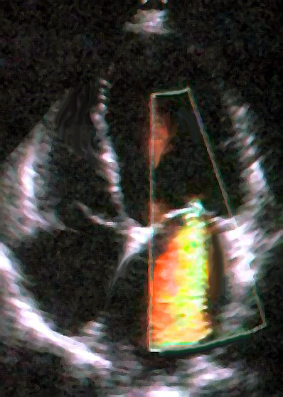

Do not grade the screenshot
From color alone ask: origin, direction, wall impingement, timing, Nyquist/gain, multiple jets. Then obtain morphology, VC/3D VCA, CW density/duration, PISA/volumetrics, pulmonary veins and chamber response.Tip & trick Before trusting any calculated area, write a one-line unit check. If the result cannot be expressed in cm² or mL, a velocity or diameter unit slipped.
C
Error propagation & memorable facts
small mistakes become big labelsSquared diameter A 10% LVOT diameter overmeasurement produces ~21% CSA/SV/AVA overestimation. DVI avoids diameter—but not a badly placed PW gate.
Squared velocity A 10% low jet velocity produces ~19% low simplified gradient. That is why alignment dominates tracing polish.
Beat matching In AF, match current and preceding R–R intervals; five representative beats beat one “pretty” beat. Post-ectopic beats exaggerate gradients/regurgitation.
Guide habit For every number say aloud: where was it sampled, when in the cycle, under what loading, what assumption converts it, and which independent measure agrees?
A
Twenty pitfalls in one page
cover the answer, recall it- ASE 2025 DD presence and LAP grading are separate algorithms.
- Grade 1 has normal mean LAP; symptoms may need exercise.
- Presence avg E/e′ is >14; pressure avg E/e′ is ≥14.
- TR velocity is not in the 2025 DD-presence count.
- Grade 2 is any E/A <2 after elevated LAP is proven.
- Normal/Grade 1 or negative stress echo does not rule out HFpEF.
- Mean LAP/PCWP ≠ LVEDP; isolated high LVEDP can occur early.
- Medial e′ preserved/high supports constriction; restriction lowers it.
- LVOT diameter error is squared; DVI avoids diameter, not PW-position error.
- No flow reserve on DSE does not exclude true severe AS.
- Current severe rheumatic MS is MVA ≤1.5 cm², not only <1.0.
- Color jet area is not a flow meter; Coanda underestimates.
- Late-systolic MVP peak PISA can overgrade whole-beat MR.
- Secondary MR: ≥30 mm²/45 mL may be severe in ESC 2025.
- Historical SMR EROA 20 mm² is prognostic, not current severe by itself.
- “Disproportionate MR” is a lens, not a TEER exclusion gate.
- Low TR Vmax can occur in torrential TR; PASP may be underestimated.
- Massive/torrential subdivide severe TR; referral starts at severe.
- Severe PR may be brief; CMR RF >40% or RVol ≥60 mL is the quantitative anchor.
- Prostheses require baseline/type/size and flow-dependent + independent evidence.
B
Comprehensive final report skeleton
mechanism · grade · consequence · confidenceSTUDY CONDITIONS — rhythm __; HR __; BP __; BSA __; image/Doppler quality __; loading/therapy __.
CHAMBERS — LV size/volumes/LVEF/GLS __; LA volume __; RV size/FAC/TAPSE/S′/FWLS __; RA/IVC/RAP __; PASP method/validity __.
DIASTOLIC — [normal / Grade 1 / Grade 2 / Grade 3 / special-population not gradeable]; mean LAP [normal/elevated/indeterminate]; evidence __; limitation __.
VALVE __ — mechanism [primary / ventricular-secondary / atrial-secondary / CIED / root-annular / mixed] __; anatomy __; severity __ by independent evidence __; chamber/venous consequence __; acute/chronic __.
DISCORDANCE — measurements disagree because __; resolving test/condition __.
COMPARISON — since __, change in grade/gradient/volume/function __ under comparable [or noncomparable] conditions.
IMPRESSION — one-line mechanism + severity + hemodynamic consequence + urgency/next imaging question.
Sample · primary MR
Severe primary MR due to Carpentier II P2 flail with a holosystolic anteriorly directed jet. VC 8 mm, EROA 45 mm², RVol 68 mL, and right-upper pulmonary-vein systolic reversal are concordant. LV is mildly enlarged with LVEF 62%; LAVI 55 mL/m²; PASP 46 mmHg. TEE is recommended for complete repair anatomy.Sample · ventricular secondary MR
Severe ventricular SMR due to Carpentier IIIb tethering under BP 112/70, sinus 68 bpm, euvolemia, and optimized GDMT/CRT. Crescentic orifice; EROA 32 mm², RVol 48 mL, biplane VC and pulmonary-venous findings concordant. LVEF 30%, LVESD 58 mm; RV mildly impaired. Valve + HF Team evaluation is appropriate.C
Final mental model
what an expert interpretation must doWhat broke?
Leaflet, annulus, ventricle, atrium, root, lead or prosthesis.
Leaflet, annulus, ventricle, atrium, root, lead or prosthesis.
How severe?
Concordant anatomy + hemodynamics + quantitative burden.
Concordant anatomy + hemodynamics + quantitative burden.
Does physiology agree?
Flow, pressure, chambers, venous signals, chronicity.
Flow, pressure, chambers, venous signals, chronicity.
What resolves doubt?
Repeat conditions, 3D/TEE, CMR, CT, stress or cath.
Repeat conditions, 3D/TEE, CMR, CT, stress or cath.
The answer that communicates “This is severe [lesion] due to [mechanism], supported by [two high-weight measures] and [consequence]. [Confounder] explains the discordant [measure]. I would [specific next test/action] to resolve [specific question].”
Teaching shelf. The guide’s organization and teaching emphasis were checked against the user-provided Otto Textbook of Clinical Echocardiography (5e/6e), Klein & Asher, Shindler et al., Pai & Varadarajan, Moradian & Alizadehasl, Sorrell & Jayasuriya, Otto & Bonow, Kaddoura’s Echo Made Easy (2e), and a local echo cheat sheet. Current ASE/EACVI/ESC/ACC documents—not older textbook tables—control published thresholds. Explanations, calculations, diagrams, and questions are original; licensed images retain the attribution shown beside each figure.