LIST
Selection principle
Evaluate expected survival, functional benefit, treatment burden, and the patient’s goals through a multidisciplinary transplant team. Optimize medical, device, and surgical HF therapy; no single exercise, age, or hemodynamic number establishes candidacy.1
Recognize advanced heart failure
Referral triggers
- Persistent severe symptoms, recurrent HF admissions, escalating diuretics, low blood pressure limiting GDMT, end-organ deterioration, recurrent appropriate ICD therapies, or dependence on inotropes/MCS should prompt advanced-HF assessment.
- Consider transplantation for selected refractory ischemia or ventricular arrhythmia even when resting EF does not convey the severity of illness. Assess recovery, durable MCS, transplantation, and palliative options together.
Interpret CPET in context
- Peak VO₂ generally ≤12 mL/kg/min on a β-blocker or ≤14 off a β-blocker supports listing consideration. A maximal study includes RER >1.05 and reaching anaerobic threshold.
- Peak VO₂ ≤50% predicted is helpful especially in women and patients ≤50 or ≥70 years. In obesity (BMI ≥30 kg/m²), lean-body-mass-adjusted peak VO₂ ≤19 mL/kg/min may help. VE/VCO₂ slope >35 is useful, especially with a submaximal test.
- Combine CPET, trajectory, hemodynamics, and validated prognostic scores. A cutoff alone should not decide transplantation.
2
Pulmonary vascular and multiorgan assessment
- When PASP ≥50 mmHg and either TPG ≥15 mmHg or PVR ≥3 WU, assess reversibility with an acute vasodilator challenge while maintaining SBP >85 mmHg. If needed, use monitored optimization over 24–48 hours and consider LV unloading with MCS.
- Severe PH that remains irreversible despite appropriate treatment makes isolated heart transplantation unsuitable; risk is continuous and cannot be reduced to one absolute PVR cutoff. Evaluate combined-organ options in selected patients.
- Assess whether renal or hepatic dysfunction is intrinsic and irreversible or secondary to congestion/low output. Nephrology/hepatology evaluation informs candidacy; allocation eligibility for heart-kidney is a separate policy question (P.9).
3
Potential barriers require individual assessment
Medical risk
- For BMI ≥35 kg/m², weight reduction below 35 is reasonable before listing. Frailty, nutrition, age, diabetes control, vascular disease, and pulmonary disease require individualized assessment.
- Active uncontrolled infection and malignancy with an unacceptable recurrence or survival risk can preclude transplantation. Controlled HIV, HBV, or treated HCV are not blanket exclusions.
Cancer and psychosocial assessment
- Use oncology input and the ISHLT stage-specific malignancy tables. Some completely treated low-risk cancers need no additional wait; others require a disease-free interval. Avoid a universal cancer waiting period.
- Assess adherence, health literacy, support needs, cognition, mental health, and access to care; address modifiable barriers. Substance-use treatment and verified abstinence are important; ISHLT recommends at least 6 months for active illicit-drug use, alcohol-use disorder, and tobacco before listing.
LIST
Evaluation purpose
Establish advanced-HF severity, operative risk, transplant benefit, and the ability to sustain post-transplant treatment. The following domains guide evaluation; tests and timing depend on history and prior results.1
Clinical record and multidisciplinary review
Reconstruct the trajectory
- Obtain prior HF admissions, procedures, catheterizations, imaging, rhythm/device data, CPET, and treatment response. Reconcile prescribed versus actually taken medications.
- Document prior sternotomies, MCS, transfusions, pregnancy, transplantation, infection, malignancy, and current functional limitations. Review patient preferences and caregiver needs.
Team assessment
- Include advanced HF, transplant surgery, nursing/coordinators, pharmacy, nutrition, psychosocial care, and financial/access support. Involve transplant ID, nephrology, hepatology, ACHD, oncology, rehabilitation, and palliative care as indicated.
- Explain alternatives, expected outcomes, lifelong immunosuppression, donor-risk choices, and the evaluation/listing process. Document informed discussion and the multidisciplinary decision.
2
Studies that answer specific candidacy questions
- Cardiovascular: ECG/device assessment, echocardiography, RHC with PH reversibility assessment when indicated, CPET when feasible, and coronary evaluation based on ischemic disease and clinical context.
- Organ reserve: renal and liver trends, urinalysis/protein assessment, glycemic and lipid status, blood counts and chemistries, pulmonary function including DLCO, and targeted vascular or neurologic studies.
- Infection and immunity: HIV, HBV, HCV, CMV, EBV, toxoplasma, TB, and exposure/geography-specific testing with transplant ID. Determine vaccination needs; evaluate and treat active infection.
- Screen for malignancy using age-, sex-, exposure-, and risk-appropriate guidance. Complete dental evaluation, nutrition and frailty assessment, and bone-risk evaluation. Prior surgery may require targeted chest imaging for operative planning.
Compatibility
Confirm ABO from the required independent samples and establish HLA typing, antibody profile, unacceptable antigens, and crossmatch strategy with the histocompatibility laboratory. Repeat or update testing when clinical events alter the risk.3
Vaccination before immunosuppression
- Use the current CDC adult schedule, prior dose history, age, and immune status. Review influenza, COVID-19, pneumococcal, hepatitis B, Tdap/Td, HPV, recombinant zoster, and other risk-based vaccines.
- Recombinant zoster vaccine is non-live. Do not group it with live MMR or varicella. Pneumococcal choices include PCV20 or PCV21, or PCV15 followed by PPSV23 when indicated; prior vaccine history changes the schedule.
- Complete indicated live vaccines at least 4 weeks and non-live vaccines ideally at least 2 weeks before transplantation. Urgency may limit this; coordinate timing with transplant ID. Live vaccines are generally avoided during post-transplant immunosuppression.
LIST
Listing is an ongoing clinical decision
After multidisciplinary approval and informed consent, register the candidate using current OPTN requirements. Reassess suitability whenever condition, support, infection, organ function, adherence, or patient preference changes.1
Reassess trajectory, not only the assigned status
Hospitalized candidates
- Review perfusion, filling pressures, vasoactive/MCS needs, kidney and liver trends, infection, nutrition, mobility, and neurologic status. Identify both deterioration and meaningful recovery.
- Repeat hemodynamic and cardiac testing when it will change management or is required for status documentation. The clinical assessment interval is separate from the OPTN form-expiration clock.
Ambulatory candidates
- Follow symptoms, congestion, exercise tolerance, blood pressure, renal function, medication tolerance, and support needs at an interval matched to stability and risk.
- Repeat RHC, CPET, imaging, and other evaluation components as indicated by change in condition and current candidate guidance. Revisit the benefit of remaining listed if medical/device therapy produces sustained improvement.
2
Sensitization and crossmatch readiness
- Review HLA antibodies and cPRA serially and after sensitizing events such as transfusion, pregnancy, or transplantation. Identify exactly what was given and when; notify the histocompatibility laboratory promptly.
- Sample timing and the need for prospective physical crossmatch are determined with the laboratory from recent exposures, antibody history, assay limitations, and donor HLA. There is no universal day-14 sample or 21-day crossmatch rule.
- cPRA estimates the fraction of donors carrying listed unacceptable antigens. It measures donor-pool restriction, not antibody concentration. Single-antigen-bead MFI is semiquantitative and assay-dependent. The 2025 consensus proposes undiluted-MFI risk bands, but dilution, complement binding, HLA locus, clinical urgency, and laboratory expertise remain essential. A band is not a universal donor-acceptance cutoff.
- Desensitization is a specialist decision balancing access to compatible organs against infection, treatment toxicity, and post-transplant rejection risk; it is not triggered by cPRA alone.
3
Maintain the ability to accept a suitable offer
Optimization
- Support nutrition and supervised physical rehabilitation as tolerated. Address frailty, immunization, dental issues, and substance-use treatment.
- If anticoagulation is needed, document the agent, last dose, renal function, and a transplant-surgery reversal plan. Choice of warfarin versus a DOAC depends on indication and program experience.
Readiness
- Keep contact information, consent, blood group, HLA risk, donor acceptance preferences, and current status documentation accurate. Retain source values and dates used in every justification form.
- When a candidate is temporarily unsuitable, make the status inactive and document the reason and reassessment plan. Inactive candidates receive no offers; reactivation follows reassessment.
STATUS
Adult allocation framework
This is a U.S. adult-heart educational summary. Policy 6 and the current OPTN justification form control assignment. An intervention or diagnosis alone does not automatically establish a status. Policy source checked 17 October 2026 against the OPTN policy effective 1 October 2026.1
Six active statuses
| Status | Principal qualifying pathways |
|---|---|
| 1 | Qualifying VA ECMO for cardiogenic shock; non-dischargeable surgically implanted non-endovascular biventricular support; MCS with defined life-threatening ventricular arrhythmia. |
| 2 | Qualifying non-dischargeable surgical LVAD; TAH/BiVAD/RVAD or single-ventricle VAD; device malfunction; qualifying percutaneous support or IABP; defined recurrent VT/VF; qualifying LVAD support ≥8 years. |
| 3 | LVAD discretionary days; qualifying inotropes with hemodynamic monitoring; specified MCS complications; eligible device pathways after their higher-status period; qualifying LVAD support ≥6 years. |
| 4 | Qualifying dischargeable LVAD; qualifying inotropes without hemodynamic monitoring; eligible CHD; intractable angina; amyloidosis/HCM/RCM with additional qualifying features; retransplant. |
| 5 | Registered for heart and at least one additional organ at the same hospital. |
| 6 | Other active candidates suitable for transplantation. |
Source:
2
Recent changes that alter the summary
- The revised IABP and percutaneous-device pathways took effect 10 September 2025. The hemodynamic pathway includes qualifying inotrope support or an actual qualifying arrhythmia after attempted initiation; alternatives for unavailable pre-device hemodynamics are defined separately (P.6).
- Time-on-LVAD escalation took effect 10 June 2026. The policy currently uses ≥8 years for Status 2 and ≥6 years for Status 3, with 180-day forms. Planned Phase 2 thresholds (7/5 years) require a later implementation step; do not apply them before it occurs.
3
Status and allocation are different questions
- Each pathway has its own evidence, duration, extension criteria, and review mechanism. Do not treat all Status 1–4 renewals as identical or assume every extension requires RRB approval.
- Congenital heart disease may support Status 4 under its policy pathway. Severe cyanosis, protein-losing enteropathy, or plastic bronchitis may support an exception request; they do not independently create automatic adult Status 2 or 3 eligibility.
- Match-run order follows the policy’s donor-age, status, blood-type, distance, and waiting-time classifications. cPRA describes sensitization and access limitations; there is no generic cPRA ≥50% bonus in adult heart allocation.
STATUS
Status 1: verify the precise pathway
The candidate must meet Policy 6.1.A and the required hospitalization/support conditions. Initial and continuing status require submission of the appropriate form, supported by original clinical data.1
VA ECMO for cardiogenic shock
- Within 7 days before support, document all three within one 24-hour period: SBP <90 mmHg, PCWP >15 mmHg, and CI <1.8 L/min/m² without inotropes or <2.0 with at least one inotrope.
- If those hemodynamics could not be obtained in that window, the alternative requires at least one within 24 hours before support: CPR, SBP <70, arterial lactate >4 mmol/L, or AST/ALT >1,000 U/L.
- Status is valid for up to 7 days from form submission. An ECMO extension requires hospitalization on support, a contraindication to durable support, and documented failure to wean within 48 hours before expiry using policy-defined hemodynamic criteria; RRB review is retrospective.
2
Biventricular support or MCS with arrhythmia
Biventricular support
- The standard pathway specifies non-dischargeable, surgically implanted, non-endovascular biventricular support requiring hospitalization because the device is not FDA-approved for out-of-hospital use.
- Do not infer Status 1 from a device trade name, RV support alone, or a TAH alone. Match actual configuration to Policy 6.1.A.ii or the relevant Status 2 pathway. Continued qualifying biventricular support is renewed every 7 days by a new form.
Life-threatening arrhythmia
- On MCS, the policy includes biventricular support placed for sustained ventricular arrhythmia, or an electrophysiologist’s determination that other treatment alternatives are unsuitable plus the specified recurrent VT/VF events.
- The recurrent-event route requires ≥3 episodes separated by ≥1 hour in the previous 7 days, with normal K/Mg and electrical cardioversion despite continuous IV antiarrhythmic therapy. Document rhythm strips, therapies, timing, and EP assessment.
3
Reassessment at each renewal
- Maintain the exact device configuration, dates, current support, inotrope/antiarrhythmic exposure, contemporaneous hemodynamics, and the pathway-specific renewal evidence.
- If an ECMO Status 1 extension is absent or not granted after 7 days, the program may assign the appropriate Status 3 pathway by submitting its form. Form expiry alone is not an automatic, safely completed Status 3 transition.
Clinical care first
Treat shock and arrhythmia according to the patient’s needs. Do not withhold rescue support or create hazardous weaning/inotrope trials to obtain allocation measurements; use the applicable alternative or exception process.STATUS
Status 2 IABP / percutaneous support
Policy 6.1.B.iv–v applies to hospitalized candidates with qualifying support for cardiogenic shock; the percutaneous pathway specifies a device without an oxygenator. Brand name alone does not define the pathway.1
Initial eligibility: two alternative evidence routes
Hemodynamic route
- Within 7 days before device placement, obtain SBP <90, CI <2.0, PCWP >15 within one 24-hour period, with qualifying inotrope support as below.
- Within this hemodynamic route, actual VT lasting ≥30 seconds or cardioversion, defibrillation, or antitachycardia pacing after inotropes were initiated in an attempt to reach qualifying doses can substitute for reaching the qualifying dose. The SBP, CI, and PCWP requirements still apply. Predicted arrhythmia risk alone is insufficient.
Hemodynamics unavailable
- If the pre-device hemodynamic measurements could not be obtained within 7 days, document at least one alternative marker within 24 hours before support: CPR, SBP <70 mmHg, arterial lactate >4 mmol/L, or AST/ALT >1,000 U/L.
- Preserve timestamps and the reason measurements were unavailable. This is a distinct policy alternative; it is not an instruction to delay emergent support for a catheterization.
2
Qualifying inotrope doses
mcg/kg/min| Agent | One high-dose agent | At least two agents, each at or above |
|---|---|---|
| Dobutamine | 7.5 | 3 |
| Milrinone | 0.50 | 0.25 |
| Epinephrine | 0.02 | 0.01 |
| Dopamine | Not a single high-dose option | 3 |
Source:
Dose thresholds are allocation criteria
These numbers describe documented therapy under OPTN policy. They are not a titration order or a reason to increase a drug that is unsafe for the patient.3
Extensions: 14-day pathway
- Initial status lasts up to 14 days from form submission. For extension, document a contraindication to durable support and the qualifying failure-to-wean or prior qualifying arrhythmia pathway.
- Within 48 hours before expiry, qualifying failure to wean while on qualifying inotropes is supported by any one: MAP <60, CI <2.0, PCWP >15, or SvO₂ <50% measured by central venous catheter.
- The alternate extension route applies when initial Status 2 qualification followed the actual qualifying arrhythmia after inotropes were initiated in an attempt to reach qualifying doses. Submit objective evidence; RRB review is retrospective.
- If Status 2 is not extended and support persists, submit the applicable Status 3 form. Other Status 2 pathways, including recurrent VT/VF, device malfunction, and long-duration LVAD support, have distinct criteria.
STATUS
Status 3: separate the pathways
Policy 6.1.C contains different qualifying routes and renewal clocks. The inotrope, discretionary LVAD, device-complication, and post-clock categories should not share a generic 14-day checklist.1
Inotropes with hemodynamic monitoring
- The candidate is hospitalized at the listing hospital with an invasive PA catheter or daily hemodynamic monitoring measuring cardiac output and LV filling pressures. A universal “PAC out >24 h means Status 4” rule is not in this criterion.
- Shock evidence in one 24-hour period: SBP <90, PCWP >15, and CI <1.8 without inotropic/mechanical support within 7 days before inotropes, or <2.2 with support. The CI threshold differs from the current Status 2 device pathway.
- Qualifying doses: one of dobutamine ≥7.5, milrinone ≥0.50, epinephrine ≥0.02 µg/kg/min; or ≥2 agents at dobutamine ≥3, milrinone ≥0.25, epinephrine ≥0.01, dopamine ≥3. “Any dose of any two agents” is incorrect.
- This pathway has a 14-day initial period and 14-day extensions. With ongoing qualifying therapy and monitoring, extension requires either CI <2.2 on the current regimen or the specified failed-wean evidence within 48 hours before expiry; inability to wean is not the sole route.
2
Durable LVAD: discretionary and time-supported routes
Discretionary 30 days
- A medically stable candidate with a qualifying dischargeable LVAD receives 30 cumulative days per implanted device across all registrations. Days need not be consecutive.
- A replacement dischargeable LVAD provides a new 30-day period; unused time on the prior device does not carry forward. This is not a once-per-lifetime allowance.
Long-duration LVAD
- Continuous qualifying dischargeable LVAD support for ≥6 years supports the current Status 3 time-on-device pathway, valid for 180 days and renewable while qualifying.
- Verify the qualifying-device list and implant/support history. The planned 5-year Phase 2 threshold is not yet substituted for the current 6-year policy.
3
MCS complications and higher-status clocks
- Hemolysis, pump thrombosis, right HF, device infection, bleeding, and aortic insufficiency each have detailed evidence and duration requirements. A raised LDH, GI bleed, positive culture, or echo label alone is insufficient.
- For example, hemolysis requires the specified repeated laboratory evidence and treatment; device infection and bleeding have their own definitions. Open the applicable subsection rather than applying one generic complication threshold.
- VA ECMO and qualifying MCS-arrhythmia support after 7 days, and specified LVAD, percutaneous support, or IABP after 14 days, can support separate Status 3 pathways. A new form is required; verify 7- versus 14-day renewal periods for the particular category.
STATUS
Status 4 is renewed, not indefinite
Policy 6.1.D has six policy subsections. Most have 90-day forms; the inotrope pathway has a 180-day form. A diagnosis must meet its additional requirements, and an active form must be maintained.1
Support-based pathways
Dischargeable LVAD
- A qualifying dischargeable LVAD without another qualifying higher-status pathway supports Status 4. Document device identity and support history.
- The form is valid for up to 90 days and may be renewed by another form while qualifying. Review discretionary days, complications, and time-on-device eligibility when relevant.
Inotropes without hemodynamic monitoring
- Requires continuous IV inotrope, CI <2.2 within 7 days before administration or while on infusion, and PCWP >15 mmHg.
- At least one qualifying dose: dobutamine ≥3, milrinone ≥0.25, epinephrine ≥0.01, or dopamine ≥3 µg/kg/min. The form is valid for up to 180 days; low-dose infusion alone is not sufficient.
2
Diagnosis-based pathways
| Pathway | Additional qualifying features |
|---|---|
| Congenital heart disease | Hemodynamically significant diagnosis on the OPTN-approved qualifying list. |
| Ischemic disease with intractable angina | CAD, CCS class IV angina not treatable by medical therapy plus percutaneous/surgical revascularization, and myocardial ischemia demonstrated by imaging. |
| Amyloidosis / HCM / RCM | At least one defined feature: medically uncontrolled CCS IV angina; NYHA III–IV symptoms with CI <2.2 or specified filling pressure >20 mmHg; or qualifying VT/VF, cardioversion, or sudden cardiac death. |
| Retransplant | Prior heart transplant with ISHLT CAV grade 2–3 or NYHA III–IV HF symptoms. |
Source:
3
Build the clinical narrative as well as the form
- For ACHD, document anatomy, prior palliation/repair, systemic and subpulmonary ventricular function, pulmonary vasculature, cyanosis, and hepatic/renal consequences. An ACHD specialist informs candidacy.
- For amyloidosis, establish AL versus ATTR and assess extracardiac involvement and disease control with the relevant specialty. A positive bone scan without excluding monoclonal protein disease does not safely establish ATTR.
- For HCM/RCM, integrate symptoms, hemodynamics, rhythm, imaging, treatment options, and extracardiac disease; preserved EF does not imply preserved prognosis.
- Allocation eligibility and transplant suitability are related but distinct. A policy diagnosis cannot replace individualized assessment of expected benefit and modifiable barriers.
STATUS
Use the current policy for each submission
Forms, source dates, qualifying periods, and exception requests are part of allocation accuracy. The policy itself controls; older copied summaries can retain obsolete subsection numbers and thresholds.1
Form mechanics and inactive status
- Renewal intervals depend on pathway. Status 1 is generally 7 days; Status 2 commonly 14 days but long-duration LVAD is 180. Status 3 includes 7-, 14-, 30-, 90-, and 180-day provisions; Status 4 commonly 90 days, with inotropes 180. Confirm the actual subsection.
- If no valid new form is submitted after expiry, Policy 6.1 assigns Status 6, or Status 5 if registered for another organ. A desired post-clock Status 3 pathway requires its own submission.
- Inactive candidates are temporarily unsuitable and receive no offers; waiting time does not accrue while inactive. Document the reason, follow-up plan, and reassessment before reactivation.
- Request an exception when comparable urgency is not captured by standard criteria. Use Policy 6.4, supporting evidence, and the current review/appeal process; an exception request is not proof of approval.
2
Blood group, sensitization, and match order
- For adult heart offers, primary matching is not always “identical first”: an O donor has O or B primary candidates and A or AB secondary candidates; A donors have A/AB primary, B donors B/AB primary, and AB donors AB primary candidates.
- The complete match order uses donor-age-specific allocation tables, urgency, blood group, distance, and waiting time. Pediatric ABO-incompatible eligibility has separate requirements; do not import kidney A2-to-B rules into adult heart allocation.
- cPRA estimates the proportion of donors with unacceptable antigens. It guides compatibility/access assessment but does not create the kidney-style cPRA bonus previously described in this guide.
3
Combined organs and kidney-after-heart priority
- Heart-kidney required sharing requires the specified registration, distance/status, and kidney criteria. CKD: GFR ≤60 for >90 consecutive days plus ESRD dialysis or qualifying CrCl/GFR ≤30. Sustained AKI: dialysis at least every 7 days or CrCl/GFR ≤25 at least every 7 days for the last 6 weeks.
- Kidney-after-heart priority: kidney-list registration before the first heart-transplant anniversary and dialysis or measured/estimated CrCl/GFR ≤20 on a date 60–365 days after heart transplant. Each qualifying report grants 30 days; 90 consecutive qualifying days preserve classification until removal. Current kidney-after-heart priority is Policy 8.4.G; verify numbering in the live policy.
- Heart-liver and heart-lung sharing have distinct rules. Do not treat Status 5 as a guarantee that another organ will accompany every heart offer; consult Policies 5, 6, and the relevant organ policy.
DONOR
Donor acceptance is recipient-specific
Weigh donor findings together with recipient urgency, pulmonary vascular burden, size, sensitization, operative complexity, preservation strategy, and expected ischemic time. Avoid a single age, weight, arrest-duration, EF, or troponin veto.1
Assess the heart in its physiologic context
Function and coronary disease
- Review serial echo images, loading conditions, rhythm, vasoactive doses, hemodynamics, ECG, and coronary anatomy when indicated. A recovered EF on heavy support is not equivalent to recovery under ordinary loading.
- Selected donors with reversible dysfunction or a history of resuscitation can be acceptable after thorough evaluation. Significant persistent dysfunction, uncontrolled arrhythmia, or clinically important coronary/valve disease requires careful reconsideration.
Match the donor to the recipient
- Use predicted heart mass and the complete donor-recipient phenotype; avoid important undersizing, particularly with elevated recipient PVR. A weight-only or sex-only rule is inadequate.
- Donor age, hypertrophy, coronary risk, ischemic time, and recipient illness interact. Extended-criteria acceptance is an individualized risk-benefit decision documented by the transplant team.
2
Infection and immunologic compatibility
- Review required donor infection tests, cultures, exposures, malignancy information, ABO, HLA, and the latest recipient antibody profile. Resolve discrepancies with the OPO and histocompatibility laboratory before acceptance.
- “Risk criteria for recent HIV/HBV/HCV exposure” and a positive pathogen test are different findings. Negative tests do not remove window-period risk. Obtain the required disclosure/consent and post-transplant monitoring.
- HCV-viremic hearts can be considered for HCV-uninfected recipients with informed consent, reliable access to treatment/retreatment, and long-term follow-up. AASLD/IDSA recommends prophylactic or preemptive pangenotypic DAA therapy for non-liver recipients; check interactions and transplant-ID/hepatology plans.
- Bacteremia, endemic infections, hepatitis B markers, or other positive testing need organism-specific assessment and a recipient treatment plan. Unexplained encephalitis is a major transmission concern; “increased-risk donors are safe” is too broad.
3
Preservation and donor-pool expansion
- In the randomized DCD trial, risk-adjusted 6-month survival was noninferior to DBD with the studied direct-procurement/ex-vivo-perfusion pathway. Moderate/severe primary graft dysfunction was more frequent with DCD. This does not prove equivalence for every donor or preservation method.
Final acceptance
Reconcile the complete donor record, onsite inspection, compatibility, recipient readiness, and the preservation/transport plan. Communicate new findings promptly; keep a clear record of the acceptance decision.DONOR
Recovery and transport: professional-team checklist
This page summarizes safety domains for trained procurement and transplant teams. The approved device instructions, OPO requirements, and treating program’s operating procedures determine solution, dose, temperature, and equipment settings.1
Before departure
Identity and compatibility
- Confirm donor ID, organ, ABO data, intended recipient, and the required verification records. Resolve discrepancies before procurement/implantation.
- Confirm availability of donor testing, imaging, cultures, authorization, and death-determination documentation. Check crossmatch requirements with the recipient laboratory.
Logistics and preservation
- Agree on recovery and implantation timing, travel contingencies, recipient operative complexity, and escalation contacts. Keep the implantation team informed when timing changes.
- Verify preservation solution, storage/perfusion system, trained personnel, consumables, expiration dates, emergency equipment, and contingency supplies. Cold static storage and commercial transport systems have distinct handling instructions.
2
At the donor hospital
- Review the latest donor physiology and imaging with the procuring surgeon. Reassess suitability after direct examination and communicate unexpected anatomy or pathology.
- Coordinate cross-clamp and recovery with the recipient and other organ teams. Confirm authorization before proceeding; document ischemic and preservation times accurately.
- Cardioplegia composition, volume, delivery pressure, temperature, and donor medications are selected by the trained surgical/perfusion team. Do not apply a copied universal heparin, steroid, or cardioplegia recipe.
- Protect the graft during removal and preparation; avoid mechanical injury, inadequate cooling, and unintended freezing. Preservation method and anticipated transport duration affect the plan.
3
Packaging, custody, and arrival
- Follow OPTN Policy 16 for sterile packaging, required organ labels, containers, accompanying specimens, and transport documentation. Verify identifiers and packaging integrity at each handoff.
- Maintain chain of custody and communication during transport. Report delays, damage, temperature concerns, or discrepancies immediately and preserve the relevant records.
- At the receiving hospital, complete the required organ/recipient/ABO verification before implantation and resolve any discrepancy. The travel deadline never justifies skipping identification checks.
Handoff content
Include donor and recipient identifiers, acceptance concerns, preservation method, all relevant timestamps, perfusion or temperature trends if used, cultures/tests pending, specimens, and the person receiving custody.DONOR
DCD and direct procurement/perfusion
Withdrawal of life-sustaining treatment, comfort care, death determination, and organ recovery are distinct responsibilities. Donation decisions and procurement must not compromise independent end-of-life care.1
Define the pathway before withdrawal
- Agree on donor/recipient selection, consent, roles, death-determination procedures, required observation interval, warm-ischemia definitions, organ-team coordination, and abort criteria. Use current OPO and jurisdictional requirements.
- Direct procurement with ex-vivo perfusion and normothermic regional perfusion are different pathways. The eligibility, equipment, ethical safeguards, and expertise for one must not be assumed for the other.
- Recipient surgery timing depends on donor progression, assessment of graft suitability, transport, and the agreed rescue/abort plan. Avoid committing irreversibly before the necessary acceptance information is available.
2
Warm ischemia: record the whole event
- The 2023 randomized DCD trial enrolled donors aged 18–49 and defined functional warm ischemia from SBP <50 mmHg or oxygen saturation <70% to aortic cross-clamp and cold cardioplegia, with a trial limit of ≤30 minutes. These are study criteria, not a universal protocol.
- Record withdrawal, physiologic thresholds, cessation of circulation, declaration of death, incision, cross-clamp/cardioplegia, and start of perfusion. A transient pressure rise does not erase accumulated injury; do not reset the ischemic clock using this guide.
3
Recovery, ex-vivo assessment, and acceptance
Trained procurement team
- Ensure a trained operator, verified device readiness, backup equipment, and a coordinated plan with abdominal/lung teams. Operate within the current device instructions.
- Premortem interventions, blood collection, cardioplegia, cannulation, and perfusion recipes require explicit approved procedures and appropriate authorization; no universal drug doses are supplied here.
Assess before implant
- Evaluate graft appearance, perfusion flow/pressure, lactate trajectory, and the complete ischemic/preservation history. One acceptable number cannot compensate for other evidence of injury.
- DCD with the trial’s ex-vivo-perfusion strategy achieved noninferior 6-month survival versus DBD, with more moderate/severe PGD. Counsel and plan postoperative support accordingly; long-term and technique-specific outcomes need their own evidence.
Stop point
Death must be independently determined and the required waiting interval completed before procurement begins. Any uncertainty about authorization, death determination, organ identity, or device readiness requires resolution by the responsible teams.OR
Operative priorities
Match the plan to anatomy, prior surgery or LVAD, sensitization, pulmonary vascular risk, preservation, and recipient stability. The surgical team determines operative technique, induction, antimicrobial therapy, and rescue support.1
Before bypass and reperfusion
- Verify identity, consent, ABO/HLA compatibility, donor suitability, ischemic timeline, blood products, and recipient readiness. Plan re-entry and anticoagulant reversal. Coordinate cardiectomy with confirmed donor suitability and arrival.
- At reperfusion, use echo and invasive measurements together to assess LV/RV function, filling pressures, pulmonary vascular load, rhythm, and oxygenation. Titrate support to the actual physiology and anticipate bleeding, vasoplegia, and pacing needs.
2
Graft dysfunction: identify cause, then classify
2026 ISHLT consensus: EGD replaces the older PGD label for unexplained dysfunction before 24 hours. sEGD describes new unexplained dysfunction at 24–72 hours.
Exclude surgical or coronary problems, tamponade/bleeding, pulmonary hypertension, rejection, and other secondary causes. Vasoplegia can coexist.
Record whether severe LV-EGD prevented separation from bypass or appeared after successful separation. Apply the complete echo, hemodynamic, and support criteria. A new IABP can count toward severe LV-EGD, while pre-existing support alone does not establish the diagnosis.
3
Support promptly and communicate the trajectory
- Persistent low output despite appropriate correction and pharmacologic support requires early multidisciplinary MCS assessment. Do not wait for a time boundary or a completed label to stabilize the patient.
- Handoff graft function, support and pacing settings, ischemic/reperfusion times, bleeding and transfusions, pulmonary vascular burden, donor cultures, pending results, and current immunosuppression/prophylaxis orders.
IMMUNO
Common maintenance framework
A calcineurin inhibitor, an antimetabolite, and corticosteroids with an individualized taper are the usual backbone. Rejection risk, kidney function, infection, toxicity, sensitization, and adherence determine the actual regimen.1
Induction and early maintenance
Induction is selective
- Routine antibody induction is not required for every recipient. It may be considered for high immunologic risk or to permit delayed CNI introduction in significant renal dysfunction.
- Choice between lymphocyte-depleting therapy and an IL-2-receptor antagonist weighs rejection benefit against cytopenia, infection, and malignancy. Verify product-specific administration, monitoring, and prophylaxis with transplant pharmacy.
Antimetabolite and steroid
- Mycophenolate is commonly preferred; adjust treatment for marrow suppression, GI toxicity, infection, and interactions with the transplant team. Formulations are not simply interchangeable milligram for milligram.
- Mycophenolate is teratogenic: provide reproductive counseling and a specialist plan before conception. Azathioprine is an alternative in selected situations; interactions and marrow toxicity need review.
- Steroid withdrawal may be achievable within 3–12 months in low-rejection-risk recipients. Prior rejection, sensitization, disease recurrence risk, and graft function can justify continued steroids; no fixed calendar guarantees safety.
2
Tacrolimus: use the right trough and context
| Setting with MPA or azathioprine | General ISHLT trough range |
|---|---|
| Early postoperative days 0–60 | 10–15 ng/mL |
| Next 3–6 months | 8–12 ng/mL |
| Stable recipients beyond 6 months | 5–10 ng/mL |
| With an mTOR inhibitor after 6 months | 3–8 ng/mL may be considered |
Source:
Ranges are not universal targets
Interpret the prescribed target in relation to time, companion drugs, rejection history, renal function, toxicity, and assay. Measure a 12-hour trough for twice-daily or a 24-hour trough for once-daily tacrolimus immediately before the next dose.3
Prevent exposure errors
- Record formulation, route, actual dose times, missed doses, and sampling time. Repeat appropriately timed levels and kidney/electrolyte testing when exposure is uncertain.
- Azoles, macrolides, diltiazem/verapamil, antiviral drugs, anticonvulsants, rifamycins, herbal products, and stopping an interacting agent can materially change CNI/mTOR exposure. Check every new or discontinued medication.
- Vomiting, diarrhea, altered GI anatomy, and NPO status can destabilize absorption. Oral, sublingual, IV, and extended-release conversions require transplant-pharmacy supervision and repeat levels; a universal 1:2 conversion is unreliable.
- Do not stop immunosuppression independently for one abnormal laboratory result. Severe toxicity, infection, or cytopenia requires prompt coordinated adjustment that preserves rejection protection.
IMMUNO
mTOR therapy is a risk-benefit decision
Sirolimus or everolimus can be useful in selected recipients with CAV, CNI nephrotoxicity, or malignancy concerns. CNI reduction/withdrawal and timing must be planned with rejection surveillance and toxicity monitoring.1
Before and after mTOR introduction
- Review wound healing, recent surgery, proteinuria, eGFR, lipids, blood counts, edema, pulmonary symptoms, infection, and rejection history. Early use can impair healing; CNI-free therapy increases rejection concerns in some patients.
- Set paired mTOR/CNI exposure targets and reassess drug interactions. Monitor troughs after steady state, kidney function, urine protein, lipids, cytopenias, mouth ulcers, edema, and pneumonitis symptoms.
- Conversion is not an automatic anniversary intervention. A biopsy requirement or proteinuria cutoff used by one program should not be presented as a universal prerequisite.
2
CAV: diffuse disease in a denervated graft
Surveillance and diagnosis
- CAV may present with exertional limitation, HF, arrhythmia, syncope, or graft dysfunction without typical angina. Assess coronary disease alongside rejection and other causes.
- Coronary angiography is used for diagnosis and ISHLT grading; annual or biennial evaluation should be considered. Frequency and alternatives depend on prior CAV, renal function, procedural risk, and expertise.
- IVUS/OCT can identify disease not apparent on angiography. PET flow quantification and other noninvasive modalities may have a role in selected surveillance strategies.
Prevention and treatment
- Use a statin after transplantation regardless of starting lipid level, with doses and interactions suited to the immunosuppressive regimen. Treat hypertension, diabetes, smoking, obesity, and CMV risk.
- Consider mTOR modification for established CAV. Focal disease may be treated with PCI; diffuse disease and microvascular involvement limit revascularization. Severe disease with graft dysfunction may prompt retransplant assessment.
- Grade disease with the complete ISHLT coronary/anatomic and graft-function definition. A single stenosis threshold cannot replace the full classification.
3
Belatacept: limited rescue role
- Belatacept after heart transplantation is an off-label option in selected circumstances such as major CNI toxicity after standard approaches are unsuitable. Heart-transplant evidence is limited; this is not routine first-line maintenance.
- The U.S. label is for adult kidney transplantation. Use only in EBV-seropositive patients; negative or unknown EBV status is contraindicated because of PTLD risk, particularly CNS PTLD. Confirm VCA and EBNA IgG evidence.
- Discuss serious infection, malignancy, PTLD, and PML risks. Transplant specialists and pharmacy must define eligibility, concomitant immunosuppression, monitoring, and infection prevention before considering use.
IMMUNO
Graft dysfunction or sepsis needs urgent assessment
New HF, low output, hypotension, arrhythmia, or deteriorating graft function requires prompt transplant-team evaluation. Obtain cultures, echocardiography, hemodynamics, DSA, and biopsy as indicated; stabilization and necessary empiric treatment must not wait for histology.1
Infection prevention is risk-specific
CMV
- Establish donor/recipient serostatus and the prevention strategy. For adult heart D+/R− recipients, prophylaxis generally uses (val)ganciclovir for 3–6 months; seropositive recipients commonly receive 3 months or an appropriate preemptive strategy.
- Dose for kidney function and monitor blood counts. Coordinate PCR surveillance, stopping prophylaxis, intensified immunosuppression, breakthrough infection, and resistance/intolerance management with transplant ID.
Other pathogens
- TMP-SMX is preferred for Pneumocystis prophylaxis, generally for at least 6–12 months after heart transplantation. Alternatives and toxoplasma coverage need deliberate selection, especially with D+/R− toxoplasma mismatch.
- Use perioperative antibacterial and fungal/HSV prophylaxis according to exposures, serostatus, colonization, graft course, and program/ID guidance. Intensified rejection treatment can require renewed or extended prophylaxis.
2
Cellular rejection: grade plus clinical severity
| Biopsy finding | Management frame |
|---|---|
| 0R | No cellular rejection on sampled tissue. Correlate with the whole clinical picture. |
| 1R, asymptomatic with preserved function | Often observation and optimization of maintenance therapy; assess adherence and risk. |
| 2R or 3R | Prompt transplant-team treatment decision; corticosteroid treatment is usual, with route/intensity based on grade, timing, symptoms, and function. |
| Hemodynamic compromise or resistant/recurrent rejection | Urgent monitored care; IV corticosteroids, consideration of lymphocyte-depleting treatment, and circulatory support as required. |
Source:
3
AMR: no universal drug ladder
- Integrate biopsy histology/immunopathology, DSA, graft function, and hemodynamics. AMR can occur without detected circulating DSA. Isolated asymptomatic DSA is not by itself an indication for treatment. Evaluate accompanying clinical or subclinical graft injury and perform biopsy when indicated.
- Treatment may include corticosteroids, plasma exchange, IVIG, and selected B-cell/plasma-cell/complement therapies. The 2025 consensus weighs graft injury, hemodynamics, DSA attributes, and biopsy findings together. A pAMR grade alone does not specify a fixed drug ladder.
- Coordinate timing because plasma exchange removes some administered therapies. Reassess graft response with appropriate biopsy/imaging/hemodynamics and renew infection prevention after augmented immunosuppression.
FOLLOW-UP
Surveillance should match risk
Early follow-up is frequent, then may become less frequent with stability. The transplant program defines a written schedule for visits, laboratories, graft assessment, rejection testing, and preventive care; there is no universal weekly-to-annual timetable.1
Core surveillance
Graft and drug safety
- Review symptoms, volume, BP, rhythm, wound recovery, adherence, actual dose times, interactions, and renal/electrolyte/blood-count trends. Ensure immunosuppressant levels are correctly timed.
- EMB remains important for rejection surveillance and diagnosis, especially early or in higher-risk recipients. Schedule and duration depend on prior rejection, graft function, antibodies, and the program’s strategy.
Antibodies and CAV
- ISHLT recommends DSA monitoring at 1, 3, 6, and 12 months, then annually, with more frequent assessment in high-risk patients. Interpret persistent/new antibodies with graft findings.
- Plan coronary/CAV surveillance and risk-factor treatment (P.15). Renal or procedural risk may justify altered modality or interval; annual angiography for exactly five years is not a universal rule.
2
Ask what each rejection test measures
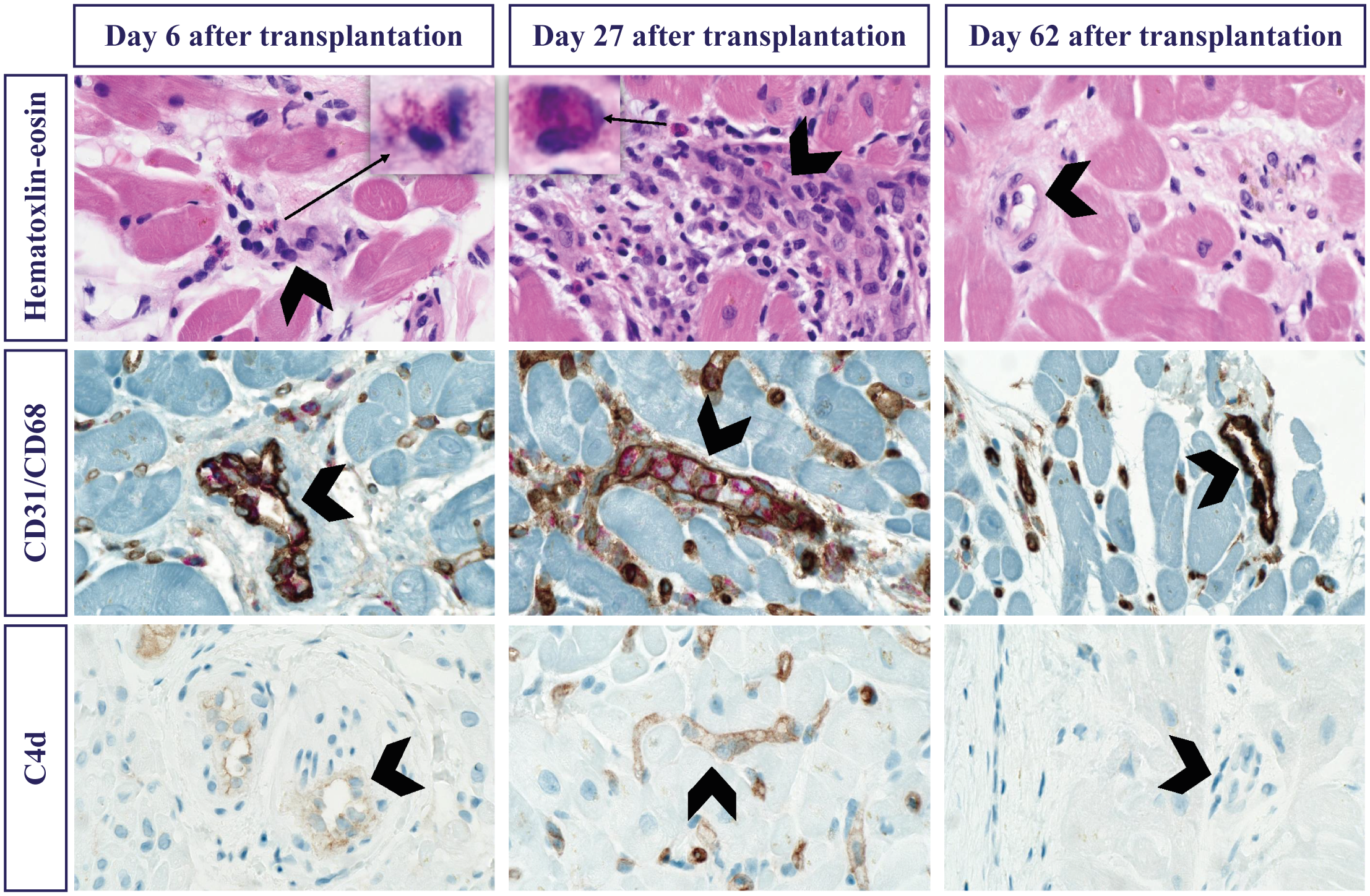
- Gene-expression profiling: helps exclude clinically important cellular rejection in selected stable, low-risk recipients during validated time windows. It does not exclude AMR.
- Donor-derived cell-free DNA: detects graft injury, not its cause. Interpret assay, timing, trajectory, graft function, and competing injury. Thresholds are not interchangeable.
- DSA and biopsy: antibody testing defines immune risk. Tissue histology and immunopathology characterize injury. Concordance is useful, but discordance needs investigation rather than automatic treatment.
Symptoms override a reassuring screening test
A low molecular score must not delay assessment of new HF, shock, arrhythmia, or falling graft function. Stabilize urgently and obtain targeted imaging, hemodynamics, biopsy, and coronary investigation as indicated.3
Prevention and procedures
- Complete required HIV, HBV, and HCV NAT at 4–8 weeks (days 28–56) after transplantation, regardless of donor risk criteria. A known pre-existing infection exempts only that virus under Policy 15.3.C; the other required tests still apply.
- Dental antibiotic prophylaxis is reasonable for qualifying dental procedures in a heart-transplant recipient with valve regurgitation due to a structurally abnormal valve (or another qualifying high-risk cardiac condition). Transplant alone is not a blanket indication.
- Before catheterization, biopsy, dental work, or surgery, agree on anticoagulant, antiplatelet, diabetes, sedation/fasting, and immunosuppression plans. Renal function, bleeding risk, indication, and current procedure guidance determine medication holds.
FOLLOW-UP
Early clinic review: discharge through year one
Use the individual discharge and surveillance plan. At each transition, reconcile completed tests, pending results, medication changes, and the next scheduled assessment; a copied visit template cannot establish that they occurred.1
Every encounter
Symptoms, examination, function
- Ask about dyspnea, congestion, palpitations, presyncope, fever, wound issues, diarrhea/vomiting, fatigue, pain, sleep, mood, and rehabilitation progress. Review BP, pulse, weight and volume trends.
- Resting pulse is often higher after denervation and chronotropic responses may be altered. New tachycardia or poor exercise tolerance still warrants evaluation in context.
Medication exposure and access
- Verify formulation, dose, last administration, trough time, missed doses, refill access, support, and all new or stopped drugs/supplements. Review kidney function, electrolytes, blood counts, glucose, and liver tests.
- Confirm the intended steroid taper and CNI targets, current rejection risk, antimicrobial prophylaxis and stop dates, and the plan after any recent rejection treatment. Do not infer doses from the postoperative month.
2
Time-sensitive first-year checkpoints
| Checkpoint | Confirm and act |
|---|---|
| First weeks | Wound/volume recovery, safe medication administration, accessible urgent contact route, appropriate laboratory and rejection surveillance, and rehabilitation referral. |
| Weeks 4–8 | Required HIV/HBV/HCV NAT regardless of donor risk (known-infection exception per Policy 15.3.C); follow up positive or missed testing. |
| Months 1, 3, 6, 12 | DSA surveillance per ISHLT, with extra testing for higher risk; align graft assessment and immunosuppression decisions with the complete findings. |
| Months 3–12 | Reassess prophylaxis duration, steroid reduction, infection and rejection history, renal function, metabolic risk, and return to function. |
| Year one | Review CAV strategy, graft/rhythm status, renal/metabolic toxicity, bone health, malignancy screening, vaccination, adherence, and goals. |
Source:
3
Do not miss the kidney safety-net window
- If kidney recovery is poor, involve transplant nephrology early. Kidney-list registration must occur before the first heart-transplant anniversary; qualifying dialysis or CrCl/GFR ≤20 must be documented on a date 60–365 days after heart transplant.
- Record the qualifying results and continued status according to current Policy 8.4.G. Do not wait for a routine annual visit to identify an approaching deadline.
4
Practical counseling
- Provide an individualized rehabilitation, driving, lifting, work, travel, pregnancy/contraception, and medication-access plan. Mycophenolate requires reproductive counseling before pregnancy planning.
- Review current influenza/COVID-19 and other indicated non-live vaccines, including recombinant zoster and pneumococcal vaccination. Coordinate timing after transplant or rejection therapy with the treating team.
FOLLOW-UP
Long-term priorities
Continue graft and CAV surveillance while preventing infection, malignancy, kidney disease, metabolic complications, bone loss, and medication-related harm. Stable years do not remove the need for ongoing immunosuppression and transplant follow-up.1
Annual and interval review
Graft, CAV, and treatment
- Review symptoms, rhythm, ventricular/valvular function, DSA/rejection history, coronary surveillance, and the current immunosuppression strategy. Individualize testing intervals to risk and prior results.
- Monitor eGFR, proteinuria, BP, lipids, glycemia, weight, and medication interactions. Consider CNI reduction or mTOR modification only with a plan to maintain rejection protection.
Prevention, function, and life goals
- Maintain dermatology and age-/risk-appropriate malignancy screening, dental care, bone-risk/BMD assessment, nutrition, exercise, and mental-health support. Evaluate new lesions, lymphadenopathy, or constitutional symptoms promptly.
- Reassess access, adherence, cognition, frailty, caregiver support, work/activity, reproductive plans, and goals of care. Coordinate the transplant and primary-care teams; avoid duplicative or unassigned follow-up.
2
Heart failure after transplant is graft dysfunction until explained
- New congestion, low output, arrhythmia, syncope, biomarker rise, EF decline, or exercise intolerance should prompt assessment for rejection, CAV/ischemia, infection, tamponade, valve disease, pulmonary vascular/RV disease, drug toxicity, and inadequate immunosuppressant exposure.
- Typical angina may be absent because the graft is denervated. Evaluate suspected ischemia/CAV and graft dysfunction promptly rather than relying on chest-pain history.
- Vomiting or diarrhea can substantially alter immunosuppressant exposure. Assess volume, renal function, interacting drugs, and correctly timed levels; severe symptoms need urgent care.
3
Escalate according to severity
| Finding | Response |
|---|---|
| Hemodynamic instability, acute neurologic deficit, syncope with instability, or sustained arrhythmia | Emergency assessment and immediate transplant-team involvement. |
| New graft dysfunction, important dyspnea/congestion, fever, or systemic illness | Prompt transplant-team assessment; echocardiography, cultures, hemodynamics, DSA, and biopsy/coronary investigation as clinically indicated. |
| Severe cytopenia, rising creatinine, electrolyte abnormality, or high CNI level | Assess severity and cause urgently; coordinate drug changes and repeat tests with transplant pharmacy/team. |
| Suspicious skin lesion, lymphadenopathy, weight loss, or other cancer signal | Arrange directed examination and tissue diagnosis when indicated; involve dermatology/oncology and the transplant team. |
Source:
Avoid reflex drug withdrawal
Holding mycophenolate, reducing CNI, intensifying steroids, or starting AMR therapy is a transplant-team decision based on the competing risks. Neither one laboratory value nor a positive viral PCR supplies the complete diagnosis.- Continue vaccination review using the current CDC schedule and immunocompromised-host guidance. Recombinant zoster is non-live; live vaccines generally remain unsuitable during transplant immunosuppression.