Counterpulsation: the balloon inflates in diastole (displaces blood → ↑ diastolic & coronary perfusion) and deflates just before systole (reduces aortic end-diastolic pressure and LV ejection load). IABP-SHOCK II found no significant 30-day mortality reduction in AMI shock with planned early revascularization. Use requires an individualized indication and an exit plan.
▤
The augmented arterial tracing (1:2)
balloon displacement · native blood flow1 Unassisted systolic peak
2 Dicrotic notch → inflation
3 Diastolic augmentation
4 Deflation → lower assisted end-diastolic pressure
5 Lower assisted systolic pressure
Idealized 1:2 tracing. Compare timing and assisted pressures, not augmentation height alone.
▤
The four timing errors
qualitative patternsEarly inflation
Inflation encroaches on systole → ↑ afterload, possible premature valve closure.
Late inflation
Gap after the notch → reduced diastolic augmentation.
Early deflation
Abrupt early pressure fall → lost support, possible retrograde coronary flow.
Late deflation
High assisted end-diastolic pressure → resistance to ejection, ↑ LV work.
A
Hemodynamic effects
why it helps| Effect | Direction | Mechanism |
|---|---|---|
| Coronary perfusion | ↑ (diastole) | Balloon inflation augments aortic diastolic pressure |
| LV afterload | ↓ | Pre-systolic deflation drops aortic end-diastolic P |
| MVO₂ / LV work | ↓ | Lower wall stress per beat |
| Cardiac output | Variable increase | Depends on native ejection, rhythm, loading and timing. No pump-flow output |
| PCWP / LVEDP | May ↓ | Response varies. Reassess congestion and forward flow |
B
Optimize timing at the bedside
dial it in on 1:2- Run 1:2 to set timing so assisted & unassisted beats sit side-by-side for comparison.
- Inflation at the dicrotic notch (sharp V). Assess the notch, assisted end-diastolic pressure and assisted systolic pressure together. Peak height alone does not prove effective support.
- Deflation just before the systolic upstroke → lowest assisted aortic EDP & reduced assisted systole.
- Re-check timing after any rate / rhythm change. Then return to 1:1 for full support.
C
Ratio & augmentation
support vs assess- 1:1 maximal support (every beat assisted).
- 1:2 / 1:3 for timing checks & weaning.
- Augmentation use the prescribed balloon-volume setting and verify its pressure effect. Less volume generally reduces support.
- Tachy/irregular reassess signal quality, trigger and timing. Treat the rhythm according to the patient’s physiology.
Pick the right patient, place it right, run it on the right trigger, and watch the limb. The IABP buys afterload reduction + coronary perfusion — escalate to an active pump when that isn't enough.
A
Indications & contraindications
who · who notIndications
- Cardiogenic-shock bridge (to recovery / decision / device).
- Mechanical complications — acute MR, VSR.
- Selected refractory ischemia or high-risk PCI after team assessment, not routine prophylaxis.
- Bridge to surgery / VAD / transplant. Weaning from CPB.
Contraindications
- Absolute Significant aortic regurgitation or aortic dissection. Confirm model-specific contraindications.
- Anatomic exclusions aortic aneurysm and severe aorto-iliac disease can preclude IABP. Check the actual catheter IFU. Assess sepsis and coagulopathy individually.
B
Position, trigger & gas
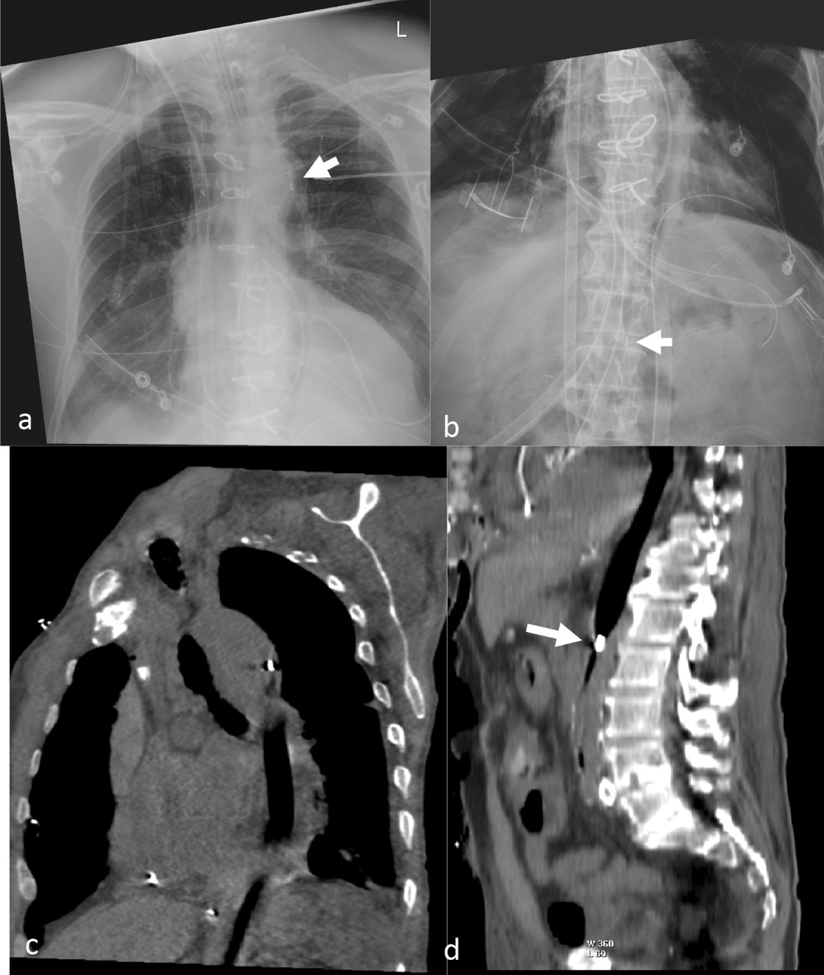
set it up right- Position upper balloon marker roughly 2 cm below the left subclavian origin in a femoral example. Assess the entire balloon and branch clearance, not only the chest marker.
- Trigger ECG or arterial pressure according to console mode. Arrhythmia can impair triggering but is not an absolute requirement for a regular rhythm. Verify on the arterial trace.
- Gas helium allows rapid inflation and deflation. A leak remains a gas-embolism and blood-loss hazard.
- Anticoagulate per protocol. Monitor platelets, limb perfusion, urine output.
- Augmentation interpret peak height with timing, assisted pressures and clinical perfusion.
C
Troubleshooting & complications
at the bedside| Problem | Cause → action |
|---|---|
| Low augmentation | Check rhythm, timing, position, gas/tubing, preload and afterload. Give fluid only when filling assessment supports it. Low augmentation alone does not diagnose hypovolemia. |
| Loss of trigger | Arrhythmia / poor ECG or arterial signal → switch trigger source. Improve signal. |
| Blood in the gas line | Suspected perforation: on Cardiosave, enter Standby, disconnect the extender from the console to deflate, then clamp the extracorporeal tubing per IFU. Notify the team and prepare removal. Do not reinflate. |
| Limb ischemia | New ischemia requires urgent device/vascular assessment, not the next scheduled check. Consider device removal or revascularization. |
| Thrombocytopenia / hemolysis | Trend platelets and hemolysis markers. Assess mechanical causes, bleeding, thrombosis, sepsis and HIT when relevant. |
| Sudden loss of augmentation | Disconnection · profound hypotension · arrhythmia · migration → check console, connections, rhythm, position. |
| "Gas leak" alarm | Inspect for blood or other perforation signs. Follow the perforation sequence above if suspected. Do not bypass leak alarms. |
D
Wean · evidence · the ceiling
when it's not enough- Wean use a supervised ratio or volume-reduction protocol. Monitor perfusion, congestion and recurrent ischemia. Tolerance is separate from the removal decision.
- Evidence No routine survival benefit established: IABP-SHOCK II in AMI-CS and ALTSHOCK-2 in selected HF-CS were neutral for their primary endpoints.
- Removal plan anticoagulation, deflation and access closure with the trained team using the actual catheter IFU. No universal hold or compression time applies.
- The ceiling IABP modifies native ejection. It does not provide independent pump flow or oxygenation. A fixed output gain is not guaranteed.
- Escalate persistent hypoperfusion → urgent shock-team reassessment of cause, LV/RV phenotype and a feasible escalation or exit plan. → Shock · MCS Weaning · ECMO cards
E
Insertion & daily care
place it · watch the limb- Access common femoral (Seldinger) — or axillary/subclavian for ambulation/bridge. Select balloon size using the catheter IFU and aortic anatomy.
- Confirm verify marker position after placement or suspected migration. Chest imaging alone may not show the lower balloon end.
- Daily frequent documented neurovascular and access checks, with platelets/Hgb and organ perfusion trends. Protect femoral access and follow ordered movement limits.
- Interruption minimize time without counterpulsation because stasis can thrombose. Suspected perforation is an exception: stop immediately and follow the IFU.
Femoral-IABP ambulation is an exception workflow—not routine bedside activity. Use only for a selected, stable adult under a unit-approved protocol with explicit HF/MCS attending clearance and PT leadership. For prolonged mobile support, consider axillary/subclavian access with the implanting team. Published experience supports feasibility, not universal safety.
1
GO / NO-GO huddle
clear every item before transferPatient readiness
- Order + team clearance HF/MCS attending order with individualized MAP, HR, SpO₂, augmentation & hip-flexion limits. PT assessment complete.
- Stable trajectory no escalating vasoactive/support needs, unstable rhythm, recent syncope, active ischemia, or symptomatic hypotension.
- Participates safely alert, follows 2-step commands, WBAT both legs, adequate balance. Cannulated-leg strength ≥4/5 or PT documents safe alternative.
- Respiratory not intubated or on rescue CPAP/BiPAP. Resting SpO₂ ≥90%.
Device / limb readiness
- Position same-day CXR reviewed. Tip position acceptable. No unresolved migration or new loss of augmentation.
- Access site dry, no expanding hematoma. Catheter, sheath, anchor & dressing secure. Tubing has safe slack and no blood in gas line.
- Perfusion distal pulse/Doppler, color, temperature, sensation and motor exam documented and unchanged.
- Do not start active bleeding, limb ischemia, device alarm/malfunction, HR >150, SpO₂ <90%, or inability to protect the femoral line.
2
Team & setup
daytime · rescue-ready| Role / item | Minimum plan |
|---|---|
| At bedside | HF/MCS physician or APP + IABP-competent RN + PT. Add second therapist/tech/RN for guarding, IV poles and oxygen. Assign one person to the patient, one to the console/line, and one to equipment. |
| Equipment | Standing bed / tilt table with 2–3 straps · rolling walker · gait belt · portable monitor/oxygen as ordered · clear return path. Console on battery only for transfer/ambulation. |
| Baseline | Record rhythm, HR, BP/MAP, SpO₂, intra-aortic & augmented pressure, symptoms, site and neurovascular exam. Secure all lines. Maintain the ordered cannulated-side hip limit (<30° in the published modified protocol). |
| On the table | Strap across abdomen and knee (± mid-thigh), avoiding all lines. Re-level invasive transducers and IABP console before tilting. Continuously display ECG, pressure waveform and SpO₂. |
3
Tilt → stand → walk → return
Ramsey mobilization sequence- Verticalize from 0° in 15° increments. Pause and reassess at least at 30° · 60° · 90°. Watch symptoms, rhythm, BP/MAP, SpO₂, augmentation, site and line tension.
- At 90° recheck site and ordered targets. A modest asymptomatic fall in BP/augmentation may occur. do not proceed if outside ordered limits. In the published protocol, augmentation <55 mmHg or HR 130–150 required provider re-clearance.
- Pre-walk loosen straps with hands-on guard. Weight shift, then march using the non-IABP leg. Keep cannulated-side hip flexion within the ordered limit.
- Step off lead with the IABP leg, then the opposite leg. Use the walker and continuous hands-on guard. RN owns console/line. Team continuously monitors.
- Progress first goal = safe stand / ≥10 ft. Increase distance only while hemodynamics, augmentation, line integrity and gait remain acceptable.
- Return back onto upright table with the non-IABP leg first, then IABP leg. Reapply straps. Lower gradually, reassessing near 45° and at 0°.
4
STOP immediately
return to table · stabilize · reassess- Chest pain/pressure, marked dyspnea, dizziness/presyncope, syncope, pallor/diaphoresis, new neurologic symptom, patient request or unsafe gait.
- Symptomatic hypotension or persistent ordered MAP violation. New significant arrhythmia. HR >150. SpO₂ <90% despite prescribed support.
- Loss of augmentation/waveform, console/device alarm, catheter or tubing tension/kink, suspected migration, blood in gas line, disconnection or malfunction.
- New bleeding/hematoma, access pain, pulse/sensation/motor change, cool/pale limb or other ischemia. Activate the device/vascular emergency pathway as indicated.
5
Close the loop
before the team leaves- Reassess supine vitals, rhythm, waveform/augmentation, alarms, site/anchor/dressing, pulses/Doppler, color/temp, sensation and motor function.
- Imaging post-session CXR per unit protocol. Obtain urgently for suspected migration or changed device function.
- Document tilt achieved, distance/time, assist level, device ratio/trigger, pre/during/post vitals & augmentation, symptoms, access/neurovascular findings and any event/action.
- Next session team reviews tolerance and sets the next mobility goal. Any adverse change requires HF/MCS re-clearance.
Evidence snapshot: a standardized femoral-IABP program reported 323 sessions in 70 selected patients, no ambulation-associated major complications, and 11 minor events across 323 sessions (3.4% per session), including migration, infection, hematoma and console dysfunction. This remains observational evidence and requires local governance, training, competency and event review.