The
Trap
Trap
Trabeculation ≠ diseaseProminent LV trabeculation is common & often benign — seen in athletes, pregnancy, and normal hearts. Morphologic criteria are sensitive but not specific: don't over-diagnose a cardiomyopathy.
Phenotype, not a verdictThe 2023 ESC framing treats hypertrabeculation as a phenotypic trait that may accompany a cardiomyopathy (DCM/HCM) — not a stand-alone diagnosis by morphology alone.
It matters when there's dysfunctionPursue it when paired with ↓ EF, arrhythmia, thromboembolism, or family history — that's when it carries prognostic weight.
Look for the real diseaseSearch for an associated cardiomyopathy, genetic syndrome, or congenital/neuromuscular cause — and treat that.
01
When to Suspect
Most are incidental- Incidental prominent trabeculation on echo/CMR done for another reason — the commonest "presentation".
- Heart failure reduced EF with deep trabeculations & intertrabecular recesses.
- Arrhythmia VT, AF, or conduction disease.
- Thromboembolism stroke/systemic embolism, esp. with low EF.
- Family screening relative of a DCM/HCM/LVNC proband.
Don't anchor on the pictures Hypertrabeculation in an athlete, pregnant patient, or person of African ancestry with a normal-functioning heart is usually a normal variant — criteria alone over-call it.
02
Diagnostic Criteria
Echo & CMR ratiosJenni (echo)
NC/C >2
Non-compacted:compacted, end-systole; 2-layer structure + colour-Doppler-perfused recesses.
Petersen (CMR)
NC/C >2.3
End-diastole; 86% sensitivity / 99% specificity in the small original selected cohort, not a population screen.
- Jenni echo maximal end-systolic NC/C >2 in a 2-layered myocardium.
- Petersen CMR end-diastolic NC/C >2.3 is a morphologic criterion, not proof of cardiomyopathy.
- Caveat different criteria (Jenni, Chin, Stöllberger, Petersen, Jacquier mass) classify different patients — criteria disagree, inflating diagnosis.
Criteria are non-specific
A positive ratio does not by itself establish a cardiomyopathy — require functional, arrhythmic, embolic, or genetic corroboration before labeling LVNC cardiomyopathy.▶
The central debate
Disease vs phenotypic traitDistinct cardiomyopathy, or a shared morphology?
Whether isolated LV non-compaction is a distinct cardiomyopathy or a morphological trait shared across normal hearts and other cardiomyopathies remains unresolved. Early cohorts described it as a discrete entity with poor prognosis, but the reported prevalence varies widely with population and imaging definition, and current criteria have low specificity. The 2023 ESC guideline therefore treats hypertrabeculation as a phenotypic trait that may accompany a cardiomyopathy rather than a stand-alone class. Practically: the prognosis lives in the associated dysfunction, arrhythmia, embolism, and genotype — not the trabeculae.
▶
The competing diagnostic criteria
They disagree — that is the problem| Criterion | Modality | Phase | Threshold | What it measures / caveat |
|---|---|---|---|---|
| Jenni | Echo | End-systole | NC/C >2 | Two-layer myocardium + colour-Doppler flow into recesses; most widely used echo rule. |
| Chin | Echo | End-diastole | X/Y ≤0.5 | Trough-to-epicardium (X) vs peak-to-epicardium (Y) at the trabecular tips; parasternal/apical short-axis. |
| Stöllberger | Echo | — | >3 trabeculae | More than three trabeculations apical to the papillary muscles, moving synchronously with myocardium. |
| Petersen | CMR | End-diastole | NC/C >2.3 | Long-axis non-compacted:compacted; sens ~86%, spec ~99% in original series. |
| Jacquier | CMR | End-diastole | >20% mass | Trabeculated LV mass >20% of total LV mass; a global, volumetric rather than single-slice measure. |
These rules classify different patients and disagree in head-to-head studies — a major driver of over-diagnosis. Apply any threshold only as a screen, then require functional, arrhythmic, embolic, or genetic corroboration. Assess global and regional function, scar and the compacted layer and exclude apical HCM, prominent normal trabeculae, false tendons, and LV thrombus as mimics.
Genetically heterogeneous, overlapping DCM & HCM When genetic, LVNC most often involves sarcomere genes — MYH7, MYBPC3, TTN account for the majority of identified variants — the same genes that cause DCM and HCM, supporting a shared molecular spectrum rather than a unique gene set.
01
Genetic Landscape
Sarcomere-predominant- Most common MYH7, MYBPC3, TTN (≈ majority of genotype-positive cases).
- Others sarcomeric & cytoskeletal genes overlapping DCM/HCM; familial in a substantial minority.
- Yield a pathogenic variant is found in about one-third in selected cardiomyopathy cohorts, not incidental healthy trabeculation.
Test & cascade when it's a cardiomyopathy Offer genetic testing & counseling when LVNC accompanies dysfunction, arrhythmia, or family disease — then cascade-screen first-degree relatives (echo/CMR + the familial variant).
02
Associations
Rarely truly "isolated"- Other cardiomyopathies coexists with DCM & HCM phenotypes (overlap syndromes).
- Congenital heart disease esp. in children (Ebstein, septal defects, complex CHD).
- Neuromuscular mitochondrial disease, Barth syndrome (TAZ, infants/boys), muscular dystrophies.
- Acquired / reversible transient hypertrabeculation in pregnancy & athletes can regress.
Find the syndrome
In children especially, LVNC is often syndromic — screen for neuromuscular/metabolic disease and congenital lesions.▶
Genes most often implicated
Same loci as DCM & HCM| Gene | Protein | Inheritance | Overlap phenotype | Note |
|---|---|---|---|---|
| MYH7 | β-myosin heavy chain | AD | HCM / DCM | One of the most frequent sarcomeric variants in LVNC cohorts. |
| MYBPC3 | Myosin-binding protein C | AD | HCM | Common sarcomeric cause; truncating variants typical. |
| TTN | Titin | AD | DCM | Truncating variants link LVNC to the DCM end of the spectrum. |
| TPM1 / ACTC1 / TNNT2 | Thin-filament sarcomere | AD | HCM / DCM | Less common; reinforce sarcomeric biology. |
| LMNA | Lamin A/C | AD | DCM + conduction | Flags early AV block / high arrhythmic & SCD risk — lowers ICD threshold. |
| TAZ (TAFAZZIN) | Tafazzin | X-linked | Barth syndrome | Infant boys: LVNC + skeletal myopathy + cyclic neutropenia + 3-methylglutaconic aciduria. |
| DTNA / mtDNA | α-dystrobrevin / mitochondrial | AD / maternal | CHD / mitochondrial | Historic isolated-LVNC locus (DTNA); mitochondrial disease a key paediatric cause. |
Reported diagnostic yield is roughly one-third of probands, with sarcomere genes predominating; a pathogenic variant, interpreted by gene and phenotype, may inform prognosis, ICD decisions and cascade testing. A negative panel does not exclude familial disease — still screen first-degree relatives by imaging.
KEY
LVNC sits on the DCM/HCM genetic spectrum — it is rarely a unique disease with a unique gene. Test when it behaves like a cardiomyopathy, and read the genotype as part of a family picture, not an isolated curiosity.
A
The three complications
When LVNC carries weightHFHeart failure — leading complication
VAVentricular (± atrial) arrhythmia
TEThromboembolism / stroke
A 2000 referral cohort of 34 adults reported HF in 18/34 (53%), VT in 14/34 (41%) and thromboembolism in 8/34 (24%) — though selection toward symptomatic referral populations inflates these rates versus incidental trait.
01
Heart Failure & Arrhythmia
Drives prognosis- Systolic dysfunction the strongest prognostic factor — outcome tracks EF, not trabecular depth.
- Ventricular arrhythmia VT/VF risk relates to scar (CMR LGE) & reduced EF.
- Atrial fibrillation common; conduction disease can occur.
Risk-stratify by the disease, not the picture Reduced EF, CMR LGE, NSVT, syncope, and high-risk genotype carry the prognosis — isolated trabeculation with normal function is low-risk.
02
Thromboembolism
A real but selective risk- Mechanism stasis within deep intertrabecular recesses, atrial fibrillation, and low-EF cavity stasis.
- Higher risk with reduced EF, prior embolism, AF, or documented LV thrombus.
- Not universal anticoagulation is not indicated for hypertrabeculation with preserved function alone (see P.4).
B
Pathologic LVNC vs benign hypertrabeculation
The high-value discriminator| Feature | Favours pathologic LVNC | Favours benign trait / physiologic |
|---|---|---|
| LV systolic function | Reduced EF, regional wall-motion abnormality | Normal EF, normal strain |
| CMR scar (LGE) | Present (mid-wall / subendocardial) | Absent |
| Context | Symptoms, arrhythmia, embolism, syndrome | Athlete · pregnancy · sickle-cell · normal variant |
| Family history | Familial DCM/HCM/LVNC or SCD | None |
| Genotype | Pathogenic sarcomere / LMNA variant | No familial disease-causing variant identified, which does not exclude genetic disease |
| Reversibility | Fixed / progressive | May regress (post-partum, detraining) |
| Deconditioning test | Trabeculation & dysfunction persist | Regression with altered loading may support adaptation, but absence of regression does not prove disease |
Loading changes may explain hypertrabeculation in pregnancy, athletes or anemia. Those settings do not exclude coexisting cardiomyopathy. Re-image after delivery or detraining before committing to a lifelong diagnosis.
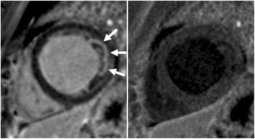
Kellman et al., Fig. 7, reproduced intact. Source · CC BY 4.0.
There is no LVNC-specific drug Management = treat the associated phenotype. Reduced EF → full HFrEF GDMT; arrhythmia → standard CM arrhythmia care + ICD by usual cardiomyopathy indications; thromboembolic risk → selective anticoagulation. Manage hypertrabeculation with normal function expectantly.
A
Anticoagulation — selective, not reflex
Indication-drivenOACAtrial fibrillation
Use current AF stroke-risk guidance and the associated cardiomyopathy phenotype. Hypertrabeculation alone does not determine the indication.
OACLV thrombus / prior embolism
Documented thrombus or prior systemic embolism → anticoagulate.
CONSIDERSignificantly reduced EF
Low EF alone is not a routine OAC indication in sinus rhythm. Consider additional thromboembolic factors and bleeding risk.
NOT routineTrabeculation + normal function
Hypertrabeculation alone is not an indication for anticoagulation.
01
Disease-Directed Therapy
As DCM / HCM- HFrEF ARNI/ACEi, β-blocker, MRA, SGLT2i; device therapy (CRT/ICD) by standard EF/QRS criteria.
- ICD primary/secondary prevention per usual cardiomyopathy & SCD risk indications (EF, NSVT, syncope, LGE, genotype).
- HCM-overlap if hypertrophic/obstructive features dominate, manage on the HCM pathway.
02
Family & Follow-up
Longitudinal- Family screening echo/CMR ± genetics for first-degree relatives when LVNC is a cardiomyopathy.
- Surveillance periodic echo (EF, function) + rhythm monitoring; CMR for scar when risk is uncertain.
- Exercise individualize; restrict only for the associated cardiomyopathy's risk, not the trait alone.
B
Surveillance & family screening cadence
Driven by phenotype, not trabeculae| Scenario | Imaging / monitoring | Interval | Genetics |
|---|---|---|---|
| LVNC + cardiomyopathy (↓EF / arrhythmia) | Echo (EF, function) + ambulatory ECG; CMR for scar | 6–12 mo or per HF/arrhythmia status | Proband testing + counselling |
| Trabeculation, normal function, no symptoms | Echo; CMR once if diagnosis uncertain | Individualize need and interval after excluding cardiomyopathy. Reassess with new symptoms | Only if family disease |
| First-degree relative of a CM proband | Echo/CMR; ECG | Typically q1–3 yr before age 60, then q3–5 yr, individualized to family and phenotype | Cascade test the familial variant |
| Child / paediatric LVNC | Echo + ECG; evaluate for syndrome | 6–12 mo (more frequent if dysfunction) | Test for syndromic / metabolic causes |
| Pregnancy / athlete (suspected physiologic) | Re-image post-partum / after detraining | Re-assess, then per result | If persists or familial |
| LMNA genotype-positive | Ambulatory ECG ± device check; lower ICD threshold | Annual | Cascade — high arrhythmic risk |
If a familial pathogenic variant is known, genotype-positive / phenotype-negative relatives still warrant periodic imaging; clinically unaffected relatives negative for the confidently causal family variant can usually leave surveillance.
SUMMARY
Confirm with criteria, but diagnose the cardiomyopathy — not the trabeculae. Treat the associated phenotype (HFrEF GDMT, indication-based ICD), anticoagulate selectively (AF, thrombus or prior embolism, with selected additional high-risk contexts), screen the family, and watch function over time. Isolated hypertrabeculation with a normal heart needs reassurance, not a label.