Myocarditis = inflammatory disease of the myocardium. Suspect with new HF, infarct-like chest pain with clean coronaries, or arrhythmia/conduction disease + a troponin rise — often after a viral prodrome. Normal troponin or EF does not exclude it. Spectrum runs subclinical → fulminant → chronic inflammatory cardiomyopathy (DCMi).
A
Etiology — three buckets
cause often remains uncertain| Class | Examples |
|---|---|
| Infectious | Infectious associations — DNA: parvovirus B19, HHV-6, adenovirus, CMV, EBV; RNA: Coxsackie A/B, influenza, HCV, HIV, SARS-CoV-2. Also bacterial (incl Lyme), Chagas (T. cruzi), fungal, rickettsial. |
| Immune-mediated | ICI & drug-hypersensitivity (PCN, sulfonamides, clozapine); systemic autoimmune (sarcoid, SLE, EGPA/Churg-Strauss, IBD, scleroderma); alloimmune (transplant rejection). |
| Toxic | Drugs/toxins can cause myocardial injury by several mechanisms. Do not equate anthracycline dysfunction, 5-FU ischemia or catecholamine stunning with proven myocarditis. |
B
Pathophysiology — three phases
why timing drives therapy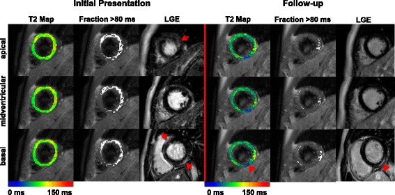
Injury and inflammation
Etiology and severity guide the next test.
Etiology and severity guide the next test.
Recovery or persistent disease
Symptoms, function, biomarkers and rhythm may recover at different rates.
Symptoms, function, biomarkers and rhythm may recover at different rates.
Residual scar
Normal EF does not erase arrhythmic risk. Persistent LGE alone does not prove ongoing inflammation.
Normal EF does not erase arrhythmic risk. Persistent LGE alone does not prove ongoing inflammation.
C
How it presents
4 syndromes + the troponin clue- Infarct-like chest pain, ST/T changes, ↑ troponin, nonobstructive coronaries — often post-viral, good prognosis.
- Heart failure new/subacute dyspnea, ↓ EF.
- Arrhythmia / block VT or high-grade AV block → think giant-cell / sarcoid.
- Fulminant rapid shock — often non-dilated LV; needs MCS.
Troponin clue: Troponin kinetics alone cannot distinguish myocarditis from MI. Nonobstructive coronaries require evaluation for myocarditis, infarction mechanisms, Takotsubo and other injury.
D
Clues to the specific etiology
narrow it early- AV block / VT (esp. younger) → giant-cell or cardiac sarcoid → biopsy.
- Eosinophilia → eosinophilic (EGPA, hypersensitivity, parasitic).
- Recent ICI → ICI myocarditis (often myositis / MG overlap).
- Drug + rash / fever → hypersensitivity; endemic exposure → Chagas / Lyme.
E
Epidemiology & natural history
who & what happens- Who often younger adults; a leading cause of sudden death in the young / athletes.
- Course most lymphocytic cases recover; a minority progress to DCMi; fulminant disease has high early risk and may leave persistent dysfunction.
F
Where it fits — the cardiomyopathy map
an acquired CM- Myocarditis is an acquired cause of cardiomyopathy; healed disease may leave a dilated CM (DCMi).
- Distinguish from genetic / familial DCM (→ Genetic CM card); sarcoid & giant-cell overlap the arrhythmogenic phenotypes (VT).
Spot the dangerous phenotypes early. Fulminant, giant-cell, eosinophilic/necrotizing, ICI, and cardiac sarcoid myocarditis change management — they need urgent etiologic assessment, often biopsy and phenotype-specific treatment (P.2–3). New HF + VT or AV block is the classic red flag.
Clinically suspected myocarditis → ECG · troponin · echo → exclude obstructive CAD → CMR (Lake Louise) for tissue characterization → EMB when the stakes are high or a treatable phenotype is suspected.
◆
The diagnostic pathway
exclude CAD → CMR → EMB if high-riskUnstable, sustained VT or advanced block → monitored care, urgent etiologic workup and early biopsy/advanced-HF assessment.
Stable → ECG, biomarkers and echo, coronary evaluation when indicated, then CMR. Use EMB when its result changes treatment.
A
First-line work-up
and rule out an MI- ECG nonspecific ST/T; watch AV block / VT (→ giant-cell/sarcoid).
- Troponin + NPs support injury; echo for global/regional dysfunction, effusion, thrombus.
- Assess for ACS/CAD choose coronary CTA or invasive angiography when presentation, age, risk, ECG, and instability make ischemia a material alternative.
- Admit & monitor telemetry for arrhythmia; serial troponin & hemodynamics.
B
CMR — Lake Louise 2018
tissue characterizationTwo-criteria rule (≥ 1 of each): a T2-based marker of edema (↑ T2 / T2-mapping) + a T1-based marker of injury (LGE, ↑ native T1, or ↑ ECV). Supportive: pericardial effusion, regional wall-motion abnormality. Typical LGE is subepicardial / mid-wall (not a coronary territory).
C
Endomyocardial biopsy — when & how it reads
don't delay in the sick patientBiopsy when: fulminant / hemodynamic compromise · new HF with high-grade AV block or VT · failure to improve · suspected giant-cell, eosinophilic, or sarcoid.
| Specimen finding | What it means |
|---|---|
| Histology + immunohistochemistry | Describe inflammation, nonischemic myocyte injury, fibrosis and cell type. Quantitative thresholds alone do not define fulminant disease. |
| Viral genome detected | Interpret viral type, amount, tissue/blood results and histology together. A positive PCR does not establish causal viral myocarditis. |
| Negative biopsy | Sampling error remains possible. Reassess targeting or repeat biopsy when suspicion is high and the result would change treatment. |
Plan samples for histology, immunohistochemistry and molecular testing with pathology before EMB. Histotype and clinical context determine treatment.
D
Pathway & the ACS look-alike
sequence the tests- Sequence presentation/risk assessment → ECG, biomarkers, echo and monitoring → CMR when feasible → EMB in selected intermediate/high-risk cases when histology or aetiology will change treatment.
- ACS look-alike infarct-like myocarditis can mimic STEMI/NSTEMI; select coronary imaging from pre-test probability and clinical stability rather than sending every patient to angiography.
- LGE pattern subepicardial / mid-wall & non-coronary → favors myocarditis over MI.
- Repeat CMR at follow-up tracks resolution vs progression to DCMi.
E
Ancillary testing
targeted, not shotgun- Serologies routine viral serology is low-yield; target HIV/HCV, Lyme & Chagas by exposure.
- Immune / rhythm ANA/ANCA, eosinophil count, sarcoid work-up (ACE, FDG-PET) if indicated; Holter; consider genetics in recurrent disease, arrhythmic phenotypes, persistent dysfunction or family history.
F
High-risk features → escalate
don't wait- Hemodynamic reduced / falling LVEF, hypotension, cardiogenic shock.
- Electrical sustained VT, high-grade AV block.
- Act → ICU + telemetry, expedite EMB, early advanced-HF / MCS referral.
- Phenotype giant-cell / sarcoid features → biopsy & treat without delay.
Everyone gets supportive care (HF GDMT, arrhythmia monitoring, exertion restriction). Layer etiology-specific immunosuppression on the EMB result, and escalate the fulminant patient to mechanical support early — recovery is possible but cannot be assumed.
◆
Treatment decision — supportive for all, IST by EMB
inflammation ± virus → phenotypeSupport physiology now → stabilize perfusion, rhythm and congestion while the cause is investigated.
Treat the established phenotype → histology, infection relevance and clinical severity guide immunosuppression. A PCR result or elapsed time alone cannot choose therapy.
A
Supportive & acute
for all- HF GDMT standard four pillars as tolerated; treat arrhythmia; telemetry.
- Physical activity restrict during the acute phase, then individualize return using clinical remission, ventricular function, biomarkers, rhythm assessment, and follow-up imaging. Anti-inflammatory therapy is phenotype- and contraindication-dependent.
- Fulminant → ICU early tMCS / VA-ECMO bridge to recovery or transplant. → VA-ECMO & Shock cards
- Devices temporary pacing for unstable block. ICD timing depends on arrhythmia, scar and histotype.
B
EMB-guided therapy
inflammation ± virus| EMB result | Therapy |
|---|---|
| Inflammation − / Virus − | Post-myocarditis or DCM → HF GDMT ± genetic evaluation / risk-adjusted therapy. |
| Inflammation + / Virus − | Specialist-selected immunosuppression — esp. giant-cell, cardiac sarcoid, eosinophilic, & virus-negative DCMi. |
| Inflammation + / pathogen signal | Confirm clinical relevance with pathology/infectious-disease expertise. Routine empiric antiviral therapy or IVIG is not established for a PCR signal alone; use organism-specific treatment only when supported. |
C
Immunosuppression — what the evidence shows
phenotype-specific| Setting | Evidence / action |
|---|---|
| Lymphocytic (unselected) | MTT: routine immunosuppression did not improve EF/survival — not for unselected acute myocarditis. |
| Virus-negative DCMi | TIMIC: prednisone + azathioprine improved EF in virus-negative inflammatory CM. |
| Giant-cell | Aggressive combination IST (steroid + cyclosporine ± others) improves survival; otherwise rapidly fatal. |
| Cardiac sarcoid | Corticosteroids ± steroid-sparing agents; device therapy for block/VT. |
| Eosinophilic / necrotizing | Corticosteroids + treat the cause (stop culprit drug, treat EGPA / hypereosinophilia / parasite). |
| ICI myocarditis | Withhold ICI and start urgent specialist-directed corticosteroid therapy; escalate immunosuppression for non-response or instability. Long-term cessation or any rechallenge is an oncology/cardio-oncology MDT decision after recovery, not an automatic bedside rule. |
D
Avoid & monitor
protect the recovering myocardium- Avoid strenuous activity during active inflammation · alcohol excess · cardiotoxins. Use NSAIDs/colchicine only when the pericardial chest-pain phenotype and patient-specific contraindications support them.
- Caution digoxin; high-dose β-blockade in the acutely decompensated.
- Monitor serial echo/EF (~1–3 mo), Holter for arrhythmia, CMR within 6 mo where appropriate. Reassess symptoms, biomarkers, ventricular function and rhythm before activity return.
- Devices individualize with EP. Sustained unstable VT/VF or high-risk histotypes may justify earlier ICD. Consider WCD selectively during reassessment.
E
Immunosuppression — practical
duration & escalation- Duration use a histotype- and aetiology-specific specialist protocol; do not apply one generic taper. Serial clinical status, ventricular function, rhythm, biomarkers, and selected CMR/EMB guide adjustment.
- Refer early giant-cell → advanced-HF / transplant evaluation; ICU + MCS for the fulminant. → LVAD / Transplant cards
- ICI myocarditis co-manage with oncology/cardio-oncology; keep ICI withheld during acute treatment and make any later rechallenge decision only through a documented MDT risk–benefit review.
Prognosis & return to activity. Lymphocytic and fulminant phenotypes may recover, while giant-cell and ICI myocarditis can be high risk. Restrict activity during active disease; timing of return is individualized after clinical remission and reassessment of ventricular function, biomarkers, rhythm, and imaging rather than a single fixed interval.