From nuclear reaction to clinical decision
A radiotracer is a physiologic question attached to an unstable atom. Its half-life governs delivery; its emission governs the camera; its molecular target governs the answer.
Actual antimatter · picogram chemistry · global logistics · quantitative physiology.
The 60-second nuclear read
sequence winsWhy image?
CAD diagnosis/risk, flow, viability, amyloid, inflammation, infection, function.
Can I trust it?
Prep, stress, counts, motion, gating, attenuation registration, curves.
What is abnormal?
Location, extent, severity, reversibility; function and ancillary markers.
What changes?
Integrate symptoms, ECG, anatomy, calcium, MBF/MFR, prior studies.
Equations to write before the stem
units firstDecay
Perfusion score
Flow & function
Four eras that built the field
invention → clinical claimHistorical cases worth remembering
boards love repurposed tracersPYP: infarct → amyloid
Originally an adjunct for recent infarction because damaged myocardium can retain bone-seeking tracer. That history explains a modern false positive: recent MI can be focally PYP-avid.Hardware changes meaning
Planar overlap could not reliably distinguish myocardium from blood pool. SPECT—and often SPECT/CT—made “uptake in the wall” a defensible claim.Quantitation changes questions
Relative MPI finds regional imbalance; PET MBF can reveal globally reduced hyperemic flow even when every territory looks equally bright.Radioactive decay
exponential, not linear| Nuclide | Physical T½ | Board association | Emission used |
|---|---|---|---|
| Tc-99m | 6.02 h | Sestamibi/tetrofosmin SPECT | 140-keV gamma |
| Tl-201 | ≈73 h | Perfusion + redistribution | Hg x-rays ≈69–83 keV; 167 keV gamma |
| Rb-82 | ≈75 s | Generator PET perfusion | positron → 511-keV pair |
| N-13 | 9.97 min | Ammonia PET; cyclotron | positron |
| F-18 | 109.8 min | FDG; flurpiridaz | positron |
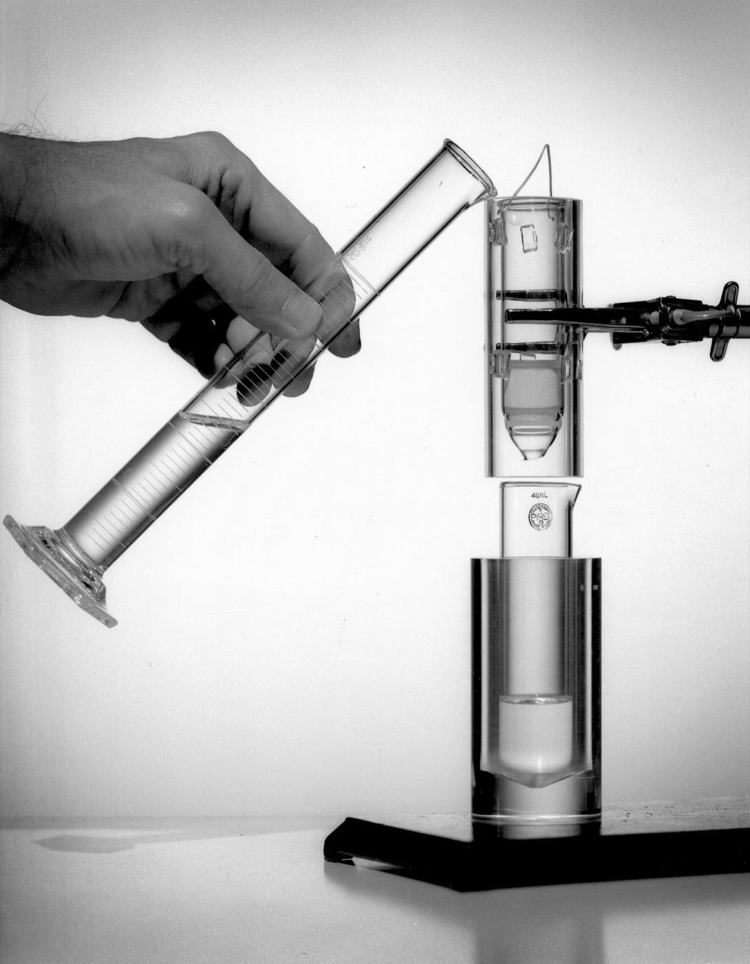

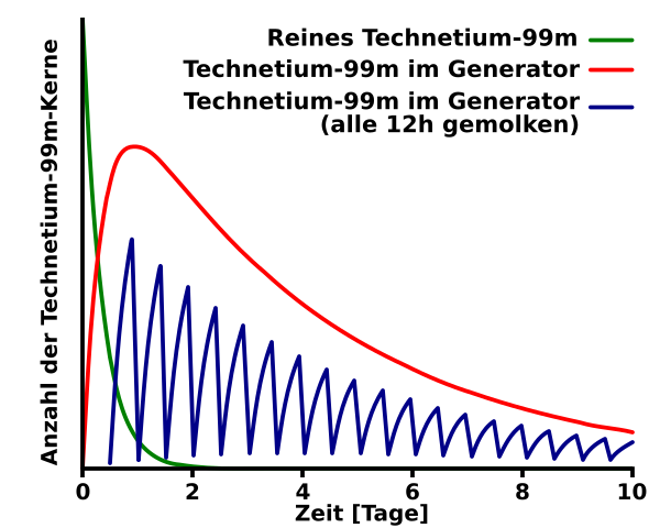

Photon interactions
contrast is a counting problemPhotoelectric
Photon is fully absorbed. Probability rises with absorber atomic number and falls strongly with photon energy; in tissue it removes useful photons and causes attenuation.Compton
Photon loses energy and changes direction. Scattered photons can enter the energy window in the wrong location, reducing contrast.Statistics
Poisson noise ≈ √N; relative noise ≈ 1/√N. Four times the counts roughly halves relative noise.Anger camera signal chain
localize, reject, countdirection selection
gamma → light
amplify/localize
energy window
matrix + image
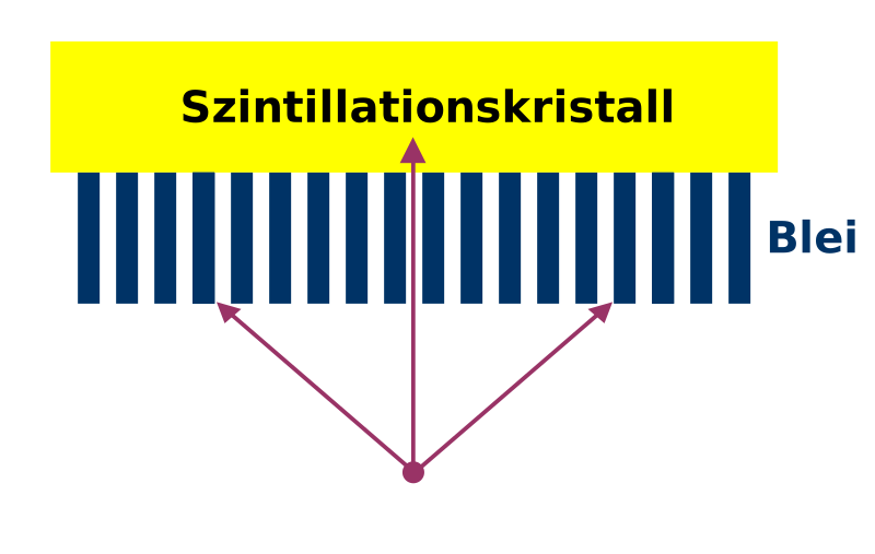

Collimator trade-offs
no free sensitivity| Type | Best use | Trade-off / trap |
|---|---|---|
| LEHR / LEGP | Tc-99m cardiac imaging | High resolution costs sensitivity; septal penetration if energy too high. |
| Low-energy high-sensitivity | Low-count/dynamic situations | More counts, less spatial resolution. |
| Medium energy | Higher-energy emissions / penetration control | Thicker septa, lower sensitivity; protocol-specific. |
| Pinhole | Magnified small-organ imaging | Not routine adult MPI; inversion and limited field. |
Energy discrimination
Center a Tc-99m window near 140 keV; a typical 20% window spans about 126–154 keV. Exact windows are camera/protocol specific.Distance law of resolution
Parallel-hole resolution worsens as source-to-collimator distance grows. Keep the detector close without compromising safety.Projection → volume
inspect before reconstruct180° or 360° validated orbit; close contour; enough views/counts.
Cine projections, motion, truncation, extracardiac activity, ECG.
FBP or iterative; filter; attenuation/scatter/resolution recovery.
True LV long axis → short axis, vertical and horizontal long axis.
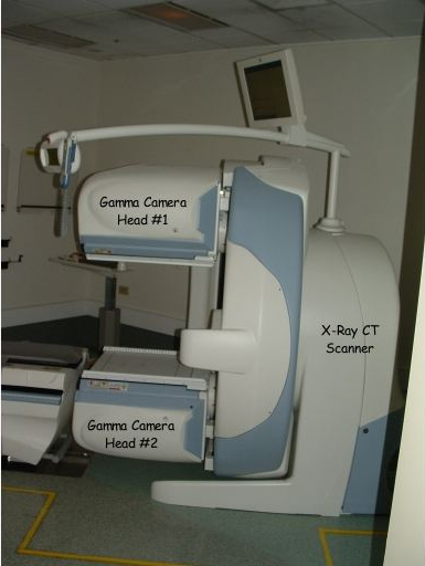

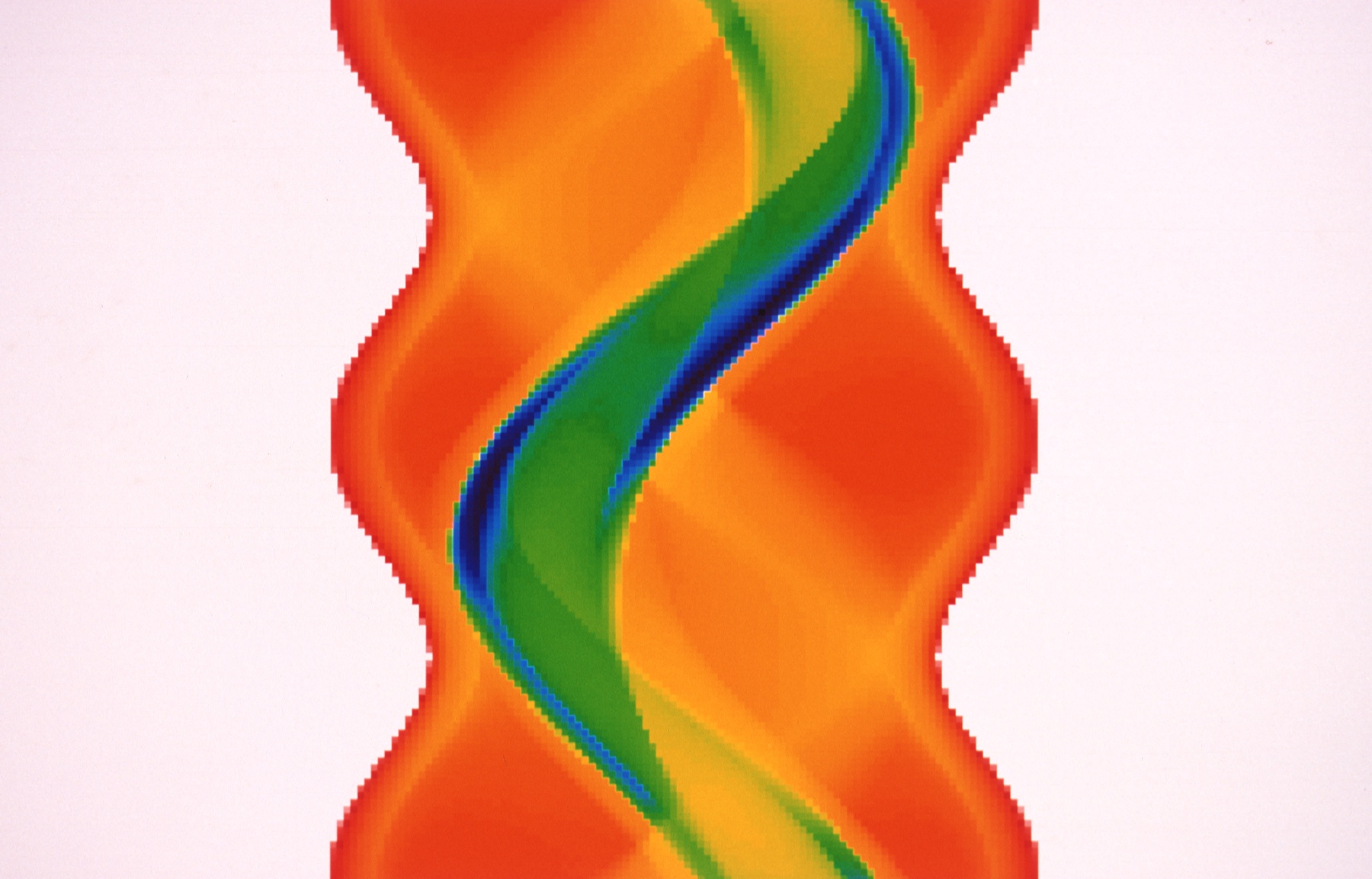

| Choice | Benefit | Failure mode |
|---|---|---|
| Iterative reconstruction | Models system physics; noise/attenuation corrections | Wrong iterations/filter create over-smoothing or edge artifacts. |
| ECG gating | Wall motion, thickening, EDV/ESV/LVEF | Arrhythmia or wrong R-wave detection produces flashing/false function. |
| Attenuation correction | Reduces breast/diaphragm attenuation | Emission–CT misregistration manufactures defects. |
| Prone imaging | Can shift inferior attenuation | Supplement, not replacement; anterior attenuation may appear. |
Coincidence imaging
electronic collimationA proton-rich nucleus emits a positron—the electron’s antimatter twin. It loses kinetic energy over a tracer-dependent range, encounters an electron, and annihilates.
e+ + e− → 2γRest mass becomes two 511-keV photons: 1.022 MeV total. PET detects the photons, not the positron.
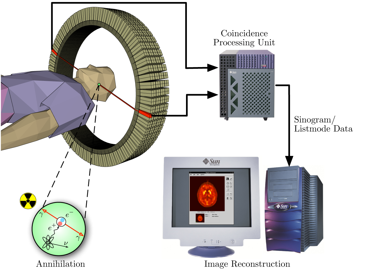

Positron travels a short range before interacting with an electron.
Two 511-keV photons depart approximately 180° apart.
Near-simultaneous detectors define a line of response; TOF narrows localization.
| Event/correction | Meaning | If uncorrected |
|---|---|---|
| True | Both photons from one annihilation, unscattered | Desired signal. |
| Scatter | One/both photons deflected | Wrong line; reduced contrast. |
| Random | Unrelated photons fall inside timing window | Background rises with count rate. |
| Attenuation | One photon absorbed removes the coincidence | Major quantitative bias; CT map needed. |
| Dead time / pile-up | System cannot process all high-rate events | Underestimated activity/flow; curve distortion. |
Spatial-resolution limits
Detector size, positron range, noncollinearity, reconstruction, motion. F-18 has shorter positron range than Rb-82.TOF
Does not alter the true line of response; it improves signal localization and image SNR, especially in larger patients.Quality control before interpretation
hardware → patient → software| Domain | Verify | Characteristic failure |
|---|---|---|
| Gamma camera | Energy peak/window, uniformity, center of rotation, detector geometry | Ring/ringlike or nonuniform defects; spatial misregistration. |
| PET | Normalization, calibration, blank/CT, timing, count-rate performance | Incorrect SUV/MBF, stripes, dead-time bias. |
| Hybrid CT | Registration, table position, breathing state, truncation/metal | Regional over- or undercorrection. |
| Patient | Identity, dose/time, position, motion, ECG leads, glucose/prep | Wrong protocol, curves, gating or interpretation. |
| Software | Correct tracer/model/database, axes, contours, beat acceptance | Precise-looking but invalid quantitation. |
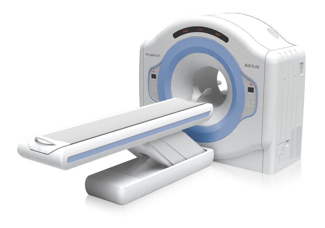
Modern detectors
benefit without immunity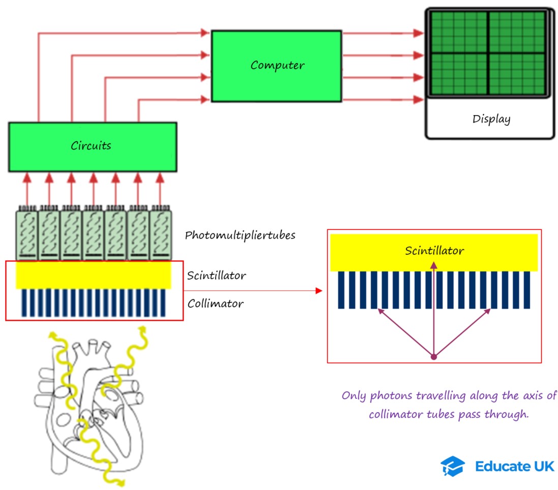


Perfusion tracer comparison
kinetics drive protocol| Tracer | Uptake/redistribution | Strength | Limitation |
|---|---|---|---|
| Tl-201 | Na/K-ATPase potassium analogue; redistributes | Delayed redistribution/reinjection can assess viability | Low photon energy, long half-life, higher dose burden, poorer counts. |
| Tc-99m sestamibi | Lipophilic cation; mitochondrial potential; minimal redistribution | High-count gated SPECT; stable distribution after injection | Hepatobiliary activity; separate rest/stress injections. |
| Tc-99m tetrofosmin | Lipophilic cation; minimal redistribution | Imaging may begin at 15 min under the current label; gated imaging | Separate rest/stress injections and protocol-specific timing still required. |
Extraction roll-off
At high coronary flow, tracer uptake rises less than linearly. Relative images can underestimate diffuse or balanced disease.Gating advantage
Tc-99m photon flux supports ventricular volumes, LVEF, motion and thickening—if rhythm and R-wave detection are valid.Choose tracer by question and logistics
perfusion ≠ metabolism| Tracer | T½ / production | Core use | Board nuance |
|---|---|---|---|
| Rb-82 chloride | ≈75 s; Sr-82/Rb-82 generator | Rapid rest/stress PET + MBF | High throughput; lower extraction and longer positron range than N-13/F-18. |
| N-13 ammonia | 9.97 min; cyclotron | High-quality perfusion + MBF | U.S. labeling describes rest/pharmacologic-stress use; exercise workflows come from professional practice and require suitable logistics. Dynamic MBF is generally pharmacologic. |
| F-18 flurpiridaz | 109.8 min; regional distribution | FDA-approved 2024 rest/exercise or pharmacologic perfusion; MBF with validated pharmacologic acquisition/software | Mitochondrial complex-I binding; rest→stress ≥30 min pharmacologic or ≥60 min exercise. At exercise inject at ≥85% APHR or ischemic signs/symptoms and continue 1–2 min. |
| F-18 FDG | 109.8 min | Glucose metabolism: viability, inflammation/infection | Viability prep stimulates uptake; inflammation prep suppresses physiologic uptake. |
Targeted imaging
phenotype firstTc-PYP / DPD / HMDP
Bone-avid tracers for ATTR cardiac amyloid pathway; SPECT confirms myocardium and monoclonal-protein testing is mandatory.FDG inflammation
Cardiac sarcoid, prosthetic/device/graft infection. Preparation and pattern are disease-specific.Emerging targets
Innervation, fibroblast activation, thrombosis and other molecular targets are not interchangeable with established routine indications.Radiation quantities
do not mix units| Quantity | SI unit | Meaning | Legacy |
|---|---|---|---|
| Activity | Bq = disintegrations/s | Amount of radioactive transformation | 1 mCi = 37 MBq |
| Absorbed dose | Gy = J/kg | Energy deposited per mass | 1 Gy = 100 rad |
| Equivalent/effective dose | Sv | Radiation/tissue weighting; population risk model | 1 Sv = 100 rem |
| Exposure rate | context-specific | Falls with distance approximately as 1/r² for a point source | Use survey meter, not mental math, operationally. |
ALARA hierarchy
quality-preserving reductionWill the answer change diagnosis, prognosis, or management?
Stress-first/stress-only when validated; weight/camera-specific activity.
Prep, IV, timing, motion prevention, attenuation/gating QC.
Time, distance, shielding, contamination controls and documentation.
Pregnancy and lactation
tracer-specific decisionsPregnancy possibility, gestational age, breastfeeding, urgency and question.
Can echo/CMR or delayed imaging answer safely without losing benefit?
If essential, select protocol/activity with nuclear medicine and medical physics.
Document dose estimate and tracer-specific breastfeeding interruption—not a generic interval.
Never improvise
Fetal dose and milk excretion differ by tracer, activity, timing and maternal clearance. Use institutional radiation-safety tables and a named expert.Risk communication
Explain expected benefit, alternatives, magnitude/uncertainty, and how protocol changes reduce exposure. Do not equate administered activity with fetal dose.Spill / contamination response
contain before cleanPrevent spread; keep people away; remove contaminated gloves/clothing safely.
Radiation Safety Officer and local emergency chain.
Mark extent; decontaminate from edge inward; resurvey people/area.
Nuclide, activity estimate, persons, readings, waste, follow-up.
Why exercise when feasible?
prognosis in the treadmillCapacity
Protocol, duration, peak workload/METs, limiting symptom and reason stopped.Hemodynamics
Chronotropic response, BP response, rate-pressure product and recovery.Electrical
ST shifts, arrhythmia, conduction and symptom–ECG concordance.| Step | Board-ready action | Common trap |
|---|---|---|
| Before | History, exam, ECG, medication/caffeine review, IV, emergency readiness | Exercise despite unstable symptoms, dangerous arrhythmia or decompensation. |
| Peak | Inject as close to maximal stress as possible; continue exercise ≈1–2 min when feasible | Inject after workload has fallen substantially. |
| Recovery | Continue ECG/BP/symptom surveillance and document resolution | Stopping observation when exercise stops. |
Rate-pressure product
Chronotropic calculation
Doses and timing
memorize exactly| Agent | Dose | Tracer timing | Key pharmacology |
|---|---|---|---|
| Regadenoson | 0.4 mg rapid IV injection (about 10 s), then saline flush | Tracer 10–20 s after the saline flush | A2A-selective agonist; fixed dose. |
| Adenosine | 140 µg/kg/min continuous IV, usually 4–6 min | Commonly 3 min after start while infusion continues | Nonselective adenosine receptor agonist; half-life <10 s. |
| Dipyridamole | 0.56 mg/kg over 4 min | About 3–5 min after infusion | Raises endogenous adenosine; longer effects. |
A2A receptor: open the resistance bed
Regadenoson directly stimulates coronary A2A receptors; adenosine stimulates several receptor subtypes. Resistance-vessel dilation raises normal flow more than flow across a stenosis, producing relative heterogeneity.Competitive blockers and amplifiers
Caffeine and theophylline are methylxanthine antagonists that can blunt hyperemia. Dipyridamole inhibits adenosine uptake/metabolism, amplifying endogenous adenosine and prolonging effect.Safety gates
screen before injectDo not proceed until resolved
Active bronchospasm; advanced AV block/sinus-node disease without a functioning pacemaker; unstable ischemia; symptomatic hypotension; or uncontrolled BP >200/110 mmHg. Verify the full agent-specific label and local emergency pathway.Relative: individualize
Severe symptomatic aortic stenosis is a relative—not absolute—contraindication to regadenoson. Weigh urgency, hemodynamic reserve, alternative imaging and monitored-lab capability; do not place it in the automatic “do not proceed” list.Dobutamine pathway
when vasodilator is unsuitable| Baseline condition | Preferred concept | Why |
|---|---|---|
| LBBB | Vasodilator MPI, preferably PET when available | Tachycardia/exercise can create septal perfusion artifact. |
| Ventricular pacing | Vasodilator rather than tachycardia-producing stress | Abnormal activation and septal artifact. |
| Active bronchospasm | Dobutamine if stress imaging is essential and appropriate | Adenosine-mediated bronchoconstriction risk. |
| High-grade AV block, no pacer | Avoid adenosine agonists; individualize alternative | Nodal suppression. |
Stop for danger
Limiting ischemic symptoms, significant ST change, sustained/severe arrhythmia, marked hypotension or severe hypertension → stop, stabilize and follow the lab emergency pathway.Stop for endpoint
Target/diagnostic endpoint or maximum validated dose is reached. “Target HR” never overrides a safety signal.Own recovery
Continue ECG, BP and symptom surveillance; treat persistent effects and document resolution. A beta-blocker may reverse dobutamine when clinically appropriate.Dose calculation
Why the ECG matters
Dobutamine may provoke ischemia, hypertension, hypotension or atrial/ventricular arrhythmia. Dose stage, endpoint, tracer time and recovery treatment belong in the report.Protocol selection
match patient + camera| Protocol | Best fit | Critical rule |
|---|---|---|
| Stress-first / stress-only | Low/intermediate risk, no known CAD, high-quality normal stress images | Omit rest only under a validated pathway with unequivocal normality. |
| 1-day rest–stress | Routine convenience | Second Tc-99m activity is generally ≈3× the first (≈3.5–4× with no inter-injection delay), using a validated camera/weight-specific protocol. |
| 1-day stress–rest | Stress-first triage | Rest only if stress abnormal/equivocal; avoid shine-through. |
| 2-day | Large body habitus or count limitations | Higher activity each day; confirm labeling/order and comparison. |
| Tl viability | Selected perfusion/redistribution question | Immediate + delayed/reinjection timing is not interchangeable with Tc. |
Three operational ledgers
quality has a costStress-only
lowest conditional burdenOne injection/acquisition if stress images are unequivocally normal after raw, gated and attenuation QC.Savings disappear if equivocal studies trigger recalls.One-day pair
throughput efficientTwo injections and acquisitions in one visit; residual first-dose counts demand adequate separation/activity ratio.Convenience trades against shine-through.Two-day pair
count-rich, time-heavyComparable higher activity each day can improve count statistics in large patients.Adds patient travel, scheduling and camera time.Before
Weight/body habitus, pregnancy/lactation, ECG/rhythm, caffeine/meds, IV and camera capacity.During
Stress adequacy, injection quality, exact activity/time, symptoms/ECG/BP, gating.After
Raw cine, counts, motion, extracardiac activity, prone/AC when validated, repeat only if needed.Shine-through arithmetic
A first Tc-99m dose of 10 mCi has ≈10 × (½)3/6 = 7.1 mCi physical activity remaining 3 h later. After a 30-mCi second dose, the first dose is still ≈7.1/(30+7.1) = 19% of combined physical activity before biologic/detection effects.Clinical consequence
Residual counts from the first study can mask a true change in the second. Waiting, an adequate second:first activity ratio, order selection and validated reconstruction reduce—but do not license ignoring—shine-through.Dynamic flow acquisition
time zero is sacredCenter heart; attenuation CT; check breathing/registration.
Accurate injection time/dose; capture arterial input and tissue phases.
Motion, curves, contours, rest MBF; consider rate-pressure product.
Synchronize hyperemia and tracer; same QC → stress MBF + MFR.
| Invalidator | Clue | Action |
|---|---|---|
| Dose infiltration | Low/late input peak; injection-site activity | Do not trust MBF; document/repeat only if justified. |
| Missed time zero | Truncated input curve | Quantitation invalid even if static perfusion is interpretable. |
| Motion | Myocardium shifts across frames | Frame-wise correction only if validated; recheck contours. |
| Inadequate stress | High caffeine risk, weak response, globally low stress flow | Integrate prep/hemodynamics; avoid labeling diffuse disease reflexively. |
| Model mismatch | Wrong tracer/model or implausible curves | Reprocess correctly; use laboratory-specific normal ranges. |
Equilibrium radionuclide angiography
reproducible serial EFArea-normalized counts
Strength
High count-based reproducibility for serial LV EF when acquisition, labeling, positioning, gating and analysis are consistent. Planar equilibrium RVEF is limited by chamber overlap; use first-pass or another dedicated validated method for RVEF.| Problem | Effect | Recognition |
|---|---|---|
| Poor RBC labeling | Background/organ activity; weak blood pool | Low target-to-background; labeling method/context. |
| Arrhythmia | Mixed cycle lengths; distorted time–activity curve | Beat histogram/rejection; flicker or temporal blur. |
| LA/LV or RV/LV overlap | Erroneous ventricular counts | Review LAO angle and septal separation. |
| Background ROI error | Can raise or lower calculated EF | Position adjacent to LV without chamber/spleen overlap. |
First-pass and shunt concepts
time–activity curvesFirst-pass RNA
Tracks a compact bolus through right heart, lungs and left heart; useful for RV function or exercise when acquisition is technically robust.Left-to-right shunt
Pulmonary recirculation after the first pass raises the fitted pulmonary-to-systemic flow ratio; severe bolus dispersion undermines the curve.Read in a fixed order
do not start at polar map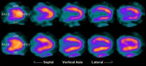

Raw cine/dynamic curves, motion, counts, gating, attenuation registration.
Location, extent, severity, reversibility; compare stress/rest side by side.
LV/RV size, EDV/ESV, LVEF, motion, thickening, post-stress change.
TID, lung/RV uptake, MBF/MFR, calcium/CT, symptoms/ECG and prior studies.
| Pattern | Perfusion | Function | Meaning after QC |
|---|---|---|---|
| Ischemia | Stress defect improves at rest | May show stunning/new dysfunction | Inducible flow limitation; quantify burden/severity. |
| Scar | Fixed defect | Reduced thickening/motion supports | Infarction likely; attenuation can mimic fixed defect. |
| Mixed | Fixed component + reversibility | Regional dysfunction common | Scar plus peri-infarct ischemia. |
| Balanced/diffuse | Relative images may look normal | TID/EF response may be abnormal | Use absolute MBF/MFR and clinical/anatomic context. |
17-segment scoring
0 normal → 4 absent| Score | Formula | Interpretive role |
|---|---|---|
| SSS | Sum of 17 stress scores | Total stress perfusion abnormality. |
| SRS | Sum of 17 rest scores | Rest perfusion abnormality—not automatically scar. |
| SDS | SSS − SRS | Reversible component; estimate ischemic burden. |
| % LV | Summed score / 68 × 100% | 0–68 scale; semiquantitative, software/lab dependent. |
Gated data: function plus artifact adjudication
verify beat histogram| Finding | What it adds | Failure mode |
|---|---|---|
| Wall thickening | Normal thickening under fixed defect favors attenuation | Small hearts, low counts, gating errors can mislead. |
| Post-stress LVEF fall | May support stunning/extensive ischemia | Timing differs by tracer; compare like with like. |
| TID | Can mark diffuse subendocardial ischemia/high risk | Cutoff is protocol, tracer, camera, sex/database and software specific. |
| RV uptake/dilation | RV pressure/volume burden or severe lung disease | Relative increase may reflect reduced LV uptake. |
| Lung uptake | With applicable tracers, severe LV dysfunction/ischemia | Timing, tracer and ROI dependent. |
Risk rises with concordance
cluster ≠ isolated numberBorderline alone
A TID of 1.23 with a laboratory limit of 1.20, normal perfusion and an obviously too-small rest LV contour should trigger reprocessing. Partial-volume/edge detection can underestimate ESV and inflate EF.First question: is the measurement real?Concordant cluster
Large/multiterritory reversibility plus a valid stress-EF fall, abnormal ventricular response and low global flow carries more weight than any single marker.Second question: what urgent consequence follows?Artifact fingerprint table
raw data are evidence| Artifact | Clue | Resolution |
|---|---|---|
| Breast / chest wall | Compare raw rest/stress cine: a shift in breast position can move a camera-fixed attenuation defect and mimic reversibility. | Match position; inspect body contour, gated thickening, and AC vs non-AC; reposition and reacquire when possible. Quantitation cannot repair a mismatched acquisition. |
| Diaphragm | Inferior fixed defect, often male/large abdomen | Prone/AC and function; confirm no true defect. |
| Motion | “Hurricane”/ring, axial shift on cine, noncoronary edge defect | Motion correction only if validated; repeat when necessary. |
| Subdiaphragmatic activity | Hot liver/bowel adjacent to inferior wall; normalization error | Delay, hydration/food or repeat per protocol; inspect unnormalized slices. |
| Gating failure | Beat-histogram tails, flashing, implausible EF/volumes | Fix leads/window; interpret nongated perfusion if valid. |
| PET/CT misregistration | Defect follows CT boundary, not coronary territory | Re-register, regenerate the attenuation map and reconstruct; moving only the display overlay does not correct AC. |
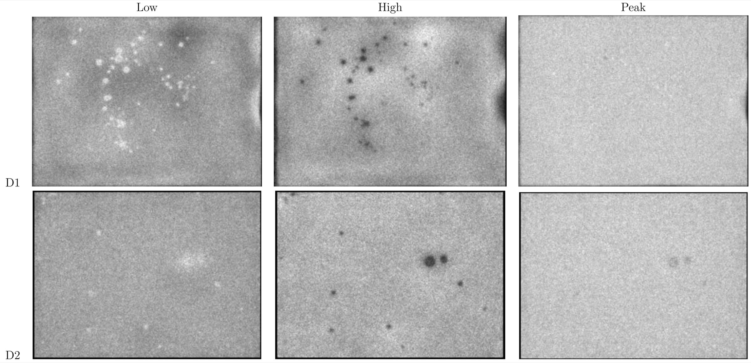

Four-question adjudicator
true defect or manufactured?Motion, attenuation, truncation, extracardiac activity?
Does wall motion/thickening agree?
AC vs non-AC; prone; CT registration?
Coronary territory, ECG, MBF, calcium, prior?
Interpret rest MBF, stress MBF and MBFR together
three numbers, one physiology| Pattern | Relative perfusion | Flow pattern | Favored physiology after QC |
|---|---|---|---|
| Focal epicardial | Regional reversible defect | Regional stress MBF/MFR reduced | Flow-limiting stenosis in matching territory. |
| Balanced multivessel | May look uniform | Global stress MBF and MFR depressed | Diffuse severe epicardial disease; integrate calcium/anatomy. |
| Microvascular/diffuse | Normal or mild diffuse heterogeneity | Global reduction without focal defect | CMD/diffuse atherosclerosis after excluding inadequate stress/technical cause. |
| High resting flow | Often normal | Low MFR but preserved stress MBF | Tachycardia, hypertension, anemia or other increased rest workload. |
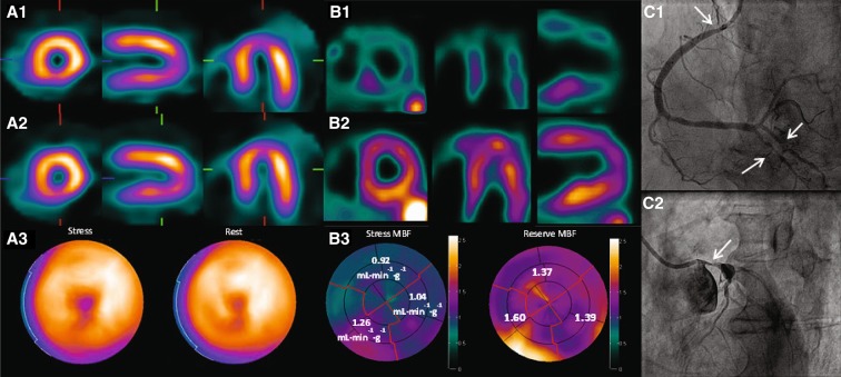

ASNC 2025 MBFR bins
Rb-82-focused practice guidance| Global MBFR | Practice interpretation | Relative risk |
|---|---|---|
| >2.0 | Normal | Low |
| 1.7–2.0 | Mildly abnormal | Intermediate |
| 1.2–<1.7 | Abnormal | High |
| <1.2 + perfusion defect | Highly abnormal | Very high |
| <1.2 without perfusion defect | Consider nondiagnostic/indeterminate | First exclude failed hyperemia/QC |
Perfusion–metabolism matrix
FDG needs uptake stimulationMake the FDG study diagnostic
stimulate viable cellsDiagnostic metabolism
Diffuse, adequately stimulated myocardial uptake with interpretable perfusion allows segment-by-segment matching.The biology is visible.Failed preparation
Globally poor or patchy uptake can mimic scar or reverse mismatch. Check glucose, insulin, timing, infiltration and motion before assigning tissue fate.Repeat may be safer than overcalling scar.Choose the test that answers the next question
patient-centered, not modality-centered| Clinical feature | Best nuclear contribution | Selection nuance |
|---|---|---|
| Exercise-capable | Symptoms, ECG, workload and hemodynamics plus perfusion | Exercise ECG alone may suffice in selected low-risk patients. |
| Prior CAD / CABG | Ischemic burden, scar, flow and serial comparison | Map graft/native anatomy; cath when high-risk actionable anatomy is the immediate question. |
| LBBB, pacing, attenuation risk | Vasodilator PET when available; modern AC/CZT SPECT where validated | Choose by local quality, body habitus, rhythm and anatomy. |
| Possible diffuse CAD / CMD | PET MBF/MBFR directly tests global and regional physiology | Requires a validated flow program; invasive coronary-function testing or stress CMR may fit the pathway. |
Three money ledgers—never substitute one for another
cost ≠ payment ≠ patient price1 · True lab cost
OPERATIONS LEDGERCapital/lease, service, facility, QC/compliance, tracer and delivery, stress drug, consumables, staff/reader time, decay/no-shows, nondiagnostic repeats and downtime.Ask: cost per completed diagnostic study at actual throughput?2 · Payer reimbursement
CONTRACT / POLICY LEDGERFacility and professional payment, HCPCS/APC packaging, site of service, payer contract, coverage and add-ons. Payment can be above or below the lab’s cost.A CMS threshold or code is not a market quote for tracer.3 · Patient out-of-pocket
BENEFIT-DESIGN LEDGERDeductible, copay/coinsurance, network, authorization, site and supplemental coverage. Estimate from the patient’s plan and performing facility.Never infer the patient bill from acquisition cost or Medicare payment.Throughput changes the fixed-cost denominator; short half-lives make schedule gaps and failed studies economically visible.
Post-PCI / CABG interpretation
territories are remodeledKnow anatomy
Native stenoses, graft origins/targets, stents, dominance, infarcts and time since procedure.Expect complexity
Competitive flow, graft/native disease, collateralization and peri-infarct ischemia blur default territories.Compare
Use identical slices/protocol where possible; state whether defect, flow, EF or volumes truly changed.Heart failure and cardiotoxic therapy
phenotype, do not over-attributeWhat ERNA does well
Count-based serial LVEF when labeling, projection, gating, background correction and software are kept constant. It is useful when acoustic windows are poor or a highly repeatable legacy series already exists.Trend one validated method.What it does not supply
No strain, valves, tissue characterization, pericardium or full HF phenotype; planar chamber overlap limits RV assessment. Integrate echo/CMR, biomarkers, symptoms and therapy timing.EF is one surveillance signal.What the landmark studies actually established
result + boundary| Study | Question and result | Practice lesson · do not overclaim |
|---|---|---|
| DIAD · 2009 n=1,123 | Screen asymptomatic type 2 diabetes with adenosine SPECT MPI? Event rates were low and screening did not reduce cardiac death/nonfatal MI. | Do not screen low-risk asymptomatic diabetes routinely. This did not test symptomatic or otherwise high-risk patients. |
| WOMEN · 2011 n=824 | In low-risk women able to exercise, initial exercise ECG versus exercise SPECT yielded similarly low 2-year event rates. | Use the lower-cost, no-radiation exercise ECG strategy when the ECG is interpretable and exercise is adequate. |
| COURAGE MPI n=314 substudy | PCI + optimal medical therapy reduced ischemia more than medical therapy alone; ≥5% ischemia reduction tracked with better outcomes in exploratory analyses. | Selected substudy, not a randomized proof that ischemia reduction itself mediates a survival benefit. |
| ISCHEMIA · 2020 n=5,179 | Moderate/severe ischemia: an initial invasive strategy did not lower the main composite or all-cause death versus a conservative strategy over the initial follow-up. | Ischemic burden alone is not an automatic PCI mandate; symptoms, anatomy, LV function and exclusions still govern. |
| PARR-2 · 2007 n=430 | FDG PET-assisted management in severe LV dysfunction did not significantly reduce the primary composite overall; benefit signals appeared with adherence and experienced-center use. | A viability test cannot help if its recommendation is ignored—and the subgroup signal is not the primary result. |
| STICH viability long-term follow-up | Viable myocardium tracked with EF improvement regardless of assignment; viability did not identify CABG survival benefit, and EF improvement was not associated with survival. | Do not use viability as a solitary yes/no gate for CABG; anatomy, scar, remodeling, surgical risk and goals remain central. |
| AURORA · 2023 578 evaluable | F-18 flurpiridaz PET had higher sensitivity than SPECT and noninferior specificity for CAD, with improved ROC performance including women and patients with obesity. | A diagnostic-accuracy trial supported the 2024 U.S. approval; it was not a randomized outcomes trial. |
A five-question trial decoder
read before quotingPrognostic evidence
“More ischemia predicts more events” can be true. It establishes risk association after adjustment and within the studied population.Useful for risk; not automatically causal.Predictive evidence
To claim imaging selects a treatment benefit, look for a randomized treatment-by-imaging interaction—not merely better outcomes among patients whose scan improved.Selection claims demand interaction evidence.Tracer passports and acquisition
do not transfer cutoffsTc-99m PYP
U.S./Canada predominantPlanar visual grade + SPECT localization. H/CL has validated adjunctive use only under a PYP protocol.
Tc-99m DPD
Europe and elsewhereVisual Perugini grade + SPECT. Do not import PYP H/CL thresholds.
Tc-99m HMDP/HDP
regional availabilityVisual grade + SPECT under tracer-specific guidance; not numerically interchangeable with PYP.
Tc-99m MDP
ordinary bone agentNot an accepted substitute for PYP/DPD/HMDP in the ATTR nonbiopsy pathway.
Perugini visual grade
compare heart with ribs| Grade | Myocardium versus ribs | Meaning |
|---|---|---|
| 0 | No myocardial uptake | Does not exclude all amyloid |
| 1 | Less than ribs | Equivocal |
| 2 | Equal to ribs | Potential ATTR only after localization + AL screen |
| 3 | Greater than ribs | Same diagnostic gates apply |
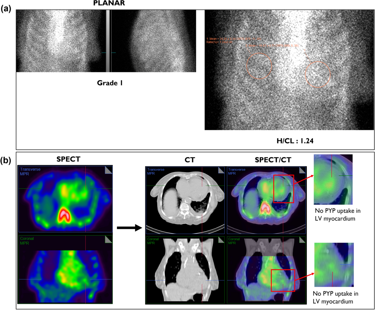
H/CL is an adjunct—not the diagnosis
For PYP only, ≥1.5 at 1 h or ≥1.3 at 3 h may support interpretation after SPECT confirms myocardial uptake. Do not calculate a diagnostic H/CL when uptake is only blood pool; a definite visual grade 2/3 does not require it.How to draw it
Place a circular/elliptical ROI over the heart on anterior planar imaging; mirror the same shape and area over the contralateral chest, avoiding sternum/ribs when possible. H/CL = mean heart count density ÷ mean contralateral count densityBlood pool masquerade
Cavity-centered activity fills LV/RV chambers, follows vessels and often diminishes on delayed imaging. Low output and renal dysfunction make it common.Activity inside the chamber—not the ventricular wall.True myocardial retention
Orthogonal tomograms show activity in the ventricular wall, circumferentially or regionally—not merely inside the cavity. SPECT/CT can resolve rib/sternal overlap.Activity in the walls.The diagnostic pathway
exclude AL urgentlyHFpEF/LVH, apical-sparing strain, low voltage relative to wall thickness, bilateral carpal tunnel, spinal stenosis, biceps rupture, neuropathy or family history.
Serum free light-chain κ/λ ratio interpreted with eGFR + serum immunofixation + urine immunofixation. SPEP/UPEP alone is inadequate. The combined screen has ≈99% sensitivity for detecting the abnormal precursor in AL, not a universal 99% negative predictive value.
PYP/DPD/HMDP with definite myocardial grade 2/3 on SPECT or SPECT-CT. Planar grade or H/CL alone does not pass.
All gates pass → ATTR-CM without biopsy; then TTR sequencing and counseling regardless of age. Discordant/equivocal → CMR and/or biopsy with mass-spectrometry typing.
When a monoclonal process is possible
Positive immunofixation, a low κ/λ ratio or a clonal/otherwise concerning FLC pattern prevents the shortcut. A borderline high ratio in CKD needs renal-range and hematology review; if clonality cannot be excluded, obtain tissue with definitive typing. Grade 2/3 uptake can occur in AL.Negative or grade 1 scan
Does not exclude early ATTR, AL, or certain TTR variants. If phenotype is convincing, proceed with expert review, CMR and biopsy rather than reassuring from scintigraphy alone.Mimics and misses
pattern predicts the trap| Pattern / cause | Why it misleads | Rescue move |
|---|---|---|
| Blood pool | Low output, renal dysfunction or early timing leaves cavity activity that overlaps myocardium on planar views. | Orthogonal SPECT/SPECT-CT; delay to 3 h when needed. |
| Injury / toxicity | Recent MI, pericarditis and hydroxychloroquine or other drug-associated myocardial injury can retain PYP. | Map pattern to anatomy, medication and timing; do not relabel focal or inflammatory uptake as diffuse ATTR. |
| Rib/sternum | Fracture, metastasis or normal bone projects over the heart. | Review rotating data and CT anatomy; redraw ROIs if used. |
| AL / rare amyloids | Bone-tracer uptake is not absolutely TTR-specific. | Complete monoclonal testing and biopsy typing when abnormal or discordant. |
| Early/variant ATTR | Some variants and early disease may have little bone-tracer retention. | Do not stop at grade 0/1 when phenotype/genotype is compelling. |
Two tracer families answer different questions
bone affinity ≠ fibril bindingBone-avid SPECT · PYP/DPD/HMDP
Best-established nonbiopsy lane for ATTR after AL exclusion; inexpensive/widely accessible in many regions. Uptake is indirect, AL sensitivity is poor, early/variant ATTR may be missed and serial burden is unreliable.Clinical strength: gated ATTR diagnosis.Amyloid-binding PET
Targets amyloid fibrils more directly and can show signal in AL as well as ATTR, with dynamic/whole-body quantitation. Blood-pool correction, tracer-specific thresholds, availability and outcome validation remain unresolved.Research promise: detection + burden + change.C-11 PiB
T½ ≈20 minClassic amyloid-binding PET research tracer; requires an on-site cyclotron and rapid synthesis.
Florbetapir / florbetaben
T½ ≈110 minBrain β-amyloid agents studied off-label in cardiac AL/ATTR; regional delivery is practical.
Flutemetamol
fibril-binding PETCardiac research results depend on tracer, subtype, timing and analytic method; do not pool thresholds.
Evuzamitide / AT-01
investigational pan-amyloidPeptide PET designed to bind systemic amyloid deposits; long-lived I-124 enables multiorgan imaging but adds dosimetry/logistics.
How PET tries to measure burden
validate every denominator| Measure | What it attempts | Main failure mode |
|---|---|---|
| SUV / SUV ratio | Static tissue concentration, normalized to injected activity/body metric; target-to-blood or target-to-background may improve contrast. | Timing, residual blood pool, body-size convention and scanner calibration change the number. |
| Retention index | Myocardial activity relative to integrated arterial input over time. | Requires valid dynamic framing, input curve, motion correction and model assumptions. |
| Kinetic binding | Separate delivery/washout from binding using compartment or graphical models. | Complexity, low counts, metabolites and no standardized clinical normal range. |
| Whole-body burden | Map heart plus soft tissue, nerve, kidney, liver or marrow deposits. | Organ-specific background and subtype biology make one universal threshold unlikely. |
Suppress physiologic myocardium
prep quality is diagnostic qualityValidated high-fat, very-low/no-carbohydrate meals—often two meals with >35 g fat and <3 g carbohydrate each.
Then fast 4–12 h; an alternative is prolonged fast >18 h per validated pathway.
UFH 50 IU/kg IV ≈15 min before FDG only if no contraindication/HIT; incremental suppression benefit is uncertain.
Document adherence/glucose; pair FDG with rest perfusion and inspect extracardiac disease.
Example prep-day menu
Meal 1: 3 eggs cooked in 2 Tbsp olive oil or butter + plain salmon; water. Meal 2: plain beef or chicken + 2 eggs, prepared/finished with 2 Tbsp olive oil; water. These portions exceed 35 g fat before counting the meat/fish; confirm <3 g carbohydrate and exact macros against the lab sheet. Then follow the lab’s fast. Avoid bread/cereal, rice/pasta/potatoes, fruit, dairy, sweets, sweetened drinks, alcohol and sugary sauces. Local instructions, allergy needs and diabetes plans supersede this example.Why optional heparin?
Unfractionated heparin acutely releases endothelial lipoprotein lipase, which hydrolyzes circulating triglycerides and raises free fatty acids. The normal myocardium shifts toward fatty-acid oxidation (the glucose–fatty-acid/Randle cycle), helping suppress physiologic FDG uptake. The incremental benefit beyond excellent diet/fasting is uncertain; screen bleeding risk and prior HIT.Read perfusion and metabolism as a pair
original pattern atlasWhere nuclear infection imaging adds value
anatomy + microbiology remain central| Scenario | FDG PET/CT value | Major limitation |
|---|---|---|
| Prosthetic valve | Perivalvular infection and extracardiac embolic/source survey | Recent surgery, adhesives and inflammation can be false positive. |
| CIED | Pocket vs lead involvement; metastatic infection | Small lead vegetations and postoperative uptake; antibiotics reduce sensitivity. |
| LVAD | Driveline, pump/pocket and cannula distribution | Metal attenuation artifact; sterile inflammation. |
| Vascular graft | Focal/intense heterogeneous graft uptake and extent | Diffuse linear postoperative uptake may persist. |
| Native valve | Extracardiac complications may help | Limited sensitivity for small mobile vegetations; TEE/blood cultures dominate. |
FDG versus labeled leukocytes
biology changes workflow and costFDG PET/CT · one perishable molecule
Labeled WBC SPECT/CT · the patient’s own cells
FDG clock
decay + courierMissed slots waste a prepared dose; nearby production and throughput matter.PET efficiency
whole bodyOne acquisition can map the cardiac focus, emboli and an extracardiac source.Cell laboratory
blood handlingSeparation, labeling, QC and reinjection add skilled labor and biosafety complexity.Serial SPECT
time + capacityMultiple acquisitions consume patient time and camera slots. Actual price/payment is site- and payer-specific.Patterns beyond epicardial CAD
nuclear phenotype needs context| Phenotype | Potential nuclear finding | Integrate with |
|---|---|---|
| Hypertrophic CM | Regional ischemia, reduced MFR, apical/midventricular scar-related defects | Echo/CMR morphology, gradients, symptoms and arrhythmia risk. |
| Dilated CM | Patchy defects, LV dilation/dysfunction, reduced global flow | CCTA/cath when CAD question remains; CMR tissue phenotype. |
| RV pressure load / PH | Perfusion-tracer RV uptake, dilation, septal flattening context | Echo and right-heart catheterization; not a PH subtype test. |
| Amyloid / sarcoid | Targeted tracer patterns | Disease-specific gates on pages 27–30. |
| Autonomic innervation | I-123 MIBG heart-to-mediastinum ratio and washout in selected settings | Protocol/system standards; not routine universal HF testing. |
I-123 MIBG sympathetic innervation
uptake-1 + vesicular storageWhat the tracer follows
Iobenguane resembles norepinephrine: presynaptic uptake-1 transport, vesicular storage and release/washout reflect cardiac sympathetic nerve integrity. Reduced uptake and faster washout are adverse signals in studied HF populations.It images nerve biology, not perfusion.Why numbers do not travel freely
Collimator energy response, scatter correction, ROI placement, acquisition timing and calibration alter H/M. Use validated camera-specific conversion/normal limits and the applicable jurisdictional indication.Standardize before serial comparison.Heart-to-mediastinum ratio
H/M = mean heart count density ÷ mean upper-mediastinal count densityDraw reproducible planar ROIs; record early/delayed time and collimator. H/M is not an ejection fraction.
Washout rate
DCF = 2Δt/13 h; Clate,corr = (Hlate−Mlate) × DCFWR = [(Hearly−Mearly) − Clate,corr] ÷ (Hearly−Mearly) × 100%
Use the same background-corrected count-density and ROI method at both times.
The newest change already in practice
approval ≠ outcome benefitAmyloid studies with data to watch
registry status, not results| Study · status | Question | What could change practice |
|---|---|---|
| CArdiag NCT05184088 completed · no posted results | F-18 florbetaben in cardiac AL; actual enrollment 244, registry completion May 2026. | Prospective diagnostic performance, standardized quantitation and incremental value versus current biopsy/imaging pathways. |
| REVEAL NCT06788535 completed · no posted results | I-124 evuzamitide cardiac amyloid imaging; actual enrollment 204, registry completion April 2026. | Subtype-aware accuracy against a credible reference standard plus reproducible burden measures. |
| Florbetaben retest NCT06790394 recruiting | Small test–retest cardiac amyloid study; estimated enrollment 15. The estimated September 2026 completion has passed, but the registry still reports recruiting. | Repeatability tight enough to distinguish biology from measurement noise—a prerequisite for therapy monitoring. |
| Evuzamitide systemic NCT05758493 recruiting | Whole-body I-124 evuzamitide across systemic amyloid; estimated enrollment 150, expected completion 2028. | Validated organ-specific interpretation and proof that quantified burden adds actionable information. |
What would earn routine adoption?
five evidence gatesOther promising targets
FAPI PET for fibroblast activation, novel infection/thrombus probes, total-body dynamic PET and digital-detector advances can reveal new biology.Interesting signal; indication-specific validation required.Board-safe language
Name tracer, target, indication, jurisdiction and study status. Say “investigational” when appropriate and date pipeline claims.Never convert an abstract or registry date into a guideline.Report skeleton
answer the indicationSTRESS — exercise protocol/workload or drug/dose; symptoms; HR/BP; ECG; adequacy; reversal __.
TRACER/ACQUISITION — tracer, activity, timing; SPECT/PET; gating; AC/CT; image quality and limitations __.
PERFUSION — location, extent, severity, reversibility; SSS/SRS/SDS or quantitative burden __.
FUNCTION — LV/RV size; EDV/ESV; LVEF; motion/thickening; post-stress change; TID using local normal __.
FLOW — rest/stress MBF + MFR by territory/global; model and technical validity; hemodynamic context __.
CT — coronary calcium when valid; actionable extracardiac findings __.
IMPRESSION — normal/abnormal; ischemia/scar burden; high-risk features; comparison; precise next implication __.
Closed-loop quality
the report is not the finishUrgent communication
Large high-risk ischemia, severe stress instability, new severe dysfunction or other critical finding → named recipient, time, read-back and action documented.Laboratory metrics
QC completion, nondiagnostic/repeat rates, dose, stress events, report turnaround, discrepancies, normal databases and peer review.Incidental CT
Define CT quality and scope. Route actionable lung, aortic, breast or other findings; do not overread an attenuation-only scan.Tc-99m
T½ 6.02 h · 140-keV γMost high-specific-activity Mo-99 comes from reactor fission → generator eluate → cold kit; alternative routes exist.
Rb-82
T½ ≈75 s · β+ → 511 keVSr-82/Rb-82 generator; no delivered daily dose. Rapid PET perfusion + dynamic MBF.
N-13 ammonia
T½ 9.97 min · β+ → 511 keVOn-site cyclotron/radiochemistry. High-extraction PET perfusion + MBF.
F-18
T½ 109.8 min · β+ → 511 keVRegional cyclotron distribution can support FDG and flurpiridaz workflows.
Last-minute board grid
numbers + guardrails| Prompt | Answer | Guardrail |
|---|---|---|
| Annihilation | Two 511-keV photons, near 180°, coincidence | Correct attenuation, scatter, randoms, motion and dead time. |
| Vasodilators | Regadenoson 0.4 mg; adenosine 140 µg/kg/min; dipyridamole 0.56 mg/kg/4 min | Screen caffeine, block, bronchospasm, hypotension and agent risks. |
| Scoring | 17 segments; 0–4; SDS=SSS−SRS; score/68×100 | Semiquantitative; adjudicate artifacts. |
| Flow | MBFR (MFR) = stress MBF/rest MBF | Inspect absolute rest + stress MBF and QC before the ratio. |
| ATTR | FLC ratio interpreted with eGFR + serum/urine IFE; then grade 2/3 myocardium on SPECT/SPECT-CT | Positive IFE or clonal FLC pattern → tissue typing; CKD-borderline ratio needs renal-range review. |
| Sarcoid | Suppress myocardium; focal FDG ± perfusion defect | Diffuse homogeneous uptake usually failed prep. |
| Infection | FDG PET/CT for prosthesis/device/LVAD/graft; WBC more specific | Post-op inflammation, antibiotics, native-valve sensitivity. |
| ASNC 2025 Rb-82-focused global MBFR guidance | Interpretation | Risk / next check |
|---|---|---|
| >2.0 | Normal | Low risk |
| 1.7–2.0 | Mildly abnormal | Intermediate |
| 1.2–<1.7 | Abnormal | High |
| <1.2 + defect | Highly abnormal | Very high |
| <1.2, no defect | Consider nondiagnostic/indeterminate | Exclude failed vasodilation/technical failure |
Source hierarchy and reference shelf
current guidance controlsClinical hierarchy
1. Current society guideline/consensus and regulator-approved protocol.2. Multisociety practice points/technical standards.
3. Primary evidence and validated normal limits.
4. Textbooks for durable physics, syllabus and board framing. When thresholds conflict, current guidance wins.
Reference shelf
Jaber & Cerqueira, Nuclear Cardiology Review: A Self-Assessment Tool, 2nd ed (2018) and 1st ed (2013). Used to audit topic coverage only; all prose, schematics, examples and review prompts here are original.Visual media: complete CC/public-domain attribution ledger.
The answer that passes
validity before certaintyPrep, stress, counts, curves, motion, gating, registration.
Perfusion/metabolism/targeted uptake with extent and severity.
Function, MBF/MFR, calcium/anatomy, symptoms and ECG.
Risk, urgent communication, next test or management question.