Acute pericarditis — inflammation of the pericardium. Viral / idiopathic in most developed-world cases. Use the 2025 ESC clinical-plus-objective criteria below. The two jobs: (1) don't miss a STEMI masquerading as pericarditis, and (2) treat with NSAID + colchicine — colchicine roughly halves recurrence.
A
Diagnose: presentation + additional evidence
ESC 2025- Presentation typically pleuritic/positional pain, sometimes dyspnea. Exclude ACS and other causes.
- Certainty definite: ≥2 additional criteria. One additional criterion supports possible pericarditis. Symptoms alone are insufficient.
- Additional criteria rub, typical ECG changes, elevated CRP, new/worsening effusion, or CMR pericardial edema/LGE.
- The familiar 2-of-4 rule is the 2015 framework. Troponin elevation prompts assessment for myocardial involvement.
▤
The four ECG stages
evolve over days–weeksI · EarlyWidespread ST elevation and PR depression
II · TransitionST/PR normalize, T waves flatten
III · RecoveryT inversion after ST normalization
IV · ResolutionT waves normalize
Not every patient passes through all stages. Shape, ST height and troponin magnitude cannot safely distinguish pericarditis from ACS on their own.
B
Pericarditis vs STEMI — the can't-miss call
when in doubt, cath| Feature | Pericarditis | STEMI |
|---|---|---|
| Pain | Pleuritic, positional (better leaning forward) | Pressure/heaviness, exertional, radiates |
| ST elevation | Often widespread and concave | Territorial, but may also be concave |
| Reciprocal ↓ | None (except aVR / V1) | Present in opposite leads |
| PR segment | PR depression (PR ↑ in aVR) | Usually normal |
| Q waves / evolution | None. Evolves over days–wks | Q waves. Evolves over min–hrs |
| Troponin | May be normal or markedly elevated with myocarditis | Can be normal early. Serial values and clinical context determine interpretation |
C
Treat — NSAID/ASA + colchicine
avoid steroids first-line- NSAID ibuprofen 600 mg TID or ASA 750–1000 mg TID (preferred after MI). Add PPI, check kidney/bleeding risk, then taper after remission.
- Colchicine 0.5 mg daily (≤70 kg) / BID (>70 kg), as in ICAP, for ≥3 mo first episode and ≥6 mo recurrent disease. Review kidney/liver function and CYP3A4/P-gp interactions.
- Avoid steroids first-line — ↑ recurrence. Reserve for contraindication / autoimmune, low-dose + slow taper.
- Refractory / recurrent colchicine-resistant, steroid-dependent inflammatory disease may warrant IL-1 blockade (rilonacept or anakinra) with a specialist. Exclude infection and review drug-specific approvals.
- Also restrict physical activity during the acute phase. Return after clinical remission. Review anticoagulation indication + bleeding/effusion context — do not stop a necessary anticoagulant automatically. Myopericardial involvement needs LV-fxn + rhythm assessment.
D
Etiology & high-risk features (admit)
triage- Causes viral/idiopathic, TB (especially endemic regions), purulent, uremic, post-cardiac-injury (Dressler, post-pericardiotomy), neoplastic, autoimmune (SLE/RA), radiation, drug (hydralazine, ICI).
- Major risk fever >38°, subacute onset, large effusion / tamponade, no ASA/NSAID response after ≥1 wk.
- Minor risk myopericarditis, immunodepression, trauma, oral anticoagulant therapy. Any → admit, seek cause.
- Workup ECG, hsTnI, CRP/ESR, echo, CXR, plus targeted infection/autoimmune/thyroid/renal testing by context. Routine viral serology is not recommended.
Tamponade is a clinical + echo diagnosis, not a size threshold. What matters is the rate of accumulation: a rapidly forming small effusion (post-procedure, dissection, trauma) tamponades on a non-compliant sac, while a slowly growing large effusion may not. Rising intrapericardial pressure equalizes with diastolic chamber pressures → impaired filling → ↓ stroke volume → shock.
▤
Tamponade physiology at a glance
compression + pulsus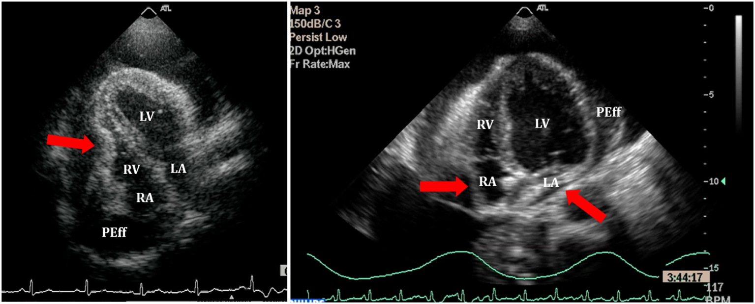
A
Recognize at the bedside
Beck + pulsus- Beck triad hypotension + ↑ JVP + muffled heart sounds (all three in only a minority — don't wait for it).
- Pulsus paradoxus inspiratory SBP fall >10 mmHg, tachycardia, narrow pulse pressure, dyspnea.
- JVP elevated with blunted Y descent (vs constriction's prominent Y).
- ECG sinus tach, low voltage, electrical alternans (swinging heart).
B
Echo & cath signs
sensitive → specific| Sign | Meaning |
|---|---|
| RA systolic collapse | Interpret with ECG timing. Collapse lasting >⅓ of the entire cardiac cycle is more specific, not one third of systole. |
| RV diastolic collapse | Supports tamponade. May be absent with pulmonary hypertension/high RV pressure or localized postoperative compression. |
| IVC plethora | Dilated IVC, <50% inspiratory collapse — sensitive, ↑ filling pressures. |
| Respirophasic inflow | Spontaneous breathing: mitral E falls and tricuspid E rises. Published thresholds are >25–30% and >40–60%. State denominator and respiratory phase. Ventilation, AF and lung disease confound. |
| RHC | Equalized diastolic pressures (RA ≈ RVEDP ≈ PADP ≈ PCWP), blunted Y, intrapericardial P tracks RA. |
C
The effusion itself — size & sampling
tap to diagnose?| Echo size (diastole) | Approach |
|---|---|
| Small <10 mm | Observe. Treat the cause. |
| Moderate 10–20 mm | Investigate cause. Follow with serial echo. |
| Large >20 mm | Drain for tamponade, significant symptoms or diagnostic need (bacterial/TB/neoplastic suspicion). Persistence alone is not an indication. Stable chronic idiopathic noninflammatory effusions may be observed. |
- Noninflammatory causes HF, renal/thyroid disease, low oncotic pressure. Fluid chemistry alone does not assign cause.
- Inflammatory / malignant integrate fluid studies with imaging and clinical evidence. Do not use pleural Light criteria.
- Hemopericardium dissection, trauma, post-procedure, free-wall rupture, OAC.
- Tap to Dx suspected purulent / TB / neoplastic, not for asymptomatic small.
D
Manage
drain itHemodynamically significant tamponade requires urgent drainage (percutaneous or surgical, selected by cause, anatomy & expertise). A cautious fluid bolus + norepinephrine may bridge selected hypotensive patients but must not delay drainage. Positive-pressure ventilation can sharply reduce venous return. If unavoidable, use a controlled plan with an experienced resuscitation/procedural team. Type A dissection or post-MI free-wall rupture generally requires immediate surgery. Controlled drainage is a specialist bridge, not routine centesis.
Constrictive pericarditis — a rigid, scarred (± calcified) pericardium fixes total cardiac volume, dissociates intrathoracic from intracardiac pressures, and drives ventricular interdependence. Hallmarks: rapid early-diastolic filling then an abrupt halt (dip-and-plateau / "square-root sign"), Kussmaul sign, and a pericardial knock. Separate it from restrictive cardiomyopathy: inflammatory/transient constriction may improve medically. Chronic permanent constriction may require pericardiectomy.
▤
The dip-and-plateau ("square-root sign")
ventricular pressureThe respiratory loop shows septal interaction inside a constrained envelope. The pressure loop shows early filling followed by an abrupt limit. Neither sign alone separates constriction from its mimics.
A
Constriction vs restriction
curable vs myocardial| Discriminator | Constriction (pericardial) | Restriction (myocardial) |
|---|---|---|
| Interdependence | Present — RV/LV systolic pressures discordant with respiration | Absent — concordant |
| Mitral e′ (tissue Dop.) | Medial e′ often ≥9 cm/s. Medial > lateral e′ = annulus reversus | Usually reduced. Mixed myocardial/pericardial disease can blur the distinction |
| Septal bounce | Respiratory septal shift supports constriction | Usually absent. Postoperative bounce alone is nonspecific |
| Pericardium | May be thickened/calcified, but normal thickness does not exclude constriction | Usually normal thickness |
| BNP | Often lower than in restriction | Often higher. Overlap and kidney function limit diagnostic use |
| Hepatic vein | Expiratory diastolic flow reversal | Often more inspiratory reversal. Interpret the full Doppler pattern |
Exam & Rx Kussmaul sign (JVP fails to fall / rises on inspiration), pericardial knock (early diastolic, after S2), prominent Y descent. Causes: post-viral/idiopathic, post-cardiac surgery, mediastinal radiation, TB. Persistent symptomatic constriction may need pericardiectomy. Inflammatory/transient disease may improve with 3–6 months of therapy. Annulus paradoxus means E/e′ loses its usual relation to filling pressure.
B
Pericardiocentesis
echo-guided| Step | Detail |
|---|---|
| Indications | Tamponade (emergent), large symptomatic effusion, diagnostic sampling (purulent / neoplastic / TB suspected). |
| Approach | Image-guided drainage by a trained team — choose the safest window from real-time effusion distribution + adjacent structures. A landmark-only needle instruction is not a substitute for imaging and local procedural protocol. |
| Confirm | Agitated-saline contrast → bubbles in pericardial space (not RV). Leave a pigtail drain. |
| Drainage | Avoid abrupt excessive decompression (usually <500 mL initially when feasible). Leave a drain for controlled completion and monitor for decompression syndrome. |
| Complications | RV/cardiac puncture, coronary / internal-mammary laceration, pneumothorax, arrhythmia, hepatic injury, vagal. |
| Choose surgery | Dissection, free-wall rupture, purulence, or inaccessible/loculated clot may require surgery. Controlled rescue drainage can bridge selected collapsing patients. Correct coagulopathy when time permits. |
C
Send the fluid · effusive-constrictive
what to orderPericardial fluid panel
- Core cell count + diff, glucose, protein/LDH, Gram stain + bacterial/fungal culture if infection suspected, cytology if cancer suspected.
- TB risk AFB smear/culture + PCR, ADA or interferon-γ using validated diagnostic guidance.
- Targeted hematocrit (hemo), triglycerides (chylo). Tumor markers do not confirm malignant disease.
- Do not apply pleural Light criteria to pericardial fluid — frequent false exudate classification.
Effusive-constrictive
- Persistent constrictive physiology after drainage. The invasive definition is failure of RA pressure to fall by ≥50% or below 10 mmHg after intrapericardial pressure normalizes.
- Exclude RV failure and severe TR as causes of persistent RA pressure. Treat active inflammation, reserve surgery for persistent constriction.
Integrate the syndrome: pericarditis requires compatible symptoms and objective evidence. Tamponade depends on pressure, rate and distribution, not size alone. Constriction requires multiple findings and an assessment of reversibility before surgery.
Cancer-associated ≠ malignant. An effusion in a patient with cancer remains etiology undetermined until the evidence supports tumor involvement, therapy toxicity, infection, or a non-cancer cause. Cancer therapy accounts for <30% of effusions in oncology cohorts. Name the syndrome, measure hemodynamic impact, then decide whether surveillance or tissue diagnosis will change care.
A
Name it correctly
association → suspicion → proofCancer-associated
- Any effusion in a patient with active/prior cancer. Cause not yet established.
- Common alternatives: renal/thyroid/HF, infection, autoimmune, post-procedure.
Suspected neoplastic
- Active metastatic/high-burden cancer + growth/recurrence or pericardial/mediastinal nodularity, mass, lymphatic obstruction.
- Imaging raises probability but may not prove malignant infiltration.
Confirmed malignant
- Positive pericardial-fluid cytology or pericardial/epicardial biopsy.
- Negative cytology does not fully exclude involvement if suspicion remains high.
B
Triage, phenotype, find the cause
same-day questions| Step | Minimum |
|---|---|
| Hemodynamics | Symptoms, BP/HR, JVP, pulsus. Urgent TTE if dyspnea, presyncope/syncope, hypotension, tachycardia or rapid interval growth. |
| TTE phenotype | Largest end-diastolic separation, circumferential vs loculated, RA/RV collapse, IVC, respiratory inflow, RV/LV fxn. Compare by the same modality when possible. |
| Cause map | Cancer trajectory + therapy timing. CT/CMR for nodules/mass, thickening, inflammation, loculation, pleural/mediastinal disease. CBC, renal/liver panel, CRP/ESR, TSH. hsTnI if chest pain/ECG change or myopericarditis concern. |
C
Observe vs sample vs drain
ESC cardio-oncology| Presentation | Action |
|---|---|
| Stable >4 to <20 mm | Repeat TTE in 7–14 d, then every 4–6 wk while evolving. Shorten for symptoms, growth or high-risk anatomy. |
| Moderate–large, uncertain | Consider diagnostic pericardiocentesis when multimodality imaging cannot establish cause and the result will change cancer therapy or procedural strategy. |
| Tamponade / unstable | Immediate image-guided pericardiocentesis. Surgery if percutaneous access is not feasible, bleeding/purulence requires operation, or anatomy is loculated. |
| If neoplastic drained | Leave extended drainage for 3–6 d to reduce recurrence. Send cytology. Remove when clinically stable and drainage is low (often <30 mL/d under a validated drainage plan). |
| Recurrent / refractory | Pleuro-pericardial window for relapsing effusion. Systemic antineoplastic therapy is first-line when malignancy is confirmed. Intrapericardial therapy or biopsy is selected with oncology/CT surgery when diagnosis or control remains unresolved. |
D
Therapy-associated disease — do not over-attribute
timing + inflammation + culprit- Known exposures chest RT, anthracycline, bleomycin, cyclophosphamide, cytarabine, ATRA/arsenic, dasatinib, IL-2/interferon, ICI.
- Isolated effusion no pain/ECG/CRP inflammation → do not empirically give NSAID/colchicine. Treat the cause. Do not reflexively stop an unrelated cancer therapy.
- ICI pericarditis severe disease or moderate/large effusion → hold ICI + high-dose steroid ± colchicine. Drain tamponade. Uncomplicated disease may use NSAID/colchicine with MDT-guided continuation/rechallenge.
- Anticoagulation reassess indication, platelet count, bleeding and procedure plan. OAC is a risk feature, not an automatic stop.
E
Decision traps
avoid the wrong label / wrong delay- Hemorrhagic ≠ malignant blood raises concern but does not replace cytology/biopsy.
- CT ≠ TTE ruler do not call growth from cross-modality size labels alone.
- Negative cytology does not close the case when imaging/suspicion remains high and tissue would change care.
- Biomarkers CRP/hsTnI phenotype inflammation or myocardial overlap. They do not diagnose malignant fluid.
- Tamponade first do not delay drainage for staging or perfect diagnostic imaging.
- After drainage use the P.3 fluid panel. Alert cytopathology to the known cancer + send a cell block when feasible.
Cardio-onc one-liner: stable >4 to <20 mm → TTE 7–14 d then q4–6 wk. Tamponade → urgent image-guided drainage. Suspected/definite neoplastic drainage → cytology + drain 3–6 d. Call it malignant only after tissue/fluid proof. If sampling will not change care, surveillance may be the better procedure.