Probability before testing In a stable patient, estimate pretest probability before ordering D-dimer or imaging. In shock or severe hypoxemia, resuscitate and pursue immediate definitive or bedside-supported diagnosis. A normal echocardiogram or negative POCUS finding does not exclude PE.
A
Stable patient: choose one validated pathway
do not mix thresholdsVery low · gestalt <15%PERC only if all 8 criteria are negative → no D-dimer or imaging.
Low / intermediateUse Wells/Geneva or another validated diagnostic pathway → D-dimer.
D-dimer positiveProceed to CTPA. Use V/Q SPECT when CTPA is unsuitable.
High probabilitySkip D-dimer → imaging. If suspected C2+ and imaging is delayed/unavailable, consider empiric AC if bleeding risk is low.
B
PERC · YEARS · age-adjusted D-dimer
FEU unitsPERC — all must be absent
- Age ≥50 years
- HR ≥100/min
- SaO₂ <95%
- Unilateral leg swelling
- Hemoptysis
- Prior VTE
- Estrogen use
- Surgery/trauma requiring hospitalization within 4 weeks
Only for very-low-probability ED patients. PERC-positive means “test further,” not “PE present.”
YEARS — 3 items
- Clinical signs of DVT · hemoptysis · PE judged most likely diagnosis.
- 0 items: PE excluded if D-dimer <1,000 µg/L FEU.
- ≥1 item: PE excluded if D-dimer <500 µg/L FEU.
Alternative validated strategy: age >50 years → cutoff age×10 µg/L FEU. Age ≤50 → 500 µg/L FEU.
UNITS
These thresholds are for fibrinogen-equivalent units (FEU). D-dimer units (DDU) are different. Confirm the assay/reporting unit. Do not use D-dimer to overrule high probability or after therapeutic anticoagulation has already reduced sensitivity.
C
Imaging and bedside interpretation
anatomy + RV physiology| Test | Use | Do not overinterpret |
|---|---|---|
| CTPA | Preferred noninvasive test. Assess clot location and alternate diagnoses. Report numeric CT RV/LV ratio. | Technical adequacy matters. A “saddle” location alone does not define severity. |
| V/Q SPECT | Preferred over planar V/Q when CTPA cannot be performed and SPECT is available. | Interpret with chest imaging and clinical probability. |
| Echo / POCUS | In instability, RV pressure overload or clot-in-transit can support urgent action. After diagnosis, defines risk. | McConnell/60-60/RV dilation are not sufficiently specific to confirm PE and normal findings do not exclude it. |
| Leg CUS | Proximal DVT establishes VTE and may avoid chest imaging when chest imaging is unsafe or pregnancy DVT symptoms are present. | A negative leg study does not exclude PE. |
D
Pregnancy-adapted diagnosis
avoid both missed PE and reflex imagingUse pregnancy-adapted YEARS
Same 3 YEARS items. If no items, D-dimer <1,000 µg/L FEU excludes. If ≥1, <500 excludes. Perform CUS first when DVT symptoms are present.
If chest imaging remains necessary
Discuss radiation/contrast, local expertise, and availability. The 2026 guideline considers a low-radiation CTPA strategy reasonable over low-dose perfusion imaging when the chest radiograph is normal.
Assign the most severe feature A–E integrates symptoms, validated clinical severity, RV imaging, cardiac biomarkers, perfusion, blood pressure, and respiratory failure. Add R when respiratory compromise crosses the thresholds below. Category is dynamic—document trajectory and reclassify after any change.
A
AHA/ACC acute PE clinical categories
A lowest → E highest| Category | Definition | Operational response |
|---|---|---|
| A | Asymptomatic, incidentally detected PE. | Assess location, DVT, recurrence, bleeding, cancer, and surveillance eligibility. |
| B1 / B2 | Symptomatic, low clinical severity: PESI ≤85, sPESI 0, or Bova ≤4. B1 subsegmental. B2 segmental or proximal. | Outpatient/early discharge may be safe only after structured suitability review. |
| C1 | Elevated clinical-severity score. RV imaging and cardiac biomarkers normal. | Hospital care. Reassess physiology and trajectory. |
| C2 | Abnormal RV imaging or ≥1 abnormal cardiac biomarker. | Monitored admission. PERT assessment and explicit rescue triggers. |
| C3 | Abnormal RV imaging and ≥1 abnormal cardiac biomarker. | Higher-acuity monitoring. PERT. Consider transfer while stable. |
| D1 / D2 | D1 transient hypotension (SBP <90 or fall >40 mm Hg for <15 min, or responsive to fluids). D2 hypoperfusion despite preserved SBP. | ICU/PERT. Advanced therapy may be considered. |
| E1 / E2 | E1 recurrent/persistent hypotension with cardiogenic shock. E2 refractory shock or cardiac arrest. | Immediate resuscitation and reperfusion/MCS decision. Stabilize before transfer. |
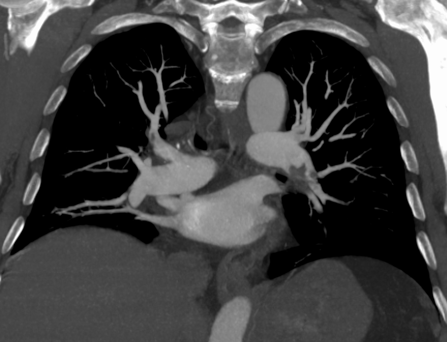
This coronal image shows a left pulmonary arterial filling defect. Clot location establishes anatomy. Assess RV size separately and combine it with perfusion, biomarkers, and respiratory status to judge severity. This view alone does not provide an axial RV/LV measurement.
James Heilman, MD, PELeft.png, CC BY-SA 4.0. Unmodified.

B
Make C–E executable
RV · biomarkers · perfusion · oxygenRV / biomarkers
CT RV/LV ≥1.0 is prognostic. Echo: RV/LV, size, TAPSE, S′, septal motion, RVSP, IVC. Troponin and BNP/NT-proBNP indicate RV stress/injury.
Hypoperfusion (D2)
Look for lactate >2 mmol/L, acute kidney injury, oliguria, altered mentation, MAP <60 mm Hg, or cardiac index ≤2.2 L/min/m². Interpret in context and trend.
Respiratory modifier R
C-R: SaO₂ <90%, RR ≥30, or supplemental O₂. D-R: >6 L/min nasal cannula or nonrebreather. E-R: hypoxemic or ventilatory failure, including NIV/invasive ventilation.
C–E
Measure lactate, trend BP/perfusion/oxygen requirement, and obtain serial clinical reassessment. Activate PERT for all C–E. A stable C3–D patient may benefit from transfer to an advanced center. An unstable E patient should be stabilized before transport.
C
Outpatient gate for A/B
score + Hestia + real-world supportsPESI — 1 point each
- Age >80
- History of cancer
- Chronic heart/lung disease
- HR ≥110/min
- SBP <100 mm Hg
- SaO₂ <90%
0 = low predicted risk, but not automatic discharge. RV injury, hypoperfusion, or oxygen need requires clinical reassessment despite a low score.
Hestia exclusion — any “yes”
- Unstable
- Lysis/embolectomy needed
- Active/high bleeding risk
- O₂ >24 h to keep >90%
- PE on anticoagulation
- IV analgesia >24 h
- Medical/social admission need
- CrCl <30 mL/min
- Severe liver impairment
- Pregnancy
- History of HIT
Discharge only when the system is ready First dose and full medication access now. Hemodynamically/respiratorily stable. Pain controlled orally. Patient/caregiver understands bleeding and recurrence warnings. Reliable phone/transport/support. And clinical follow-up arranged within 1 week (in person or by telehealth).
D
Isolated subsegmental PE: surveillance is selective
B1 is not “no treatment”Before withholding anticoagulation
Confirm a true isolated SSPE with radiology. Exclude proximal DVT with bilateral CUS. Require minimal symptoms, good cardiopulmonary reserve, low recurrence/progression risk, and reliable structured follow-up.
Favor anticoagulation
Proximal DVT, active cancer, pregnancy, hospitalization/reduced mobility, no reversible provoking factor, marked symptoms, low reserve, multiple SSPE, or unreliable follow-up. If observing, use serial CUS and explicit return precautions.
Start promptly when indicated Before the first dose: CBC/platelets, Cockcroft–Gault CrCl, hepatic function, PT/INR/aPTT, pregnancy status, active bleeding, prior HIT, interacting drugs, absorption/food issues, and likely procedure or reperfusion. Confirm actual body weight.
A
Agent-selection gates
one question at a timePregnant / breastfeedingLMWH preferred in pregnancy. UFH for selected periprocedural/renal situations. No DOAC in pregnancy or breastfeeding.
Thrombotic APSWarfarin generally preferred over a DOAC. Involve hematology, especially with triple positivity or arterial events.
Need parenteral therapy · C1–E1LMWH is preferred over UFH, including after thrombolysis/endovascular therapy. Reserve UFH for a specific renal/procedural/ECMO/local-protocol reason.
Eligible, stable, reliable oral routeDOAC over VKA. Apixaban/rivaroxaban need no heparin lead-in. Dabigatran/edoxaban require 5–10 days parenteral treatment.
B
Acute and maintenance doses
adult VTE — not AF dosing| Agent | Treatment dose | Operational notes |
|---|---|---|
| Apixaban | 10 mg PO BID ×7 d → 5 mg BID | No parenteral lead-in. AF age/weight/creatinine dose-reduction criteria do not apply to acute VTE. |
| Rivaroxaban | 15 mg PO BID ×21 d → 20 mg daily | No lead-in. Take 15- and 20-mg doses with food. Avoid CrCl <15 mL/min. Data are limited at CrCl 15–29. |
| Edoxaban | After 5–10 d parenteral: 60 mg PO daily | 30 mg daily if CrCl 15–50 mL/min, weight ≤60 kg, or specified P-gp inhibitor. Avoid CrCl <15. |
| Dabigatran | After 5–10 d parenteral: 150 mg PO BID | VTE treatment requires CrCl >30 mL/min. Swallow capsule whole. |
| Enoxaparin | 1 mg/kg SC q12h | CrCl <30: labeling uses 1 mg/kg q24h. Pharmacy/hematology input and anti-Xa strategy when accumulation/extremes matter. |
| Fondaparinux | <50 kg 5 mg · 50–100 kg 7.5 mg · >100 kg 10 mg SC daily | Avoid CrCl <30. No routine laboratory titration. |
| UFH | Weight-based IV bolus/infusion per institutional anti-Xa or aPTT nomogram | Short half-life/reversible, but less predictable and higher HIT burden than LMWH. Do not default to UFH solely because PE is severe. |
| Warfarin | Overlap therapeutic parenteral AC for ≥5 d and until INR ≥2 for ≥24 h | Target INR 2–3 for most VTE. Account for diet, interactions, and frequent early INR checks. |
C
Transitions and failure-prevention checks
most “breakthrough” is explainableParenteral → oral
Start apixaban/rivaroxaban when the next parenteral dose is due or infusion stops. Count the full lead-in regimen. Do not abbreviate the loading phase without a deliberate expert plan.
Interactions / absorption
Review strong P-gp/CYP3A modifiers, bariatric or malabsorptive surgery, enteral-tube site, vomiting, adherence, and rivaroxaban food requirements.
Renal / hepatic trajectory
Recalculate CrCl when kidney function changes. Recheck liver function and CBC. A dose that was safe on admission may not remain safe.
D
Initial treatment guardrails
before “send”- Imaging delayed: empiric therapeutic AC is reasonable for suspected C2 or higher when bleeding risk is low and definitive imaging is delayed/unavailable.
- After lysis/procedure: use the PERT/procedural hemostasis plan. The 2026 guideline favors LMWH over UFH in C1–E1 once parenteral therapy is appropriate.
- Possible HIT: calculate 4Ts. Stop all heparin if intermediate/high probability and begin an appropriate non-heparin anticoagulant while testing.
- Medication reconciliation: record agent, indication, phase, dose, last dose, renal function, weight, planned duration, and prescriber/follow-up owner.
VERIFY
Doses above are adult reference doses, not a substitute for the current FDA label, pharmacy review, renal/hepatic restrictions, neuraxial/procedure timing, or a validated anticoagulation pathway.
Reperfusion is a physiology decision Organize stabilization and definitive reperfusion in parallel for D/E. Do not perform routine advanced therapy for stable A–C2. The guideline leaves C3 selection uncertain. Later HI-PEITHO supports a selected distressed intermediate-risk population, not routine intervention for all C3. Define rescue triggers with PERT.
A
Category-to-modality matrix
2026 guideline direction| Category | Systemic thrombolysis | Catheter lysis / thrombectomy | Surgical embolectomy | VA-ECMO |
|---|---|---|---|---|
| A–C1 | Do not use | No routine benefit | No benefit | Not indicated |
| C2 | Harm outweighs routine use | Benefit uncertain | No benefit | Not indicated |
| C3 | Uncertain. No routine use | Uncertain. PERT trial/selected rescue strategy | No routine benefit | Not indicated |
| D1–D2 | May be considered | May be considered | May be considered | Prepare if trajectory worsens |
| E1 | Reasonable | Reasonable | Reasonable | Consider if shock persists |
| E2 | Reasonable during refractory shock/arrest | Usually not feasible during arrest | No routine benefit during arrest | Reasonable bridge in capable center |
Right-heart clot-in-transit: for C3–E2, an advanced strategy rather than anticoagulation alone is reasonable. Choice depends on mobility/location, PFO, shock, bleeding risk, and immediately available expertise.
B
Systemic thrombolysis
screen before drug leaves pharmacyReference regimen
Alteplase 100 mg IV over 2 hours. Coordinate anticoagulant interruption/restart with PERT, pharmacy, and the active protocol. Accelerated or lower-dose regimens are off-label/center-specific—do not describe 0.6 mg/kg as a bolus.
Before and during
Document neurologic baseline, last anticoagulant/antiplatelet dose, CBC/platelets, PT/INR/aPTT, fibrinogen if available, BP, recent procedures/trauma, pregnancy/postpartum status, and a hemorrhage rescue plan.
| Absolute | Do not give routinely | Relative | Risk–benefit / PERT decision |
|---|---|---|---|
| Intracranial / CNS | Prior hemorrhagic stroke or stroke of unknown origin. Ischemic stroke within 6 months. CNS neoplasm. | Recent neurologic | TIA within 6 months. |
| Bleeding / trauma | Active bleeding. Bleeding diathesis. Major trauma, surgery, or head injury within 3 weeks. | Bleeding-prone | Oral anticoagulation, noncompressible puncture, traumatic CPR, active peptic ulcer. |
| — | In arrest or otherwise near-certain death, contraindications must be weighed against immediate mortality. | Systemic | Pregnancy/first postpartum week. Refractory SBP >180. Advanced liver disease. Infective endocarditis. |
C
Catheter, surgery, ECMO: choose the constraint
not one universal hierarchyCatheter-directed
Local lysis or mechanical thrombectomy when rapid debulking is desired and anatomy/access/bleeding risk fit. Requires an experienced team and a plan for vascular/hemorrhagic complications.
Surgical embolectomy
Consider E1 or selected D when lysis is contraindicated/failed, clot is surgically favorable, or intracardiac thrombus/PFO/other surgical target changes the balance.
VA-ECMO
For E2 refractory shock/arrest in a capable system. It is a bridge to RV recovery or definitive strategy—not proof that every patient needs an additional clot-removal procedure.
D
PERT activation packet and evidence guardrails
one call · complete data- State: A–E/R category and trend. BP/pressors. Oxygen/ventilation. Lactate/urine/mental status.
- Show: CTPA clot/anatomy, CT RV/LV. Echo RV/LV/TAPSE/septum. Troponin/BNP. Clot-in-transit/PFO.
- Safety: last AC/antiplatelet, Hgb/platelets/coagulation, stroke/bleed/surgery history, access, pregnancy, renal function.
- PEITHO: systemic lysis reduced deterioration but increased major bleeding and hemorrhagic stroke.
- PEERLESS: thrombectomy versus catheter lysis, with benefit driven by deterioration/bailout and ICU use. It does not establish benefit over anticoagulation.
2026 randomized evidence
HI-PEITHO (n=544): RV/LV ≥1.0 + troponin elevation + ≥2 distress signs (SBP ≤110, HR ≥100, RR >20). Ultrasound-facilitated catheter lysis plus AC versus AC alone reduced the 7-day composite of PE death, decompensation/collapse, or symptomatic recurrence: 4.0% vs 10.3%, driven by decompensation. Major bleeding was 4.1% vs 2.2% (P=0.32). This does not prove a mortality benefit or equivalent bleeding risk.
STORM-PE (n=100): vacuum thrombectomy improved 48-hour RV/LV reduction versus AC, a surrogate endpoint. The October 90-day report found longer walk distance, but only 65/100 completed testing. Clinical survival benefit remains unproven.
Support systemic pressure and oxygenation while arranging reperfusion. Excess fluid, hypoxemia, acidosis, and high intrathoracic pressure can worsen RV function. A preserved SBP can coexist with hypoperfusion.
Original mechanism schematic. Highlights show causal order, not measured timing or inevitable progression.Static sequence
A
Run two rails in parallel
minutes matterRail 1 · Stabilize oxygenation and perfusion
- Continuous ECG/SpO₂. Arterial line when feasible. Large-bore IV access. Trend lactate, urine, mentation, renal/hepatic function.
- HFNC for significant hypoxemia. Treat fever, pain, agitation, acidosis, and arrhythmia without causing hypotension.
- If hypotensive, start norepinephrine. Add a second vasopressor rather than unlimited catecholamine escalation when needed.
- Give only a small fluid challenge (≤500 mL) when low preload is likely. Stop if congestion or no response. Diurese cautiously when overloaded.
Rail 2 · Remove obstruction / build rescue
- Confirm current D/E category, bleeding contraindications, clot/anatomy, and fastest available reperfusion option.
- Mobilize systemic lysis, catheter, surgery, or E2 VA-ECMO pathway—do not wait for supportive measures to fail sequentially.
- For C3/D, define objective rescue triggers: falling BP/MAP, rising lactate/pressors/O₂, oliguria, altered mentation, worsening RV or arrest.
- Notify receiving OR/cath/ECMO, blood bank, pharmacy, anesthesia, perfusion, and transport with a single handoff owner.
B
Hemodynamic tools
bridge, then reassess| Tool | When useful | Guardrail |
|---|---|---|
| Norepinephrine | Preferred first vasopressor for systemic hypotension. Restores RV coronary perfusion pressure. | Titrate to perfusion, not a number alone. Excessive dose can increase pulmonary vascular load. |
| Dobutamine | Adjunct for low cardiac output when systemic pressure is supported. Typical ceiling 10 µg/kg/min. | Can lower SVR and provoke tachyarrhythmia. Do not use as sole therapy in hypotension. |
| Inhaled pulmonary vasodilator | Inhaled nitric oxide or epoprostenol may transiently reduce RV afterload in severe failure. | Bridge only. No proven mortality benefit. Prevent interruption/rebound and monitor oxygenation/BP. |
| Volume / diuresis | Small bolus only for likely low preload. Diuresis if venous congestion is impairing RV mechanics/organ function. | Avoid reflex liters. Reassess BP, JVP/echo, oxygenation, urine, and lactate after every intervention. |
C
Airway is a hemodynamic procedure
avoid intubation when safeBefore induction
- Senior airway + PERT/critical care present.
- Preoxygenate. Vasopressor running. Rescue boluses and arterial monitoring ready.
- Reperfusion/VA-ECMO plan active for D/E. Anticipate peri-intubation arrest.
After the tube
- Minimize apnea, hypotension, mean airway pressure, and excessive PEEP.
- Use lung-protective ventilation. Avoid severe hypercapnia/acidemia.
- Immediately reassess BP, ETCO₂, SpO₂, RV/perfusion, and pressor need.
AVOID
Deep sedation, NIV, and invasive positive-pressure ventilation can precipitate collapse in C–E. Use only when required for gas exchange/work of breathing and with full hemodynamic preparation.
D
E2 arrest or refractory shock
activate the whole systemRecognizePE known or strongly suspected. Pulseless electrical activity is common. Continue high-quality CPR and identify hemorrhage/alternate causes.
ReperfuseSystemic thrombolysis is reasonable when PE is the arrest mechanism and no faster definitive option exists. Continue prolonged resuscitation as clinically appropriate.
BridgeVA-ECMO is reasonable for refractory shock/arrest in a capable center. Initiate systemic parenteral AC if bleeding does not preclude it.
ReassessOn ECMO, decide recovery versus additional clot therapy individually. Evidence does not mandate routine thrombectomy/embolectomy.
DVT treatment is not one-size-fits-all First identify limb threat, proximal versus distal location, upper-extremity/catheter involvement, provoking factors, and PE symptoms. Anticoagulant choice and dosing generally follow the PE/VTE regimen on Page 3.
A
Suspected lower-extremity DVT
2-level Wells → D-dimer / ultrasoundWells DVT score
- Active cancer +1
- Paralysis/cast +1
- Bed ≥3 d or surgery <12 wk +1
- Deep-vein tenderness +1
- Entire leg swollen +1
- Calf ≥3 cm larger +1
- Pitting edema +1
- Collateral superficial veins +1
- Prior DVT +1
- Alternative diagnosis as likely −2
≥2 likely · ≤1 unlikely. Adjust for the validated diagnostic workflow and special populations.
Test sequence
- Unlikely: high-sensitivity D-dimer. Negative excludes, positive → proximal or whole-leg CUS.
- Likely: CUS first. If initial proximal CUS is negative but suspicion/D-dimer remains positive, repeat proximal CUS in about 1 week or obtain whole-leg imaging.
- Positive: document proximal/distal extent and symptoms of PE. Do not use D-dimer to monitor treatment.
- Discordant: consider iliac/pelvic imaging when swelling is extensive but standard CUS is negative.
B
Treat by VTE phenotype
location changes urgency and duration| Phenotype | Initial plan | Key nuance |
|---|---|---|
| Proximal leg DVT | Therapeutic anticoagulation unless contraindicated. Minimum treatment phase usually 3–6 months. | Assess PE symptoms, limb threat, cancer/pregnancy, recurrence, bleeding, and need for extended therapy. |
| Isolated distal DVT | Severe symptoms or extension risk → anticoagulate 3 months. Low risk → serial CUS, typically weekly for 2 weeks. | Anticoagulate if extension, proximal propagation, or surveillance cannot be completed reliably. |
| Upper-extremity DVT | Anticoagulate axillary or more proximal DVT for at least 3 months. | Assess catheter, thoracic outlet, cancer, device infection, and PE symptoms. |
| Catheter-associated | Anticoagulate. Catheter may remain if needed, functioning, positioned, and uninfected. | Remove if unnecessary, infected, malfunctioning, or symptoms progress. Treat at least 3 months and often while catheter remains. |
| Upper saphenous SVT >3 cm from SFJ ≥5 cm long | Fondaparinux 2.5 mg SC daily ×45 d. Rivaroxaban 10 mg daily ×45 d is an alternative. | ≤3 cm from the saphenofemoral junction (SFJ) → full anticoagulation for ≥6 weeks. Specialist review may extend to 3 months for anatomy or persistent risk. |
| Unusual-site VTE | Cerebral/splanchnic/renal/ovarian thrombosis requires site-specific specialty review. | Confirm acuity and provoking disease. Bleeding, recanalization benefit, and duration differ by site. |
C
Limb threat: phlegmasia
vascular emergencyAct now
RecognizeMassive swelling, cyanosis, severe pain, sensory/motor deficit, weak pulses, shock.
ImmediateElevate limb, therapeutic parenteral anticoagulation if possible, analgesia, IV access, labs/type-cross.
EscalateUrgent vascular/IR consultation for thrombus removal. Evaluate compartment syndrome and venous gangrene.
Avoid delayDo not wait for routine clinic follow-up or serial ultrasound when the limb is threatened.
D
Structured surveillance pathways
withholding AC requires more follow-up, not lessDistal DVT surveillance
Document why extension risk is low, schedule serial CUS before discharge, and give return precautions for proximal swelling, chest pain, dyspnea, syncope, or bleeding. Start AC if extension occurs.
Isolated SSPE surveillance
Radiology confirmation + bilateral CUS + low recurrence risk + adequate reserve + minimal symptoms + reliable serial follow-up. Cancer, pregnancy, immobility, no reversible trigger, low reserve, or DVT favors treatment.
Make two decisions, not one Most treated PE/proximal DVT receives a 3–6 month primary-treatment phase. Before it ends, separately decide whether to stop or continue extended therapy by recurrence risk, bleeding risk, preference, drug burden, and whether the provoking factor truly resolved.
A
Phases of therapy
document the calendar date0–21 d
Initial phaseagent-specific lead-in
3–6 mo
Primary treatmentminimum therapeutic course
Beyond
Extended preventionfull or reduced dose
Every visit
Reassessbleeding · renal · preference
B
Duration by provoking context
risk-factor taxonomy matters| Context | Examples | After 3–6 months |
|---|---|---|
| Major reversible | Surgery with GA ≥30 min. Hospitalization with bed confinement ≥72 h. Lower-limb fracture. Cesarean delivery. | Stop is usually appropriate when the factor has resolved and the course is complete. |
| Minor reversible | Shorter surgery/hospitalization, out-of-hospital bed rest ≥72 h, estrogen exposure, peripartum state, trauma with reduced mobility. | Shared decision: recurrence reduction versus bleeding/burden. Confirm the factor is gone. |
| Persistent | Active cancer, chronic inflammatory/autoimmune disease, IBD, chronic immobility, persistent thrombophilia with management implications. | Extended therapy is generally favored while risk persists and bleeding risk is acceptable. |
| No major reversible factor | No clear trigger, or trigger too weak to explain the event. | Extended therapy is generally beneficial. Revisit risk and patient preference periodically. |
| Recurrent VTE | Especially recurrent unprovoked/persistent-risk events. | Usually indefinite treatment unless bleeding risk or goals of care outweigh benefit. |
C
Extended-dose options after ≥6 months
only after full primary treatmentApixaban
2.5 mg PO BID is an evidence-based extended-prevention dose after ≥6 months. Use full dose if the clinical situation requires therapeutic intensity.
Rivaroxaban
10 mg PO daily is an evidence-based extended-prevention dose after ≥6 months. 20 mg daily may be selected for higher recurrence risk.
Before reducing
Check APS, breakthrough VTE, adherence, organ function, and interactions. Active cancer alone does not exclude dose reduction after ≥6 months. See API-CAT on Page 8.
ASPIRIN
Aspirin is less effective than anticoagulation for secondary VTE prevention. Use it only when anticoagulation is stopped and the separate cardiovascular/recurrence context supports it—not as an equivalent substitute.
D
Do not let tests drive unnecessary treatment
test only if it changes a decision- Thrombophilia: do not interrupt required AC solely for testing. Consider selective testing with hematology/lab input when age, family history, unusual site, or the stop/continue decision could change.
- Assay interference: acute thrombosis, pregnancy, heparin, DOACs, and warfarin alter several tests. Plan timing before ordering.
- Cancer: use history, examination, basic labs, and age-appropriate screening. Do not order routine CT/PET solely for an otherwise unexplained VTE.
- Residual clot / D-dimer: do not use one residual ultrasound defect or D-dimer result as an automatic stop/continue command. Integrate the whole recurrence/bleeding profile.
Use the same VTE principles, then add the constraint Confirm the indication and treatment phase first. Next account for pregnancy/lactation, malignancy and drug interactions, APS, kidney/liver function, body size, platelet count, intracranial lesions, menstrual bleeding, procedures, and patient goals.
A
Pregnancy, postpartum, breastfeeding
coordinate hematology + MFM + anesthesiaDuring pregnancy
- LMWH preferred. UFH for selected severe renal or imminent-procedure situations.
- Avoid DOACs and warfarin for VTE treatment in pregnancy.
- Make a written delivery/neuraxial interruption and restart plan. Do not improvise at labor.
Postpartum / breastfeeding
- Treat pregnancy-associated VTE through at least 6 weeks postpartum and for a minimum total of 3 months.
- LMWH, UFH, and warfarin are compatible with breastfeeding. Avoid DOACs.
- Address contraception and future-pregnancy prophylaxis before discharge.
B
Cancer-associated thrombosis
clot risk and bleeding risk both change| Constraint | Practical approach |
|---|---|
| Agent | Apixaban, rivaroxaban, edoxaban, or LMWH are options. Choose by GI/GU lesion bleeding, tumor site, interactions, nausea/absorption, thrombocytopenia, renal function, procedures, cost, and preference. |
| Duration / extended dose | Continue while cancer or recurrence risk remains active. API-CAT: after ≥6 months without recurrence, apixaban 2.5 mg BID was noninferior to 5 mg BID for recurrence (2.1% vs 2.8%) and reduced clinically relevant bleeding (12.1% vs 15.6%) over 12 months. Individualize for bleeding, interactions, and breakthrough VTE. |
| Platelets / brain lesion | No universal threshold fits every patient. Coordinate oncology/hematology for dose modification, transfusion support, temporary hold, or LMWH strategy. Urgent review for intracranial symptoms. |
| Recurrence on LMWH | After confirming the event and adherence, specialists may increase LMWH above the therapeutic dose or continue the therapeutic dose. Evidence is very low certainty, so avoid an automatic fixed escalation. |
C
APS, kidney, liver, and body size
avoid false certaintyThrombotic APS
Warfarin is generally preferred over DOACs, particularly with triple-positive antibodies or arterial events. A single low-risk antibody is not the same as established APS. Confirm persistence and involve hematology.
Stage 4–5 CKD / ESRD
Evidence comparing apixaban and VKA is uncertain. Use Cockcroft–Gault CrCl, current labeling, dialysis status, trajectory, interaction/bleeding risk, and pharmacy/hematology input. Avoid a reflex “apixaban or UFH” answer.
Hepatic disease
DOAC use is often reasonable in Child–Pugh A, agent-specific/limited in B, and generally avoided in C. Check synthetic function, varices/bleeding, thrombocytopenia, and label restrictions.
Obesity / low weight
Standard-dose apixaban or rivaroxaban is reasonable for many patients with obesity. At >150 kg, extreme low weight, altered absorption, or uncertain renal estimates, individualize with pharmacy/hematology. Do not empirically underdose.
D
Bleeding-prone and preference-sensitive situations
solve the cause. Preserve protection- Abnormal uterine bleeding: ask proactively. Check CBC/ferritin, gynecologic causes, and contraception. Do not advise skipped doses or abrupt AC cessation. Coordinate hormonal/procedural options and agent change.
- Estrogen exposure: stopping/changing estrogen is a shared decision based on indication, alternatives, and whether therapeutic AC continues—not a one-line universal command.
- Falls / frailty: quantify actual bleeding risk, cognition, support, renal function, interacting drugs, and goals. Fall risk alone is not an automatic contraindication.
- Procedures / neuraxial: use a drug-, dose-, renal-, and bleeding-risk-specific interruption plan. Avoid routine bridging for DOACs. Identify who owns restart.
CONSULT
Pregnancy, APS, ESRD, severe liver disease, active cancer with bleeding, severe thrombocytopenia, intracranial tumor/bleed, HIT, and recurrent VTE deserve specialist and pharmacist review rather than a pocket-card-only decision.
Life-threatening bleed Stop anticoagulant, resuscitate, control the source, document drug/dose/last dose and renal function, send CBC/PT/INR/aPTT/fibrinogen/type-cross as appropriate, and activate pharmacy plus the relevant procedural/stroke/bleeding pathway. Reversal does not replace source control.
A
Reversal matrix
major / life-threatening bleeding| Drug | Preferred targeted action | Critical notes |
|---|---|---|
| Apixaban / rivaroxaban | 4-factor PCC under the major-bleeding pathway (off-label for Xa-inhibitor reversal). U.S. Andexxa sales ended December 2025 after FDA judged its risks to outweigh benefits. | Use agent, last dose, renal function and drug-calibrated testing when available. Normal PT/aPTT cannot reliably exclude Xa-inhibitor effect. Consult pharmacy. |
| Edoxaban / factor-Xa effect | 4-factor PCC per major-bleeding protocol. | PCC use for Xa-inhibitor reversal is off-label. Verify current jurisdictional guidance. Do not extrapolate an antidote indication across agents. |
| Dabigatran | Idarucizumab 5 g IV. | Consider drug level/thrombin-time context and dialysis when clinically relevant. Monitor for rebound in renal failure. |
| Warfarin | 4-factor PCC + vitamin K (up to 10 mg IV for life-threatening bleeding). | Recheck INR and clinical hemostasis. Avoid FFP when PCC is appropriate and available. |
| UFH / LMWH | Protamine. Calculate from heparin type, amount, and time since last dose. | UFH reversal is more complete. LMWH reversal is partial. Avoid empiric excess protamine. |
| Fondaparinux | No specific antidote. Consider PCC-based specialist pathway. | Long half-life and renal clearance matter. |
| Thrombolytic bleed | Stop lytic/AC. Activate stroke/massive-bleed pathway. Replace fibrinogen with cryoprecipitate when low. | Consider antifibrinolytic only for life-threatening bleeding with expert guidance. Repeat fibrinogen and imaging/source control. |
RESTART
Once hemostasis is secured, reassess thrombotic indication and restart timing every day. Recent PE/proximal DVT has high early recurrence risk. Use a multidisciplinary plan instead of leaving anticoagulation off indefinitely.
B
Suspected recurrent VTE on therapy
confirm before declaring failure1 · ConfirmObtain objective CTPA/V/Q or venous imaging and compare with baseline. Residual defects are common.
2 · ExplainCheck adherence, correct phase/dose, CrCl/weight, food/absorption, interactions, interruptions, cancer, APS, HIT, and catheter/anatomic driver.
3 · IntensifyReduced-dose DOAC → full treatment dose. Therapeutic-intensity recurrence → switch drug class. For cancer on LMWH, consider specialist-guided escalation.
4 · Re-planHematology/PERT input. Treat the underlying driver. Review duration, filter indication, and bleeding mitigation.
Do not “stack” anticoagulants or add antiplatelet therapy without a defined indication. A new symptom plus unchanged chronic imaging is not proven breakthrough thrombosis.
C
IVC filter lifecycle
indication → retrieval ownerPlace selectively
Acute proximal DVT/PE with an absolute contraindication to anticoagulation. Selected recurrent PE despite confirmed optimal AC may justify expert consideration.
Do not place routinely
A filter added to effective anticoagulation does not improve routine outcomes and introduces DVT, migration, fracture, penetration, and thrombosis risk.
Retrieve deliberately
Before placement, record filter type, indication, date, retrieval target, and named owner. Resume AC and retrieve as soon as bleeding risk falls/AC is tolerated.
D
HIT contingency
thrombosis + falling platelets- Calculate 4Ts. Low probability generally needs no HIT testing. Intermediate/high: stop UFH, LMWH, heparin flushes, and heparin-coated exposures.
- Send immunoassay ± functional confirmation without delaying a non-heparin anticoagulant when thrombosis risk is high.
- Select argatroban, bivalirudin, fondaparinux, or a DOAC by acuity, renal/hepatic function, procedure need, and current guidance. Avoid starting warfarin before platelet recovery.
- Avoid routine platelet transfusion unless bleeding/procedure demands it. Document HIT prominently for future care.
Every discharge needs a closed loop The patient should leave with medication in hand, exact dose/phase change date, planned minimum and likely total duration, bleeding/recurrence instructions, filter plan if present, and clinical follow-up within 1 week, in person or by telehealth.
A
Discharge checklist
read back · do not just printMedication
- Agent, indication, exact dose and time.
- Lead-in end date / next-dose transition.
- Food, missed dose, interactions, cost/refill access.
Safety
- Bleeding signs and when to call 911.
- No NSAID/antiplatelet addition without review.
- Procedure/dental/neuraxial contact plan. Medical alert/list.
VTE plan
- Provoking factors and duration decision date.
- Pending cancer/thrombophilia questions—not reflex panels.
- Filter retrieval, pregnancy/contraception, mobility/travel plan.
B
Follow-up timeline
symptoms + function + treatment| When | What to assess | Decision |
|---|---|---|
| Within 1 week | Dyspnea/chest pain/syncope, oxygen, bleeding, adherence, access/cost, renal function when unstable, pending tests, filter, pregnancy/cancer needs. | Confirm correct dose/phase and whether outpatient status remains safe. |
| By 3 months | Functional recovery, exercise tolerance, RV/oxygen course, recurrence/bleeding, provoking factor, medication burden and preference. | Set the stop/extend plan. Standard reduced-dose apixaban/rivaroxaban regimens start after ≥6 months. Investigate persistent symptoms. |
| Every visit ≥1 year | Ask directly about dyspnea, exercise limitation, chest symptoms, syncope, edema, cognition/mood, work and quality of life. | Do not normalize persistent limitation. Evaluate post-PE syndrome/CTEPD and rehabilitation needs. |
| Extended AC | CBC, renal/hepatic function, bleeding, interactions, adherence, falls/frailty, procedures, cancer status, goals. | Continue, reduce, switch, or stop through shared decision. Document next review date. |
C
Persistent dyspnea or exercise limitation after ≥3 months
CTEPD with or without PHScreenHistory/exam, ECG, CBC/chemistry, TTE + perfusion imaging (V/Q or V/Q SPECT). Echo alone cannot exclude CTEPD.
ClarifyConsider CPET. Evaluate lung disease, HF, anemia, deconditioning, ischemia, dysautonomia, and mood/sleep contributors.
ReferPersistent perfusion defect or PH/RV concern → expert CTEPD/PH center for CTPA/angiographic mapping and RHC when indicated.
TreatContinue AC during workup unless bleeding risk precludes. Expert center determines PEA, BPA, PH therapy, and rehabilitation pathway.
NO ROUTINE SCAN
Do not repeat CTPA, V/Q, echo, or leg ultrasound solely to prove clot resolution in an asymptomatic patient with low CTEPD suspicion. Image for symptoms, recurrence concern, or another defined question. Severe or progressive symptoms need earlier review, not a mandatory 3-month wait.
D
Recovery, travel, and return precautions
patient-facing essentials- Mobilize early once anticoagulated and stable. Prescribe graded activity/rehabilitation for persistent deconditioning. Compression can relieve selected DVT symptoms but is not routine prevention for every patient.
- Travel: for C2–E, avoid long-haul travel for about 4 weeks or until symptoms resolve. Prior VTE travelers on trips ≥5 h may use mobility/hydration and properly fitted compression.
- Call 911: new/worsening dyspnea, chest pain, syncope, hemoptysis, focal neurologic deficit, uncontrolled bleeding, black/bloody stool, or rapidly increasing painful/cyanotic limb swelling. Head injury on anticoagulation needs urgent medical assessment, even without visible bleeding.
- Call the anticoagulation team: missed/extra doses, pregnancy, new drug/supplement, planned procedure, persistent heavy menses, falls, or inability to obtain medication.
Evidence review: all 10 chapters read and source-checked 2026-10-17. The 2026 multisociety guideline, its July echo correction, current labels, HI-PEITHO, API-CAT, and October STORM-PE follow-up inform this educational update. Before use, verify anticoagulant nomograms, thrombolysis, reversal, PERT activation, transfer, ECMO, pregnancy, and procedure timing against current labels, guidelines, and a validated institutional pathway.