Start
Here
Here
PH is a hemodynamic findingmPAP >20 mmHg at rest by RHC. “PAH” is only Group 1 pre-capillary PH—not a synonym for PH.
Classify before treatingThe same pressure may arise from pulmonary arteriopathy, left-heart disease, lung disease, chronic thromboembolism, or mixed mechanisms.
V/Q + RHC are pivotalUse V/Q imaging to exclude chronic thromboembolic disease and RHC to confirm physiology before disease-specific therapy.
Therapy is indication-specificPAH drugs are standard for confirmed arterial Group 1 phenotypes. PVOD/PCH is an exception: therapy can cause edema and needs cautious expert use. Selected therapies also apply to CTEPH and PH-ILD.
A
Hemodynamic definitions
Resting RHC · interpret PAWP in contextPulmonary hypertension
mPAP >20
mmHg at rest
Pre-capillary PH
PAWP ≤15 · PVR >2
Plus mPAP >20 mmHg · PVR in WU
Isolated post-capillary
PAWP >15 · PVR ≤2
IpcPH
Combined post + pre
PAWP >15 · PVR >2
CpcPH
Unclassified PH
PAWP ≤15 · PVR ≤2
Often high flow. Define cause
Exercise PH
mPAP/CO slope >3
mmHg·min·L⁻¹. Normal rest
The cutpoints are not self-interpreting. PAWP varies with volume, intrathoracic pressure, obesity and measurement quality. In a phenotype suggestive of HFpEF, a resting PAWP ≤15 does not end the evaluation. Verify the wedge and consider fluid or exercise provocation.
B
Clinical classification — five groups
Dominant mechanism drives management1 · Pulmonary arterial HTNPAH
TypesIdiopathic, heritable, drug/toxin. CTD, HIV, portal HTN, CHD, schistosomiasis. PAH with venous/capillary involvement.
RxRisk-based combination therapy at a PH center.
2 · Left-heart diseasePOST-CAP
TypesHFrEF/HFpEF, valvular disease, congenital/acquired inflow or outflow obstruction.
RxTreat heart/valve/volume. PAH drugs are not routine.
3 · Lung disease / hypoxiaPARENCHYMAL
TypesCOPD/emphysema, ILD, mixed disease, hypoventilation syndromes, developmental lung disease, chronic high altitude.
RxOptimize lung disease. Selected PH-ILD may receive inhaled treprostinil.
4 · Pulmonary artery obstructionCTEPD
TypesCTEPH/CTEPD, tumor, arteritis, congenital stenosis or parasites.
RxCTEPH team: PEA, BPA and/or riociguat.
5 · Unclear / multifactorialMIXED
TypesHematologic, systemic (e.g., sarcoid), metabolic, CKD, fibrosing mediastinitis.
RxMechanism-specific, multidisciplinary care.
Scope & languageADULTS
UseThis guide is for adult care. Pediatric PH needs a pediatric PH team.
AvoidDo not label “PAH” until Group 1 physiology and phenotype are established.
C
Core formulas & high-flow states
Calculate · then explainSame pressure, different mechanism
PVR = (mPAP − PAWP) ÷ CO. These illustrative cases all have mPAP 30 mmHg. Read the wedge, flow and resistance together.
High resistance: pre-capillary PH. Identify the cause before choosing therapy. This alone does not establish Group 1 PAH.
| Mechanism | PAWP | PVR | CO | Classification |
|---|---|---|---|---|
| High resistance | 10 | 5 | 4 | Pre-capillary PH |
| High wedge | 22 | 2 | 4 | Isolated post-capillary PH |
| High flow | 10 | 2 | 10 | Unclassified PH |
Fast
Track
Track
Urgent PH-center referralSyncope/presyncope on exertion, WHO FC III–IV, rapidly progressive symptoms, hypotension, low output, marked RV dysfunction, or signs of RV failure.
Emergency featuresResting chest pain, cyanosis, altered mentation, oliguria, lactate rise, new arrhythmia, severe hypoxemia, or decompensation after interruption of PAH therapy.
Do not wait for every testStabilize and contact the PH center while the diagnostic workup proceeds. Acute RV failure is covered on Page 11.
A
From unexplained dyspnea to a hemodynamic diagnosis
Parallel testing shortens delay1 · Suspect Dyspnea, fatigue, reduced exercise capacity, chest pain, presyncope/syncope, edema/ascites. Loud P2, RV heave, TR murmur, elevated JVP. Review risk conditions and drugs.
2 · Detect ECG + BNP/NT-proBNP + transthoracic echo probability. A normal ECG/BNP does not exclude early PH. Echo is a probability tool, not confirmation.
3 · Characterize Groups 2 and 3 CXR, PFTs with DLCO, resting/exertional oximetry ± ABG, high-resolution CT, sleep/hypoventilation evaluation and full cardiac/valvular assessment.
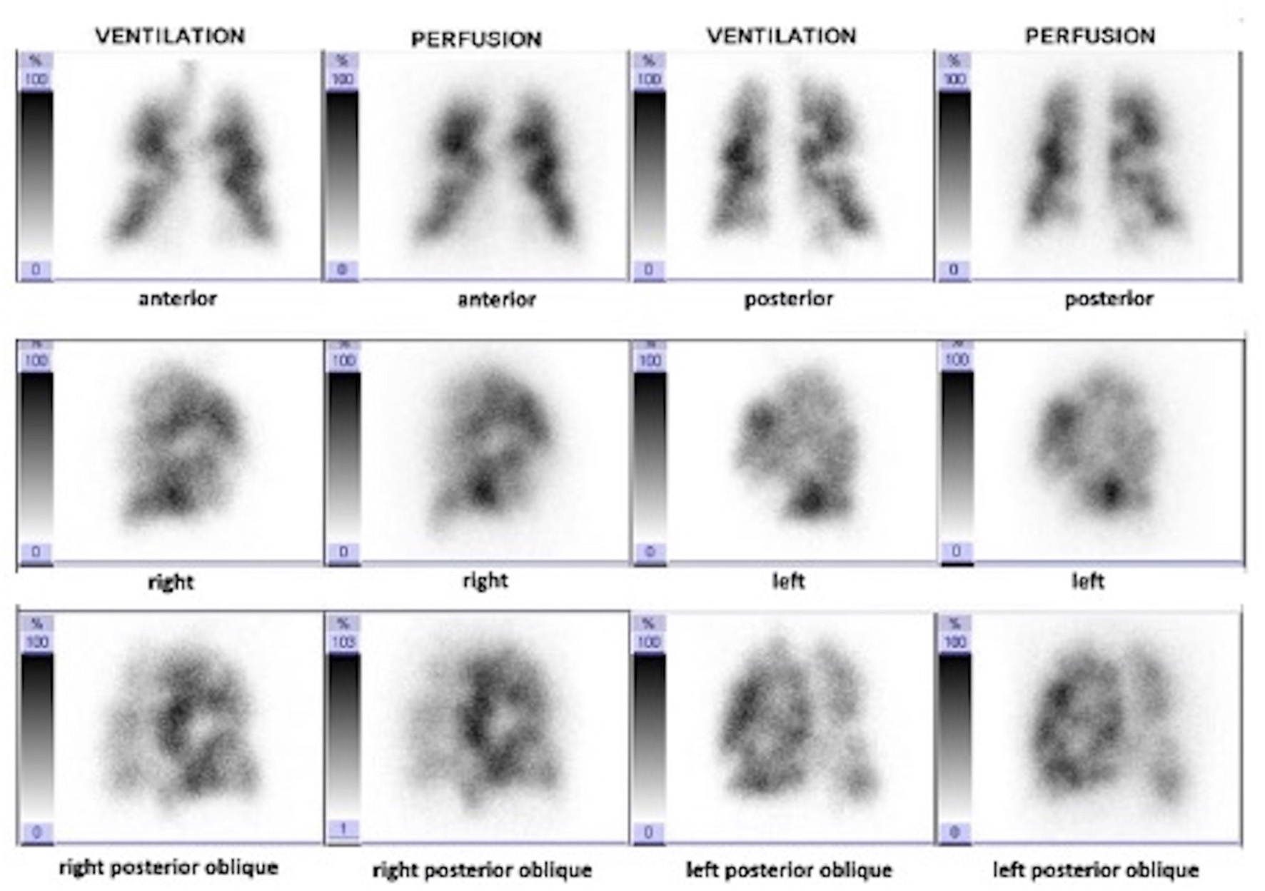
4 · Exclude CTEPD/CTEPH Obtain V/Q imaging (planar or SPECT) in unexplained PH whenever CTEPH has not been confidently excluded—do not wait for RHC proof of pre-capillary physiology. A normal perfusion study makes CTEPH very unlikely.
5 · Confirm and classify Perform RHC at an experienced center when results will establish diagnosis or change treatment—especially suspected PAH/CTEPH, severe PH, uncertainty, or transplant evaluation.
B
Minimum etiologic workup
Choose additional tests from phenotype| Domain | Core assessment | What it helps identify |
|---|---|---|
| Cardiac | ECG, BNP/NT-proBNP, echo. Consider CMR, coronary/valve assessment. | RV burden, Group 2 disease, CHD/shunt, alternative cardiac cause. |
| Pulmonary | PFTs + DLCO, HRCT and pulse oximetry/ABG. Add sleep and hypoventilation testing when indicated. | Group 3, PVOD clues, chronic hypoxemia/hypercapnia. |
| Thromboembolic | V/Q. If abnormal, CTPA/dual-energy CT and catheter angiography as directed by CTEPH team. | CTEPD/CTEPH anatomy and intervention planning. |
| Laboratory | CBC, CMP/LFTs, creatinine/eGFR, TSH, iron indices, HIV and ANA with disease-directed CTD serology. | Anemia/high flow, hepatic/renal/thyroid disease, HIV, CTD. |
| Portal / abdominal | History, exam, LFTs and abdominal ultrasound/portal assessment when suspected. | Portopulmonary PH, schistosomiasis, volume/high-flow physiology. |
| Exposure | Lifetime medication/substance history: anorexigens, methamphetamine, dasatinib, mitomycin-C, carfilzomib and other implicated agents. | Drug- or toxin-associated PAH/PVOD. A potentially removable cause. |
01
Interpret key patterns
Phenotype first- Disproportionately low DLCO consider SSc-PAH, PVOD/PCH, emphysema or combined fibrosis-emphysema.
- Orthopnea, LA enlargement, AF, LVH raise suspicion for occult HFpEF even if resting PAWP is borderline.
- Mismatched perfusion defects are not “just old PE.” Send images to a CTEPH center.
- Cyanosis/clubbing assess CHD, shunt, advanced lung disease and PVOD.
02
Common diagnostic traps
Avoid delay and harm- Starting PAH therapy from echo-estimated PASP without RHC.
- Using CTPA alone to “exclude” chronic thromboembolic disease.
- Accepting a poor-quality wedge or estimated Fick output.
- Calling mild PH in an older patient “idiopathic PAH” without testing for LHD, lung disease and mixed phenotypes.
 Open full-size V/Q image
RULE
RHC is not a formality. It prevents inappropriate PAH treatment, identifies occult left-heart physiology, quantifies severity, enables vasoreactivity testing in the correct subgroup, and supplies a baseline for risk-based care.
A
Echo probability—not an echo diagnosis
Peak TRV + signs from ≥2 categories| Peak TR velocity | No other PH signs | Other PH signs present | Action in clinical context |
|---|---|---|---|
| ≤2.8 m/s or unmeasurable | Low | Intermediate | Low/unmeasurable TRV does not exclude PH. Severe TR may underestimate the gradient. Pursue workup when risk, symptoms or RV/RA/PA signs warrant. |
| 2.9–3.4 m/s | Intermediate | High | Complete etiologic evaluation. RHC if it will change diagnosis or treatment. |
| >3.4 m/s | High | PH-center evaluation and RHC unless an exceptional clinical reason argues otherwise. | |
Other signs: ventricles / PA
- RV/LV basal diameter ratio >1 or septal flattening.
- RVOT acceleration time <105 ms and/or midsystolic notching.
- Early-diastolic PR velocity >2.2 m/s or PA diameter >25 mm.
Other signs: IVC / RA
- IVC >21 mm with reduced inspiratory collapse.
- RA area >18 cm². Abnormal RV-PA coupling or RV function supports severity assessment.
- “Other signs present” requires findings from at least two of three ESC/ERS categories.
B
RHC acquisition checklist
Experienced center · clinically optimized state| Setup | Document BP, HR, rhythm, oxygen/ventilatory support and volume status. Zero at the mid-thoracic level. Record during spontaneous breathing and at end-expiration without breath-holding. |
| Pressures | RAP, RV, systolic/diastolic/mPAP and PAWP. Record waveforms. Use mean over several cycles, especially with AF or respiratory variation. |
| Validate PAWP | Confirm typical a/v waves, fluoroscopic position and wedge saturation when uncertain. If unreliable, repeat in another branch and consider LVEDP. PAWP and LVEDP are related but not interchangeable. |
| Cardiac output | Average ≥3 thermodilution measurements with <10% variation when suitable. Use direct Fick for significant shunt or when thermodilution is unreliable. Avoid estimated oxygen consumption when precision matters. |
| Shunt study | Obtain stepwise oximetry when saturation “step-up,” congenital disease or high-output physiology is suspected. Calculate Qp/Qs and PVR with appropriate pulmonary flow. |
| Report | Include RAP, mPAP, PAWP, CO/CI, PVR, SvO₂, systemic BP, method and tracings—not merely “pre-capillary” or “post-capillary.” |
C
Provocative testing & vasoreactivity
Answer a defined question| Test | Use | Interpretation / guardrail |
|---|---|---|
| Fluid challenge | Suspected occult HFpEF with normal/borderline resting PAWP. | 500 mL saline over 5–10 min. ESC/ERS uses post-fluid PAWP ≥18 mmHg as suggestive of HFpEF. Validation is limited. Interpret borderline values with phenotype and measurement quality. |
| Exercise RHC | Unexplained exertional dyspnea. Distinguish pulmonary vascular from left-heart limitation. | Exercise PH: mPAP/CO slope >3. PAWP/CO slope >2 mmHg·min·L⁻¹ supports a post-capillary contribution. |
| Acute vasoreactivity | At index RHC only for idiopathic, heritable or drug/toxin-associated PAH. | Inhaled NO or iloprost preferred. IV epoprostenol alternative. Do not use adenosine. ESC/ERS positive response = mPAP fall ≥10 mmHg to ≤40 mmHg with unchanged/increased CO. |
| CCB response check | After a positive test and high-dose CCB initiation. | Reassess at 3–4 months including RHC. CCB-only care requires WHO FC I–II plus near-normal hemodynamics (mPAP <30 and PVR <4 WU). |
PITFALL
Do not perform vasoreactivity testing to choose CCBs in Groups 2–5 or other PAH phenotypes. A nonspecific pressure fall is not a “positive” test, and empiric high-dose CCB therapy can cause hypotension, RV failure or pulmonary edema.
A
Risk-group screening
Screening intensity follows absolute risk| Population | Recommended approach | Escalate to |
|---|---|---|
| Systemic sclerosis | Annual multimodal screening with symptoms/exam, PFT/DLCO and BNP/NT-proBNP ± echo. Use DETECT if disease >3 years, FVC ≥40% and DLCO <60%. | RHC when algorithm/echo or unexplained symptoms indicate. |
| BMPR2 carrier / first-degree HPAH relative | Annual clinical, ECG, BNP, echo and functional assessment in an expert program. Individualize additional testing. | Genetic counseling and early RHC for abnormalities. |
| Portal hypertension | Echo before liver transplantation or TIPS and when symptoms/signs suggest PH. | For LT candidates, RVSP >40 mmHg, >mild RV enlargement/dysfunction or intermediate/high PH probability prompts PH consultation and consideration of RHC. |
| Other CTD, HIV, CHD | Symptom- and risk-driven evaluation. Do not apply blanket annual echo to every low-risk patient. | Complete PH pathway when unexplained dyspnea, biomarker or imaging abnormality appears. |
| After acute PE | Ask about persistent/new dyspnea or exercise limitation at 3–6 months. No routine CTEPH imaging in an asymptomatic patient. | V/Q and CTEPH referral if symptoms or imaging suggest chronic obstruction. |
B
Genetic evaluation
Counsel before testing01
Offer testing
Specialist genetics pathway- Heritable/familial or idiopathic PAH regardless of age.
- Anorexigen-associated PAH. Other drug-associated PAH when phenotype or family history suggests inherited susceptibility.
- Suspected PVOD/PCH, congenital/developmental features, pediatric-onset or syndromic disease.
- Discuss incomplete penetrance, sex differences, insurance/privacy implications and family cascade testing.
02
Genes to recognize
Panels evolve- BMPR2 most common heritable PAH. Also TBX4, ACVRL1/ENG, SOX17, ATP13A3, KCNK3, GDF2 and others.
- EIF2AK4 biallelic variants strongly support heritable PVOD/PCH.
- A negative panel does not exclude inherited susceptibility. Re-review as variant classification changes.
C
Group 1 etiologic map
Association changes prognosis and practical care| Phenotype | Clues / tests | Immediate implication |
|---|---|---|
| Idiopathic / heritable | No secondary cause. Family history may be absent. | Vasoreactivity testing, genetics and the standard risk-based algorithm. |
| CTD | SSc, SLE, MCTD. Assess ILD, LHD and PVOD overlap. | Low threshold for combination therapy, but phenotype precisely. |
| Portal HTN | Portal disease ± cirrhosis. May have high CO and volume overload. | RHC before transplant decisions. Specialist PoPH therapy. |
| HIV | Confirm HIV. Assess viral control, liver disease and drug interactions. | Antiretroviral therapy plus PAH treatment. Interaction review. |
| CHD | Pre-/post-tricuspid shunt, repaired defect, Eisenmenger physiology. | Qp/Qs and PVR. Closure decisions only in expert congenital/PH team. |
| Drug / toxin | Methamphetamine, anorexigens, dasatinib. Chemotherapy/solvent exposures relevant to PVOD. | Stop exposure, treat PAH if persistent, provide substance-use care. |
| Schistosomiasis | Endemic exposure, portal/hepatic disease. | Antiparasitic and liver care plus PH-center assessment. |
| PVOD / PCH | Marked hypoxemia/low DLCO. CT septal lines, centrilobular GGO, adenopathy. | PAH drugs may trigger edema. Cautious expert use and early transplant referral. |
Exposure history needs detail. Ask about prescribed and non-prescribed stimulants, weight-loss agents, dasatinib, alkylating chemotherapy, interferons and occupational solvents. Mitomycin-C and carfilzomib are definite associations in the 2024 WSPH classification. Review the strength of association for other agents.
Risk assessment is multiparametric and serial. Use the full 3-strata assessment at diagnosis, the validated 4-strata model at follow-up, and always overlay RV imaging, hemodynamics, trajectory, end-organ function and clinical judgment.
A
Baseline 3-strata model
Estimated 1-year mortality: <5% · 5–20% · >20%| Variable | Low risk | Intermediate risk | High risk |
|---|---|---|---|
| Clinical course / syncope | Stable. None | Slow progression. Occasional | Rapid progression. Recurrent |
| WHO functional class | I–II | III | IV |
| 6MWD | >440 m | 165–440 m | <165 m |
| BNP / NT-proBNP | <50 / <300 | 50–800 / 300–1100 | >800 / >1100 ng/L |
| RAP | <8 | 8–14 | >14 mmHg |
| Cardiac index | ≥2.5 | 2.0–2.4 | <2.0 L/min/m² |
| Stroke-volume index | >38 | 31–38 | <31 mL/m² |
| SvO₂ | >65% | 60–65% | <60% |
B
Follow-up 4-strata model
Score available non-invasive variables| Variable | Low (1) | Intermediate-low (2) | Intermediate-high (3) | High (4) |
|---|---|---|---|---|
| WHO FC | I–II | — | III | IV |
| 6MWD | >440 | 320–440 | 165–319 | <165 m |
| BNP | <50 | 50–199 | 200–800 | >800 ng/L |
| NT-proBNP | <300 | 300–649 | 650–1100 | >1100 ng/L |
Assign 1–4 to WHO FC, 6MWD and one natriuretic peptide. Average available values and assign the closest stratum. Do not let a mathematically low score override severe RV dysfunction, pericardial effusion, low output or rapid deterioration.
C
Visit cadence & treatment goals
First reassessment at 3–4 months01
Every PH follow-up
Usually every 3–6 months- Symptoms/trajectory, syncope, WHO FC, 6MWD or CPET as appropriate.
- BNP/NT-proBNP, renal/hepatic function, CBC/iron and drug-specific labs.
- Volume status, oxygen need, adherence, side effects, access and pump/line safety.
- Echo/RV assessment as indicated. The 2026 ERS update suggests follow-up RHC for intermediate-low or higher risk when the result can change therapy.
02
Targets & escalation
Goal = low risk- Low risk continue effective regimen and surveillance.
- Intermediate-low add-on sotatercept is recommended by the 2026 ERS update for treated PAH. Individualize other pathway escalation.
- Intermediate-high / high urgent escalation—parenteral prostacyclin if absent, consider sotatercept, and initiate transplant pathway.
- Re-phenotype when response is discordant. Exclude PVOD, CTEPH, LHD, lung disease and adherence/administration failure.
GOAL
“Better” is not the endpoint—low risk is. Track both patient-centered outcomes and RV recovery. A patient who walks farther but remains congested, with poor RV function or low output, still needs escalation.
Before
Rx
Rx
Confirm the algorithm fitsIt principally covers clear IPAH, HPAH, drug/toxin PAH and CTD-PAH with no major vasoreactivity. PoPH, HIV, CHD, schistosomiasis and PVOD/PCH require association-specific expert care.
Threshold ≠ treatment evidenceFor mPAP 21–24 and/or PVR 2–3 WU, evidence for approved PAH therapy is limited. Use close expert follow-up or trials. Individualize any off-label treatment.
Assess comorbiditiesOlder patients with HFpEF/lung-disease phenotype may tolerate combination therapy poorly. Start deliberately and re-evaluate the diagnosis if congestion or hypoxemia worsens.
A
Initial therapy after full baseline risk assessment
Clear-cut PAH · generally mPAP ≥25 and PVR >3 in trials01
Not high risk
Low or intermediateInitial ERA + PDE5 inhibitor for most treatment-naive patients without major cardiopulmonary comorbidities—commonly macitentan or ambrisentan plus tadalafil/sildenafil.
- Fixed macitentan/tadalafil is an adherence option for adult Group 1 PAH WHO FC II–III. Convenience does not establish superiority over equivalent separate components.
- Initial oral triple therapy with ERA + PDE5i + selexipag is not recommended. It did not improve the primary endpoint in TRITON.
- Despite a non-high composite score, consider initial IV/SC prostacyclin + ERA + PDE5i for severe hemodynamics and/or poor RV function.
- In a comorbidity-heavy phenotype, selected monotherapy with careful early reassessment may be safer.
02
High risk / severe low output
Do not delay parenteral therapyInitial triple therapy: IV epoprostenol or IV/SC treprostinil plus ERA + PDE5i, with urgent expert-center management.
- Start transplant referral at diagnosis. Correct congestion, hypoxemia, iron deficiency and precipitating factors.
- Sotatercept is not a substitute for parenteral prostacyclin in shock or severe low-output RV failure.
- If parenteral therapy is declined or impossible, document the limitation and pursue the strongest feasible regimen with very close follow-up.
B
Reassess at 3–4 months—or sooner if worse
Use the 4-strata model| Follow-up stratum | Preferred next step | Additional actions |
|---|---|---|
| Low | Continue current therapy. | Maintain surveillance, adherence and drug-specific monitoring. |
| Intermediate-low | The 2026 ERS update recommends add-on sotatercept for treated PAH at this stratum. Other options include selexipag, prostacyclin-pathway therapy or selected PDE5i→riociguat switch. No head-to-head ranking is established. | Never overlap. Before riociguat: stop sildenafil ≥24 h or tadalafil ≥48 h. Before a PDE5i: stop riociguat ≥24 h. Monitor BP. |
| Intermediate-high | Start IV/SC prostacyclin if absent. The 2026 ERS update recommends add-on sotatercept for treated PAH at this stratum. | Expedite transplant evaluation. Investigate why the target was missed. |
| High | Prioritize IV/SC prostacyclin if absent. Add-on sotatercept is also recommended for treated high-risk PAH, with specialist monitoring. | Hospitalize if unstable. Transplant/bridge planning at an expert center. |
C
Sotatercept in the modern sequence
Add-on activin-signaling inhibitorEvidence & role
- STELLAR (n=323, FC II–III): add-on therapy improved 24-week 6MWD by a placebo-adjusted 40.8 m (95% CI 27.5–54.1).
- ZENITH (n=172, high risk): death/transplant/PAH admission was 17.4% vs 54.7%, HR 0.24. Stopped early for efficacy.
- HYPERION (n=320, diagnosis <1 year, already on double/triple therapy): worsening 10.6% vs 36.9%, HR 0.24. Stopped after external evidence changed equipoise. Deaths were 7 vs 6.
Do not miss
- It does not replace volume management, parenteral prostacyclin, transplant referral or confirmation of the correct PH group.
- New/worsening hypoxemia can rarely reflect intrapulmonary right-to-left shunting despite improved hemodynamics. Evaluate rather than assuming disease progression.
- Composite results do not establish an isolated mortality benefit in each trial. The 2026 ERS update gives no recommendation for adding sotatercept at low risk. See Page 8 for current safety safeguards.
HARD STOP
Never combine a PDE5 inhibitor with riociguat. Do not abruptly stop epoprostenol or treprostinil infusion. New pulmonary edema after PAH therapy should trigger urgent reassessment for PVOD/PCH or occult left-heart disease.
A
Associated PAH
Treat PAH and the association| Phenotype | PAH strategy | Key modifications / hazards |
|---|---|---|
| CTD-PAH | Risk-based combination therapy for clear PAH. | Quantify ILD, LV disease and PVOD clues. Manage CTD, reflux/aspiration and digital limitations. Bleeding/anemia and drug tolerability may be amplified. |
| Portopulmonary PH | PAH therapy can improve hemodynamics and transplant eligibility. Coordinate with hepatology. | Distinguish PAH from high-output/volume physiology. U.S. MELD-exception post-treatment profiles include mPAP <35 with PVR <5 WU, or mPAP ≥35 but <45 with PVR <3 WU. These are selection criteria, not proof of safety. Severe RV dysfunction, PVR >5 WU or mPAP >45–50 preclude LT in ILTS guidance. |
| HIV-PAH | Antiretroviral therapy plus PAH treatment. Combination therapy when indicated. | Review CYP interactions, adherence, liver disease, infections and thromboembolic risk. |
| Drug/toxin PAH | Stop the causal exposure. Observe mild low-risk cases closely or treat persistent/high-risk disease as PAH. | Dasatinib-associated disease may improve after withdrawal. Methamphetamine-associated PAH requires nonjudgmental substance-use treatment and relapse prevention. |
| Schistosomiasis | PAH therapy may be used at expert centers. | Treat active infection, portal/hepatic disease and anemia. Recognize mixed high-flow physiology. |
B
Congenital heart disease
Anatomy · shunt direction · operability01
Classify the CHD-PAH phenotype
Adult congenital + PH team- Eisenmenger syndrome, PAH with prevalent systemic-to-pulmonary shunt, PAH with small/coincidental defect, or PAH after defect correction.
- Measure Qp/Qs, PVR and systemic resistance with accurate oxygen consumption. Define anatomy and extracardiac causes.
- Do not close a defect in established Eisenmenger physiology or when closure would remove a needed RV “pop-off.” Operability is individualized—not a single PVR number.
- Repaired-CHD PAH generally follows the standard risk-based PAH algorithm.
02
Eisenmenger safety
Avoid destabilizing balance- PAH therapy (often ERA-based, then sequential combination) can improve symptoms and outcomes. Use specialist protocols.
- Avoid dehydration, abrupt systemic vasodilation, excessive diuresis and unfiltered IV air. Use meticulous line de-airing/air filters.
- No routine phlebotomy. Treat iron deficiency. Consider isovolumic phlebotomy only for severe hyperviscosity after excluding dehydration and iron deficiency.
- Anticoagulation is not routine. Individualize for atrial arrhythmia, thrombosis or pulmonary artery thrombus against bleeding/hemoptysis risk.
C
PVOD/PCH and true CCB responders
Two uncommon phenotypes with high stakesPVOD
Venous/capillary involvement
Early transplant referral- Clues: hypoxemia, very low DLCO, occult alveolar hemorrhage. CT triad of smooth septal thickening, centrilobular ground-glass opacities and mediastinal adenopathy.
- EIF2AK4 biallelic variants support heritable disease. Lung biopsy is generally avoided.
- PAH drugs can cause life-threatening pulmonary edema. If used, start one agent cautiously with close monitoring and aggressive transplant planning.
CCB
Sustained vasoreactive responder
IPAH/HPAH/drug PAH only- Use high-dose nifedipine, diltiazem or amlodipine only after a positive test. Avoid verapamil because of negative inotropy.
- Choose diltiazem for relative tachycardia and nifedipine/amlodipine for relative bradycardia. Titrate carefully.
- At 3–4 months, repeat RHC. If not WHO FC I–II with near-normal mPAP/PVR, add standard PAH therapy rather than simply increasing CCB.
PEARL
Association is not destiny, but it changes the hazards. Use the Group 1 treatment framework only after accounting for lung/left-heart overlap, shunts, hepatic function, drug interactions, bleeding, pregnancy and transplant strategy.
Doses below are common adult regimens, not a substitute for prescribing information. Adjust for renal/hepatic function, interactions, pregnancy potential, formulation/device and PH-center protocol. Confirm agent-specific indication. A class effect cannot be assumed.
A
Oral agents
Combine across pathways—not within cGMP pathways| Pathway / agent | Typical adult regimen | High-yield safety |
|---|---|---|
| PDE5i · sildenafil | 20 mg PO TID, titrate by response/tolerance up to 80 mg TID | No nitrates or riociguat. Hypotension, headache, visual/hearing effects. |
| PDE5i · tadalafil | 40 mg PO daily | No nitrates or riociguat. Renal/hepatic and CYP3A considerations. |
| sGC · riociguat | 0.5–1 mg TID, titrate by 0.5 mg at ≥2-wk intervals to 2.5 mg TID | PAH or inoperable/residual CTEPH. No PDE5i/nitrates. Increase only if SBP >95 without hypotension. Pregnancy contraindicated. REMS removed August 2026. |
| ERA · ambrisentan | 5 mg daily → 10 mg daily | Edema, anemia, embryo-fetal toxicity. Avoid in IPF. |
| ERA · macitentan | 10 mg daily | Anemia, edema, embryo-fetal toxicity. Interaction review. |
| ERA · bosentan | 62.5 mg BID ×4 wk → 125 mg BID | Baseline/monthly LFTs. Anemia. CYP induction. Hepatotoxicity REMS. Lowers hormonal contraceptive exposure. |
| Fixed macitentan/tadalafil | Naive/from ERA: 10/20 mg daily ≥1 wk → 10/40. Already on tadalafil 40 ± macitentan 10: start 10/40 | Adult Group 1 PAH FC II–III. All ERA/PDE5i warnings apply. |
| Selexipag | 200 mcg BID. Weekly +200 mcg to max 1600 mcg BID | CYP2C8: gemfibrozil contraindicated. With clopidogrel use once daily. Headache, jaw pain, diarrhea. |
| CCB—true responder only | amlodipine 15–30 mg/d · nifedipine 120–240 mg/d · diltiazem 240–720 mg/d | Titrate gradually. Edema/hypotension. Avoid verapamil. Repeat RHC at 3–4 months. |
B
Prostacyclin-pathway therapy & sotatercept
Training and backup systems are treatment| Agent / route | Use & dosing principle | Do not miss |
|---|---|---|
| Epoprostenol IV | Continuous central infusion. Initiate/titrate in expert program. | Very short half-life: never interrupt. Dedicated line, backup pump/drug, line-infection plan. |
| Treprostinil IV/SC | Continuous, weight-based titration. SC avoids central line but causes site pain. | Never abruptly interrupt. High-output state may reflect excessive dose. |
| Treprostinil inhaled/PO | Device/formulation-specific titration. Inhaled forms for PAH and PH-ILD where labeled. | Do not substitute devices or formulations dose-for-dose. Cough/throat irritation, hypotension. |
| Iloprost inhaled | Frequent daily inhalations. PAH indication. | Administration burden, bronchospasm, hypotension. |
| Sotatercept SC | 0.3 mg/kg q3wk → 0.7 mg/kg q3wk if Hgb/platelets acceptable | Check Hgb/platelets before first 5 doses (longer if unstable), then periodically. |
C
Sotatercept holds, bleeding & reproductive safety
Current U.S. Label · September 2026- Contraindicated after serious hypersensitivity to sotatercept/excipients. Anaphylaxis or angioedema: discontinue and provide emergency treatment.
- Do not initiate if platelets <50 ×10⁹/L.
- Hold ≥3 weeks for platelets <50 ×10⁹/L, Hgb >2 g/dL above the prior value and above ULN, Hgb >4 g/dL above baseline, or Hgb >2 g/dL above ULN. Recheck before every restart and resume only after resolution to an acceptable level.
- If interrupted >9 weeks, restart at 0.3 mg/kg. Do not give during serious bleeding.
- Bleeding risk rises with thrombocytopenia, prostacyclin infusion or antithrombotics. Consider endoscopy for angiodysplasia in unexplained/recurrent GI bleeding.
- Pregnancy test before treatment. Effective contraception during and for ≥4 months after the last dose.
- No breastfeeding during treatment and for 4 months after. Counsel regarding possible impaired fertility.
- Monitor for telangiectasia, epistaxis, erythrocytosis, thrombocytopenia, increased BP and serious bleeding.
- Evaluate new hypoxemia for rare intrapulmonary R→L shunting. Pericardial effusion has been reported postmarketing.
REMS UPDATE
U.S. REMS changes do not remove pregnancy risk. FDA removed ambrisentan/macitentan pregnancy REMS in 2025 and riociguat REMS on August 4, 2026. Bosentan retains its hepatotoxicity REMS. Pregnancy remains contraindicated. For riociguat, exclude pregnancy before starting and use effective contraception through 1 month after stopping.
A
Group 2 — PH due to left-heart disease
The most common PHDX
Recognize & phenotype
IpcPH vs CpcPH- Consider HFpEF/HFrEF, valve disease, infiltrative cardiomyopathy and congenital/acquired inflow or outflow obstruction.
- PAWP is a continuum. Values around 12–18 mmHg require measurement scrutiny and phenotype. Use fluid/exercise testing when occult HFpEF remains likely.
- PVR >5 WU indicates a severe pre-capillary component and should prompt expert referral and reconsideration of alternate/mixed causes.
RX
Treat heart, valve & volume
Not a PAH algorithm- Optimize guideline-directed HF care, BP, rhythm, ischemia, valve disease, sleep-disordered breathing and diuresis.
- PAH drugs are not recommended routinely. Trials have been neutral or harmful, including fluid retention/pulmonary edema.
- Selected CpcPH therapy is a research question, not an indication for empiric outpatient sildenafil.
B
Group 3 — PH due to lung disease / hypoxia
Severe PH = PVR >5 WU| Step | What to do | Clinical detail |
|---|---|---|
| Confirm phenotype | Define ILD, COPD/emphysema, mixed disease, hypoventilation, developmental lung disease or high-altitude exposure. | RHC when it will change therapy, classification, transplant listing or trial eligibility. |
| Optimize basics | Maximize lung-disease therapy, oxygen when indicated, sleep/hypoventilation treatment, smoking cessation, vaccination and pulmonary rehab. | Refer early for lung transplant when disease severity warrants. |
| PH-ILD | Inhaled treprostinil improves exercise capacity and is approved in the U.S. Use specialist-guided titration. | Not restricted to a “severe PH” subgroup. Selected severe PH-ILD may receive a PDE5i off-label at a PH center. |
| PH-COPD | No approved PAH therapy. Treat COPD, oxygen need and transplant candidacy. | Do not use inhaled treprostinil: PERFECT stopped early for excess serious adverse events and possible mortality. |
C
Specific no-go therapies & Group 5
Avoid harmful extrapolation| Setting | Avoid | Why / alternative |
|---|---|---|
| Idiopathic pulmonary fibrosis | Ambrisentan | ARTEMIS-IPF showed harm/lack of benefit. Treat IPF and evaluate PH separately. |
| IIP-associated PH | Riociguat | RISE-IIP terminated early for increased serious adverse events and mortality. |
| PH-COPD | Inhaled treprostinil | Unfavorable benefit-risk in PERFECT. Do not infer benefit from PH-ILD. |
| Sickle-cell disease | Routine PAH drugs, specifically sildenafil | Walk-PHaSST stopped early for excess serious adverse events, mainly pain hospitalizations. Confirm mechanism by RHC and prioritize sickle-cell/cause-specific care. |
| Other Group 5 | Routine PAH-drug treatment | First identify dominant mechanisms: sarcoid vascular/compressive disease, hematologic disease, CKD, metabolic disease, tumor obstruction or fibrosing mediastinitis. |
PRINCIPLE
“Pre-capillary” does not automatically mean “PAH treatment.” In Groups 2, 3 and 5, benefit and harm depend on the underlying mechanism, disease subtype and specific agent—not merely PVR elevation.
Every
Case
Case
Send to a multidisciplinary CTEPH teamOperability and BPA candidacy cannot be determined from a radiology report alone. Review anatomy, hemodynamics, comorbidity and center expertise.
PEA is first-line when operableProximal disease is not required. Experienced centers can operate on selected segmental and subsegmental disease.
Use multimodal treatmentPEA, BPA and medical therapy are complementary. Do not let pretreatment delay a potentially curative operation.
A
Definitions & diagnosis
Defined after ≥3 months anticoagulation · refer sooner if severe| CTEPD | Symptoms plus mismatched perfusion defects and chronic organized fibrotic clot on CTPA/angiography after adequate anticoagulation, with or without resting PH. |
| CTEPH | CTEPD with PH confirmed by RHC. “Chronic clot on CT” without hemodynamic PH is not CTEPH. |
| Imaging | V/Q is the screening test. Expert CTPA looks for webs, bands, ring stenoses, pouch defects, eccentric wall-adherent material and mosaic perfusion. Catheter angiography helps intervention planning. |
| Differential | Pulmonary artery sarcoma/tumor, arteritis, in-situ thrombosis, congenital stenosis and fibrosing mediastinitis may mimic chronic thromboembolic obstruction. |
B
Treatment sequence
Anatomic cure when possible| Therapy | Best use | Key points |
|---|---|---|
| Anticoagulation | All CTEPH, lifelong. | Test for antiphospholipid syndrome. Use a VKA for APS. Choice outside APS is individualized because comparative CTEPH data are limited. |
| Pulmonary endarterectomy | Operable CTEPH. | Potentially curative. Assessment depends on accessible anatomy, PVR-anatomic concordance, comorbidity and experienced surgical judgment. |
| Balloon pulmonary angioplasty | Technically inoperable disease, unfavorable surgical risk, or persistent/recurrent PH after PEA. | Staged procedures at expert centers. Risks include vascular injury, hemoptysis and lung injury. |
| Riociguat | Symptomatic inoperable CTEPH or persistent/recurrent PH after PEA. | May precede BPA when PVR is high. Do not combine with PDE5i or nitrates. |
| Other medical therapy | Selected expert-center cases. | SC treprostinil may be used for inoperable/residual CTEPH where locally approved. Other agents are evidence-limited/off-label. Use only after CTEPH-team review and never delay PEA/BPA. |
C
CTEPD without resting PH & follow-up
Symptoms must be attributable to obstructionNO PH
Selected intervention
Expert decision- Exclude deconditioning, cardiac/lung disease and other causes of dyspnea. Consider CPET/exercise hemodynamics.
- Selected symptomatic patients may benefit from PEA or BPA when lesions are anatomically suitable and symptoms are convincingly obstruction-related.
- Balance benefit against procedural risk. No blanket intervention rule.
AFTER RX
Structured reassessment
Residual disease matters- Reassess symptoms, functional capacity, echo/biomarkers and hemodynamics 3–6 months after PEA or the final BPA session.
- Then review at least annually. Investigate recurrent dyspnea, rising BNP or RV dysfunction.
- Residual PH may require BPA, riociguat or multimodal therapy. Recurrent obstruction demands adherence and thrombophilia review.
DO NOT MISS
A distal label is not the same as inoperable. Get an expert surgical opinion before defaulting to BPA or lifelong medical therapy. Conversely, not every residual perfusion defect explains symptoms—link anatomy, hemodynamics and functional limitation.
Acute
RV Failure
RV Failure
Call the PH/ICU team nowShock, low output, escalating oxygen need, rising lactate/creatinine, syncope, severe congestion or new arrhythmia require expert critical care and transfer planning.
Never interrupt PAH infusionImmediately inspect pump, drug cassette, tubing, central access and backup supply. Give parenteral prostacyclin only through trained protocols.
Avoid the intubation spiralInduction, positive pressure and systemic hypotension can precipitate arrest. If unavoidable, use an experienced team with vasopressors ready and the least destabilizing strategy.
A
Acute decompensated RV failure
Preload · pressure · perfusion · afterload| Priority | Action | Pitfall |
|---|---|---|
| Find trigger | Infection, arrhythmia, PE, anemia/bleeding, hypoxemia, medication interruption, pregnancy, surgery, renal failure or excess salt/fluid. | Do not attribute every deterioration to “PAH progression.” |
| Optimize preload | Most congested patients need IV loop diuresis ± thiazide synergy/ultrafiltration. Track urine, renal function, lactate and perfusion. | Avoid reflex large fluid boluses. A small monitored challenge is reserved for clear underfilling. |
| Support pressure | Norepinephrine is commonly first-line. Low-dose vasopressin may support systemic pressure. Maintain RV coronary perfusion. | Avoid pure systemic hypotension and excessive tachycardia. |
| Improve output | Dobutamine or milrinone for low output with adequate pressure. Pair with vasopressor when needed. | Both may cause hypotension/arrhythmia. Monitor invasively in shock. |
| Lower RV afterload | Correct hypoxemia, acidosis and hypercapnia. Use inhaled NO or inhaled epoprostenol as selective rescue. Never interrupt continuous prostacyclin. | Continue other PAH drugs when feasible. Adjust/hold in profound hypotension or acute organ failure only with PH-specialist input. |
| Bridge | VA-ECMO or other support only at an expert center as a defined bridge to recovery, intervention or transplantation. | Mechanical support without a destination plan is not definitive therapy. |
B
Pregnancy & contraception
Very high risk · shared decision-making- Pregnancy in PAH carries very high maternal and fetal risk. There is no hemodynamic cutoff that makes it “safe.” Provide preconception counseling and reliable contraception.
- If pregnancy occurs, refer urgently to a Pregnancy Heart Team/PH center. Discuss all options, including termination, while respecting patient autonomy.
- If continuing, stop ERAs, riociguat, selexipag and sotatercept. PDE5 inhibitors and prostacyclin analogues may be used by expert teams. Continue CCB only in a true responder.
- Plan serial maternal/fetal surveillance, delivery at an expert center, individualized anesthesia and proactive RV support.
- The early postpartum period is especially dangerous from fluid shifts and RV failure. Monitored/ICU care is often required.
- Sotatercept: contraception during treatment and ≥4 months after. Avoid breastfeeding during and 4 months after the last dose.
C
Procedures, supportive care & transplant
Plan before the day of surgery| Situation | Practical approach |
|---|---|
| Noncardiac surgery | Defer elective procedures if unstable. Coordinate PH, anesthesia and surgeon. Continue PAH therapy without interruption. Avoid hypoxia, hypercapnia, acidosis, pain, hypothermia and systemic hypotension. Use monitoring appropriate to severity. Titrate regional anesthesia gradually. |
| Supportive care | Use diuretics for congestion and oxygen for documented hypoxemia (generally PaO₂ <60 mmHg). Treat iron deficiency and offer vaccination, supervised rehabilitation, psychosocial and palliative support alongside disease-directed therapy. |
| Anticoagulation | Not routine for all PAH. Individualize for another indication and bleeding risk. CTEPH requires lifelong anticoagulation. Eisenmenger and portopulmonary patients need special caution. |
| Transplant | Refer early for persistent intermediate-high/high risk despite optimized therapy, progressive RV failure, recurrent hospitalization, hemoptysis or PVOD/PCH. Lung transplant is usual. Heart-lung transplant is reserved for selected structural/cardiac situations. |
BOTTOM LINE
Classify precisely, confirm hemodynamics, treat the correct phenotype and reassess early. In emergencies, protect RV perfusion, reduce avoidable afterload, preserve life-sustaining PAH therapy and involve the expert center before irreversible collapse.