The structural-heart question is not only "how severe?"Define mechanism, hemodynamic burden, chamber response, symptoms, reversibility, anatomy, procedural risk, and the lifetime sequence of future interventions. Every severe lesion being considered for intervention belongs in a multidisciplinary Heart Team.
◆
Four valves - four load states
stenosis = pressure · regurgitation = volume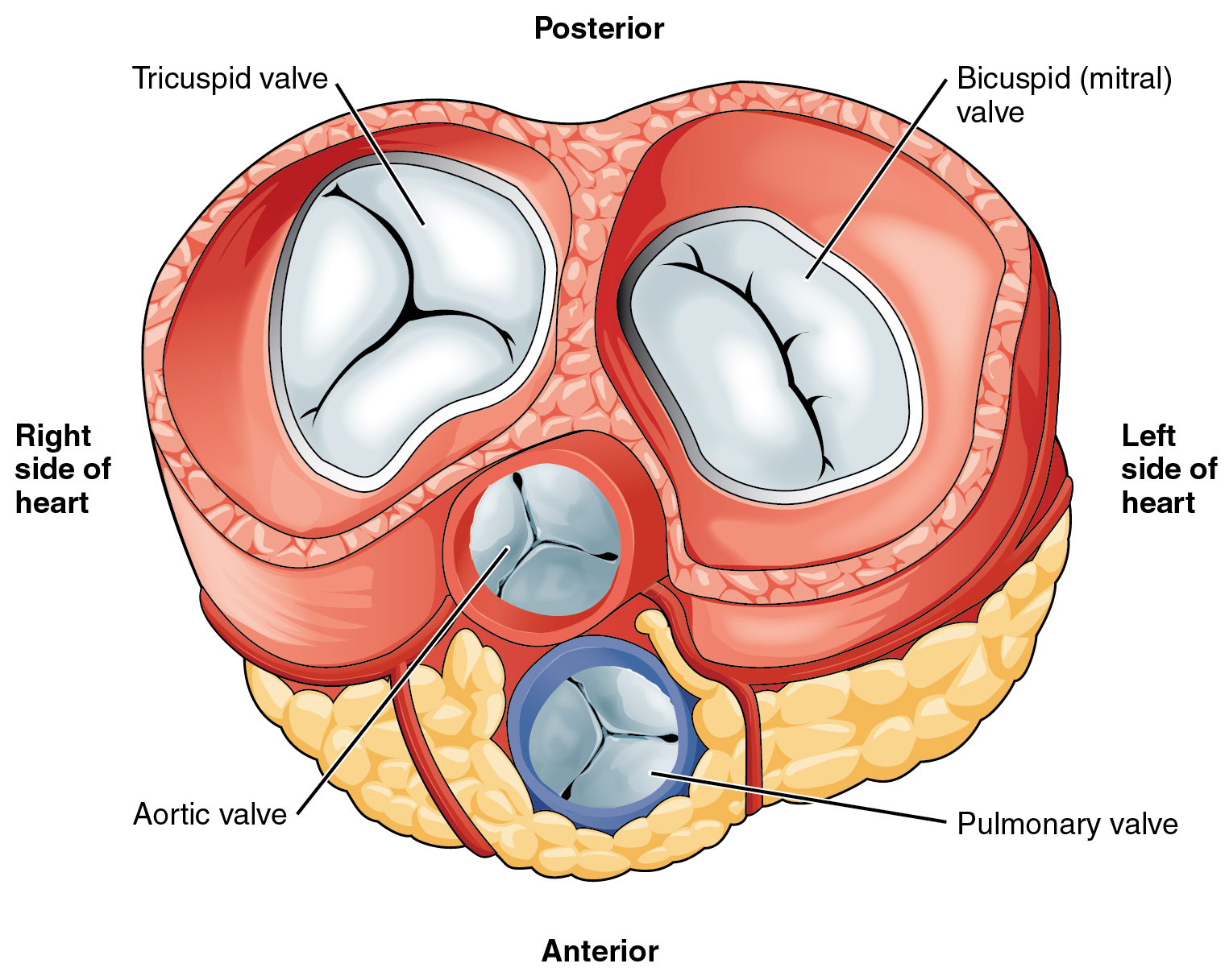

A
Diagnostic sequence
do not jump from murmur to device1 · Clinical phenotypeSymptoms, tempo, murmur, pulse, congestion, frailty, goals.
→
2 · TTE severityValve anatomy + integrated Doppler + LV/RV/LA/PA response.
→
3 · Resolve discordanceRepeat measurements. Stress echo, TEE, CT, CMR or cath.
4 · Stage and optimizeCAD, HF GDMT/CRT, rhythm, BP, volume, endocarditis/dental.
→
5 · Procedural anatomyCT/3D TEE. Access, landing zone, LVOT/coronaries/leads.
→
6 · Lifetime planRepair vs replacement. Surgical vs transcatheter. Next valve.
B
What each modality answers
integrate - never single-parameter grade| Tool | Best question | Structural use | Trap |
|---|---|---|---|
| TTE | Severity, hemodynamics, chamber response | First-line for every valve. Surveillance | Loading conditions and Doppler alignment |
| TEE / 3D | Mechanism, leaflet/scallop anatomy, thrombus | TEER, LAAO, ViV, endocarditis. Intraprocedural guidance | Sedation lowers BP and can reduce MR/TR |
| CT | Calcium, annulus/landing zone, access, coronary/LVOT risk | TAVR, TMVR/ViMAC, TTVR, LAAO planning | Contrast, rhythm, phase, virtual-valve assumptions |
| CMR | Accurate RV/LV volumes and regurgitant fraction | Discordant AR/MR/PR. Repaired TOF/RVOT | Device artifact. Less direct leaflet anatomy |
| Exercise / cath | Symptom attribution. Dynamic gradient/PH. True filling pressures | "Asymptomatic" severe disease or noninvasive discordance | Resting study may miss exertional physiology |
SAFETY
This guide is a reference, not an order set or device IFU. Verify current labeling, antithrombotic regimen, local outcomes, imaging measurements, and procedural candidacy with the active structural-heart program.
Severe AS is a hemodynamic syndrome, not an isolated AVA.Use Vmax, mean gradient, AVA, dimensionless index, stroke-volume index, valve calcium, LV function and symptoms together. Recheck LVOT diameter and Doppler windows before labeling low-gradient disease.
◆
Pathophysiology and phenotype
fixed afterload → myocardial diseaseLeaflet calcification / fusionDegenerative trileaflet · BAV · rheumatic
→
Pressure overloadConcentric LVH · diastolic dysfunction · LA/PH
→
DecompensationFibrosis · ischemia · low flow · reduced EF · RV failure
Clinical translation
Angina can occur without obstructive CAD. syncope reflects inability to augment output/vasodilation. dyspnea reflects elevated LV filling pressure. Once symptoms or LV decompensation are attributable to severe AS, valve intervention is the disease-modifying therapy.A
Severity anchors
normal flow · integrated grading| Measure | Severe anchor | How to use / common error |
|---|---|---|
| Peak velocity | Vmax ≥4.0 m/s | Search all windows with Pedoff. Underalignment undercalls severity. |
| Mean gradient | ≥40 mmHg | Flow-dependent. High flow can raise and low flow can hide gradient. |
| Aortic valve area | ≤1.0 cm² or ≤0.6 cm²/m² | Continuity equation magnifies LVOT measurement error. Index cautiously in obesity. |
| Dimensionless index | <0.25 | Useful when LVOT diameter is unreliable. Still flow and signal dependent. |
| Low flow | SVi ≤35 mL/m² | Measure rhythm/BP. Identify MR, RV failure, small LV, AF and infiltrative disease. |
Same narrowed valve, different forward flow
B
Discordant / low-gradient algorithm
AVA ≤1.0 but gradient <40Quality checkBP, rhythm, LVOT location/diameter, Doppler alignment, BSA.
→
Classify flow + EFReduced EF vs paradoxical preserved EF. SVi ≤35.
→
Confirm true severeDSE if reduced EF. Noncontrast CT calcium if still discordant. >2,000 AU in men or >1,200 in women supports severe AS.
| Pattern | Definition | Next move |
|---|---|---|
| Classic low-flow | Gradient <40 · AVA ≤1 · LVEF <50 · SVi ≤35 | Low-dose dobutamine: true severe if gradient rises with persistently small AVA. CT calcium if no flow reserve/indeterminate. |
| Paradoxical low-flow | Gradient <40 · AVA ≤1 · LVEF ≥50 · SVi ≤35 | Exclude measurement error, small-body indexing, uncontrolled HTN. CT calcium and symptoms. |
| Normal-flow low-gradient | Gradient <40 · AVA ≤1 · LVEF ≥50 · SVi >35 | Often moderate or measurement discordance. Review flow rate, CT calcium, serial trajectory. |
C
When to intervene
refer before decompensation- Symptomatic severe high-gradient AS intervention recommended.
- Confirmed severe low-flow AS intervene when symptoms are attributable, after Heart Team confirmation.
- LVEF <50% without another cause is an intervention trigger even if symptoms are denied.
- Asymptomatic high-risk signals very severe AS, rapid Vmax rise, markedly elevated BNP, abnormal exercise BP/symptoms, progressive EF decline.
- No expected benefit if predicted survival is short or symptoms/QoL will not improve. Integrate palliative goals.
Asymptomatic severe AS
EARLY TAVR enrolled 901 selected patients (mean age 75.8): death, stroke or unplanned cardiovascular hospitalization occurred in 26.8% with early TAVR vs 45.3% with surveillance (HR 0.50, median 3.8 years). Death alone was 8.4% vs 9.2%, and 87% of the surveillance arm ultimately underwent AVR. ESC supports discussing early intervention for severe high-gradient AS with EF ≥50%, a normal exercise test when feasible and low procedural risk (IIa A). SAPIEN 3/Ultra/Ultra RESILIA gained an FDA asymptomatic severe calcific AS indication in April 2025 for Heart Team-selected patients. This does not cover moderate AS or all TAVR systems.ACUTE
Shock with severe AS: stabilize perfusion, avoid unmonitored vasodilation or abrupt preload loss, involve structural + surgery early. Balloon valvuloplasty is a bridge in selected unstable or urgent noncardiac-surgery patients - not durable therapy.
Do not equate low STS risk with a TAVR mandate.Age, life expectancy, valve morphology, coronary access, aortopathy, CAD, annulus/root, vascular access, conduction risk, patient goals and the feasibility of the next procedure matter as much as perioperative risk.
◆
Side-by-side
TAVR = transcatheter · SAVR = surgical AVR| Domain | TAVR | SAVR |
|---|---|---|
| Access / recovery | Usually transfemoral. Conscious sedation. Shorter stay and faster early recovery. | Sternotomy or minimally invasive access. Cardiopulmonary bypass. Slower recovery. |
| Early tradeoffs | Less major bleeding, AKI and new AF in many trials. More vascular complications, PVL and pacemaker with some systems. | More bleeding/AF/AKI. Lower PVL and lower pacemaker rate. Direct visual debridement. |
| Anatomy | Best with suitable annulus, coronaries and transfemoral access. CT-defined. | Handles unfavorable coronary height, bulky LVOT calcium, root enlargement and complex BAV/aorta. |
| Other disease | PCI may be staged. Isolated-valve solution. | Best if CABG, aortic/root/arch work or another valve operation is needed. |
| Lifetime issues | Coronary reaccess, commissural alignment, sinus sequestration, future TAV-in-TAV, residual gradient in small annulus. | Mechanical valve option. Root enlargement. Future valve-in-valve planning. Surgical durability track record. |
| Do not use | Active endocarditis. Anatomy/access outside safe IFU. Futility. | Prohibitive surgical risk or no expected surgical benefit. Patient refusal after informed decision. |
A
Decision anchors
U.S. and European age cut points differFactors favoring transfemoral TAVR
- Older age / limited life expectancy. ESC 2025 recommends TAVI at ≥70 for suitable tricuspid AS. ACC/AHA 2020 favors TF-TAVR at >80 or life expectancy <10 years.
- High/prohibitive surgical risk with expected survival >1 year and meaningful QoL gain.
- Prior chest radiation, porcelain aorta, hostile chest, prior CABG with patent grafts, frailty that will improve after valve treatment.
Factors favoring SAVR
- Younger patient / long life expectancy. Desire for mechanical valve.
- BAV with unfavorable anatomy. Ascending/root aneurysm. Active endocarditis.
- Need CABG, other valve surgery, septal myectomy, root enlargement, or anatomy that threatens coronaries/access.
B
CT planning - the non-negotiables
annulus to femoral arteries| Question | Measure / inspect | Failure prevented |
|---|---|---|
| Will it anchor and seal? | Annular area/perimeter, calcium distribution, LVOT, BAV raphe, sizing range | Rupture, embolization, PVL, residual gradient |
| Are coronaries safe? | Coronary heights, sinus/STJ, leaflet length, prior surgical valve, virtual valve-to-coronary | Coronary obstruction / sinus sequestration |
| Can it get there? | Minimal iliofemoral lumen, calcium, tortuosity, aorta, alternate access | Dissection, rupture, bleeding, conversion |
| What comes next? | Commissural alignment, coronary reaccess, patient-prosthesis mismatch, future TAV-in-TAV | Lifetime therapeutic dead end |
C
Evidence at 5 years - what it does and does not prove
low-risk randomized cohorts22.8%
PARTNER 3 TAVR death/stroke/rehospitalization27.2%
PARTNER 3 SAVR composite15.5%
Evolut TAVR death/disabling stroke16.4%
Evolut SAVR compositeAt 5 years, randomized low-risk trials found no significant difference in major composite outcomes and low reintervention rates. These cohorts averaged the mid-70s and do not settle lifetime durability, coronary access, or redo strategy for a 50-year-old.
POST
Before discharge: neurologic/vascular check, rhythm/ECG, hemoglobin/renal function, baseline post-implant TTE, medication reconciliation, endocarditis prevention and a written plan for conduction monitoring, antithrombotic therapy and surveillance.
AR management is driven by symptoms, LV response and aortic phenotype.Vasodilators treat hypertension and reduce afterload. They do not replace surgery when severe AR has produced symptoms or adverse LV remodeling.
◆
Pathophysiology
acute and chronic AR are different diseases| Phenotype | Hemodynamics | Clinical picture | Management implication |
|---|---|---|---|
| Acute severe AR | No time for LV dilation. LVEDP rises sharply. Premature MV closure | Pulmonary edema, hypotension/shock, soft/short murmur | Emergency surgery. Suspect endocarditis, dissection, trauma |
| Chronic severe AR | Combined preload + afterload. Eccentric LV dilation preserves output until decompensation | Wide pulse pressure, exertional dyspnea, bounding pulses | Serial LV size/EF. Intervene before irreversible dysfunction |
A
Integrated diagnosis
multiple concordant signs| Domain | Severe AR anchors | Pearl / limitation |
|---|---|---|
| Valve / jet | Flail/perforation. Large central or eccentric jet. VC >0.6 cm | Jet area alone is unreliable, especially eccentric jets. |
| Quantitation | EROA ≥0.30 cm² · regurgitant volume ≥60 mL · fraction ≥50% | Use volumetric CMR when echo is discordant or windows are poor. |
| Aortic flow | Holodiastolic flow reversal in descending/abdominal aorta | Strong supportive sign. Affected by compliance and heart rate. |
| LV response | Progressive LV dilation, falling EF | A normal EF can mask early myocardial injury in severe AR. |
Echo grading anchors follow integrated ASE principles. Do not grade from one numeric cutoff or a visually impressive jet alone.
B
When to intervene
chronic severe AR- Symptoms attributable to severe AR surgery recommended regardless of EF.
- Asymptomatic LV dysfunction ACC/AHA 2020 recommends surgery at LVEF ≤55% without another cause. ESC 2025 Class I uses LVEF ≤50%.
- Severe LV enlargement LVESD >50 mm or indexed >25 mm/m² supports surgery. ESC 2025 also permits earlier surgery at low risk with LVEF ≤55%, LVESDi >22 mm/m² or LVESVi >45 mL/m² (Class IIb).
- Concomitant surgery treat severe AR when another cardiac operation is planned.
- Valve repair consider in expert centers for suitable cusp prolapse/root disease. Durability and aortic procedure are linked.
- Dedicated TAVR for native AR Trilogy received FDA approval in March 2026 for symptomatic severe native tricuspid AR in patients at high-or-greater SAVR risk. Exclude acute endocarditis, rheumatic disease and acute dissection and confirm suitable anatomy.
C
Bicuspid aortic valve - always image the aorta
valve phenotype + root/ascending phenotype| Question | What to assess | Implication |
|---|---|---|
| Valve | Raphe, cusp number, calcification, AS/AR, repairability | Complex asymmetric calcium can increase TAVR PVL, stroke, rupture and coronary risk. |
| Aorta | Root, STJ, ascending, arch. Growth. Coarctation | May require surgical aortic replacement. TAVR does not treat aortopathy. |
| Family | First-degree relative TTE for BAV and aortic dilation | Escalate to CT/MR if echo cannot see the ascending aorta. |
| Procedure | Age, aortic size, coronary anatomy, annulus, durability | SAVR often favored in younger/complex BAV. TAVR selected by expert Heart Team. |
EMERGENCY
New severe AR with fever, chest/back pain, hypotension, new AV block, emboli or pulmonary edema: urgent TEE/CT as appropriate and immediate endocarditis/aortic-surgery pathway. Do not wait for a loud diastolic murmur.
Ask Carpentier before asking Clip.Define leaflet motion and lesion, then decide whether MR is primary valve disease or secondary ventricular/atrial disease. The mechanism determines whether durable surgical repair, HF/rhythm treatment, TEER or replacement is rational.
◆
Mechanism map
lesion → motion → jet → treatment| MR type | Mechanism | Typical imaging | Therapeutic center |
|---|---|---|---|
| Primary valve disease | Leaflet/chordal disease: prolapse, flail, cleft, endocarditis, rheumatic | Excess or restricted leaflet motion. Eccentric jet. Focal lesion | Durable surgical repair if operable. TEER if high/prohibitive risk |
| Ventricular secondary | LV dilation/infarct displaces papillary muscles. Tethering + annular dilation | Apical tethering, tenting, central/posterior jet. LV disease dominates | GDMT + CRT + revascularization, then COAPT-like TEER if persistent |
| Atrial secondary | LA/annular dilation, often AF/HFpEF. Relatively preserved LV | Large LA/annulus, shallow coaptation, central jet | Volume/BP/rhythm. Surgery + AF/LAA treatment or selected TEER |
| Acute severe | Papillary rupture/dysfunction, chordal rupture, endocarditis | Hyperdynamic nondilated LV. Pulmonary vein reversal. Jet may look small | Shock pathway and urgent surgery. Temporary support individualized |
A
Grade severity integratively
load, rhythm and blood pressure matter- Direct signs flail leaflet, large coaptation defect, dense triangular CW, large VC, pulmonary-vein systolic flow reversal.
- Quantitative primary MR anchors EROA ≥0.40 cm², regurgitant volume ≥60 mL, regurgitant fraction ≥50% support severe MR.
- Chamber consequence interpret LA/LV remodeling by mechanism and chronicity. Atrial secondary MR may occur with a relatively normal LV.
- Secondary MR crescentic orifice and low flow can underestimate PISA. ESC 2025 allows contextual severe anchors of EROA ≥0.30 cm² (elliptical orifice) or regurgitant volume ≥45 mL (low flow). Integrate all findings.
- TEE maps scallops and suitability. Anesthesia can reduce MR.
- CMR resolves echo discordance and quantifies LV volume/regurgitant fraction.
B
Primary MR - surgery before the ventricle fails
repair preferred when durable| Trigger | Action |
|---|---|
| Symptoms + severe PMR | Mitral surgery if operable. Repair at an expert center when durable result is likely. |
| LVEF ≤60%, LVESD ≥40 mm or LVESDi ≥20 mm/m² (ESC 2025) | LV dysfunction in MR despite an apparently "normal" EF - intervene before further decline. |
| Preserved LV, high-risk features | ESC recommends early durable repair at low risk with preserved LV function when ≥3 are present: AF, SPAP >50, LA enlargement, ≥moderate secondary TR (I B). ACC/AHA supports elective early repair when durability is >95% and expected mortality <1%. |
| High/prohibitive surgery risk | TEER for symptomatic severe anatomically suitable PMR when meaningful benefit is expected. |
C
Secondary MR - treat the ventricle first
reassess after real optimizationHF optimizationARNI/ACEi/ARB · beta-blocker · MRA · SGLT2i · diuresis
→
Correct driversCRT if indicated · revascularize · AF/rhythm · BP
→
Persistent severe symptomatic MRHeart Team: TEER vs surgery vs LVAD/HTx/palliation
PEARL
Do not call secondary MR "fixed" until congestion, BP, GDMT, CRT eligibility, ischemia and rhythm have been addressed. Conversely, do not delay referral until severe RV failure, pulmonary vascular disease or stage D HF removes the chance of benefit.
MitraClip is a device. TEER is the procedure class.FDA-labeled MitraClip use includes prohibitive-risk symptomatic degenerative MR and selected symptomatic secondary MR despite maximally tolerated GDMT (LVEF 20%-50%, LVESD ≤70 mm). PASCAL Precision is another FDA-approved M-TEER system for prohibitive-risk significant degenerative MR.
◆
Selection - clinical benefit before clip feasibility
COAPT-like secondary MR| Domain | Favors benefit | Concern / futility signal |
|---|---|---|
| Clinical | NYHA ≥II. Recurrent HFH or elevated NP. Symptoms despite optimized therapy | Symptoms mainly noncardiac. Severe frailty not reversible. Short survival |
| LV / HF | LVEF 20%-50%. LVESD ≤70 mm. Not stage D. GDMT + CRT optimized | End-stage LV dilation, inotrope dependence, active LVAD/HTx pathway |
| Right heart | SPAP ≤70. No severe RV failure. TR addressed in plan | Severe precapillary PH or irreversible RV/end-organ failure |
| Valve result | Anatomy likely to achieve MR ≤2+ without important MS | Residual ≥3+ expected. Baseline small MVA/high gradient. Poor leaflet tissue |
A
Anatomy - grasp, reduce, do not stenose
3D TEE + hemodynamicsMore favorable
- Central A2-P2 jet. Localized prolapse or central secondary MR.
- Adequate mobile leaflet tissue and coaptation. Posterior leaflet long enough to grasp.
- Mitral valve area usually >4.0 cm² and low baseline mean gradient.
- No prohibitive calcification at the grasping zone. Transseptal access feasible.
Complex / unfavorable
- Large flail gap/width, wide coaptation gap, commissural lesion, multiple jets.
- Short/restricted posterior leaflet, cleft/perforation. Rheumatic MV disease is contraindicated in U.S. MitraClip/PASCAL labeling.
- Leaflet/annular calcium in grasping zone. Baseline gradient or small valve area.
- Prior repair/device, severe tethering, thrombus or active endocarditis.
B
Procedure
femoral vein · transseptal · TEE1 · TransseptalPosterior-superior height tailored to target. Heparinize.
→
2 · Align and graspPerpendicular to coaptation. Verify both leaflets in multiple views.
→
3 · Optimize resultMR ≤2+, stable leaflets, acceptable gradient, no major complication.
| Before release | Confirm |
|---|---|
| Leaflet insertion | Both leaflets captured with adequate tissue. No single-leaflet device attachment. |
| Residual MR | Multiple views, pulmonary veins, 3D. Balance additional device against stenosis risk. |
| Mitral gradient | Mean gradient at recorded HR and BP. A post-TEER mean gradient >5 mmHg is concerning, especially at normal HR. |
| Hemodynamics | LA pressure/V waves, iatrogenic ASD, pericardium, LV/RV and oxygenation. |
C
Evidence and follow-up
benefit is population-specific0.53
COAPT 5-y HFH hazard ratio0.72
COAPT 5-y mortality hazard ratio96.5%
CLASP IID PASCAL MR ≤2+ at 6 mo96.8%
CLASP IID MitraClip MR ≤2+COAPT showed fewer HF hospitalizations and lower mortality through 5 years in carefully selected secondary MR after GDMT. CLASP IID found PASCAL noninferior to MitraClip for prohibitive-risk degenerative MR. Device choice is anatomical and operator/program specific.
Why the trials differ
MITRA-FR did not reduce death or HF admission at 2 years. RESHAPE-HF2 randomized 505 patients with HF and moderate-to-severe functional MR: recurrent HF hospitalization or cardiovascular death was 37.0 vs 58.9 events/100 patient-years at 24 months (rate ratio 0.64). This does not establish a mortality benefit for every MR phenotype. Match patient, GDMT, anatomy and endpoint to the trial. An EROA/LV-volume ratio alone is not a validated treatment rule.AFTER
TTE before discharge and at structured follow-up: residual MR, mean gradient/HR, leaflet-device attachment, iatrogenic ASD, LV/RV remodeling and pulmonary pressure. Recurrent dyspnea demands reassessment for residual/recurrent MR, stenosis, HF, TR and pulmonary disease.
Do not balloon a calcific nonfused valve as if it were rheumatic MS.Rheumatic MS is a commissural-fusion disease suited to percutaneous mitral commissurotomy when morphology is favorable. Degenerative MS from MAC is a rigid annular/leaflet disease with different imaging, risks and interventions.
◆
Rheumatic vs degenerative obstruction
same gradient · different mechanism| Feature | Rheumatic MS | Degenerative MS / MAC |
|---|---|---|
| Pathology | Commissural fusion, leaflet thickening, chordal shortening. "hockey stick" | Posterior-predominant annular calcium extends into leaflets. No commissural fusion |
| Hemodynamics | LA pressure → AF/thrombus → PH → RV/TR | Obstruction plus stiff LA/LV/HFpEF. Gradient highly flow/HR dependent |
| Quantitation | Planimetry, mean gradient at HR, pressure half-time with caveats | 3D planimetry if possible. Continuity equation. CT calcium. PHT often misleading |
| Intervention | PMC if suitable. Surgery if unfavorable or other disease | Surgery, dedicated TMVR, selected off-label ViMAC, or palliation at expert center |
A
Diagnose the hemodynamic syndrome
record heart rate and flow- Clinically severe rheumatic MS MVA ≤1.5 cm². Symptoms may be exertional only.
- Mean gradient changes with HR, flow, pregnancy, anemia and MR. Never report without HR.
- Exercise echo exposes exertional gradient/PH when symptoms and rest data disagree.
- TEE before PMC to exclude LA/LAA thrombus and define MR/commissures.
- Cath if noninvasive data remain discordant. Avoid substituting wedge for LA pressure without waveform scrutiny.
- MAC burden CT defines calcium distribution and procedural landing/LVOT risk.
B
Rheumatic MS management
commissurotomy pathwaySymptomatic MVA ≤1.5Control rate/volume. Assess valve, MR, LA thrombus, PA pressure.
→
Favorable for PMCCommissural fusion. No LA thrombus. No more than mild MR.
→
Unfavorable / other surgerySevere calcification, no fusion, thrombus, significant MR, CAD/other valve.
| Medical issue | Management |
|---|---|
| Congestion / tachycardia | Diuretics and rate control improve filling time/symptoms but do not relieve obstruction. |
| AF / embolic risk | Use VKA rather than a DOAC for rheumatic MS with AF. Prior systemic embolism or LA thrombus also warrants anticoagulation even in sinus rhythm. |
| Rheumatic fever | Secondary antibiotic prophylaxis when indicated. Pregnancy planning at valve center. |
C
MAC - why every option is hard
calcium + small LV + frailty + CKD| Problem | Surgical risk | Transcatheter risk |
|---|---|---|
| Rigid annulus | AV-groove disruption, circumflex injury, PVL, difficult sutures | Inadequate/irregular landing zone, embolization, PVL |
| Anterior leaflet / septum | May require complex resection/reconstruction | Neo-LVOT obstruction - often catastrophic |
| Mixed disease | MS + MR + small LV. Multivalve/CAD burden | TEER can worsen stenosis. ViMAC/TMVR requires CT simulation |
| Patient substrate | Advanced age, CKD, frailty, HFpEF and pulmonary vascular/RV disease can limit benefit despite technical success. | |
REFER
Symptomatic severe MAC-associated dysfunction needs an experienced valve center with complex mitral surgery, CT simulation, TEER/TMVR, LAMPOON/septal strategies and advanced HF/palliative expertise. "Inoperable" does not automatically mean "transcatheter-benefit eligible."
Terminology prevents dangerous conflation.Dedicated TMVR: Tendyne is FDA-approved for selected symptomatic severe MAC-associated dysfunction unsuitable for surgery or TEER. SAPIEN M3 gained FDA approval in December 2025 for selected MR and MAC-associated disease unsuitable for surgery or TEER. ViMAC: implantation of a balloon-expandable aortic THV in native MAC remains off-label and highly selected.
◆
Options in severe MAC
selection by anatomy + center capability| Strategy | Best fit | Advantages | Signature limitations |
|---|---|---|---|
| Complex surgery | Operable patient. Need CABG/other valve. Reconstructable annulus | Direct calcium management, leaflet resection, myectomy, durable options | AV groove/circumflex injury, PVL, bleeding, bypass burden |
| Tendyne TMVR | FDA-labeled severe MAC dysfunction. Unsuitable for surgery/TEER | Dedicated D-shaped valve. Tether fixation. Retrievable/repositionable | Transapical access. Bleeding, apical injury, anticoagulation, screen failure |
| SAPIEN M3 TMVR | Symptomatic disease: ≥3+ MR, or MAC with ≥3+ MR, severe MS or mixed moderate MR/MS. Unsuitable for surgery/TEER | Transseptal valve with a separate chordal-encircling dock | Device-specific screening, docking and LVOT risk, anticoagulation. Distinct from SAPIEN 3 ViMAC |
| Balloon-expandable ViMAC | Extreme-risk, circumferential landing calcium, acceptable neo-LVOT | Transseptal or hybrid access. Established THV platform | Off-label. LVOT obstruction, embolization, PVL, high early mortality |
| TEER / palliation | Predominant MR, adequate valve area and grasping tissue. Or no procedural benefit | Less invasive / goal-concordant symptom strategy | TEER raises gradient. May not treat mixed MS/MR |
A
CT simulation
virtual valve before real valve1 · Landing zone3D annulus, calcium arc/thickness, trigones, skirt contact.
→
2 · Neo-LVOTVirtual valve + AML. Systolic neo-LVOT and "skirt" neo-LVOT.
→
3 · Access + bailoutTransseptal/transapical/transatrial. Surgery/MCS. Circumflex/coronaries.
| Risk | Predictors | Mitigation |
|---|---|---|
| LVOT obstruction | Small predicted neo-LVOT, acute aorto-mitral angle, long AML, small/hyperdynamic LV, basal septum | LAMPOON, pre-emptive septal ablation, surgical leaflet resection/myectomy, different device, no implant |
| Embolization / PVL | Incomplete/noncircumferential MAC, poor sizing, conical annulus, inadequate skirt contact | Dedicated TMVR, hybrid fixation, careful oversizing, alternate strategy |
| Hemodynamic collapse | Severe PH/RV failure, small LV, iatrogenic ASD, bleeding/apical injury | Invasive plan, RV optimization, MCS/surgical bailout strategy, avoid futile case |
Neo-LVOT thresholds are not universal. Values around 170-200 mm² are commonly used as high-risk anchors for balloon-expandable TMVR, but device, phase, AML behavior and center-specific modeling matter.
B
LAMPOON
intentional anterior leaflet laceration- Purpose split the anterior mitral leaflet before TMVR so blood can flow through valve-frame cells into the LVOT.
- Use patients predicted to obstruct LVOT with ViMAC/valve-in-ring. Anatomy and operator expertise are decisive.
- Does not solve skirt-mediated obstruction, poor landing zone, PVL, embolization or patient futility.
- Evidence prospective feasibility with 5-year follow-up. Survival reflects extreme comorbidity despite technical prevention of obstruction.
C
Outcomes - set expectations honestly
dedicated device vs legacy ViMAC cohorts94.2%
SUMMIT-MAC Tendyne technical success6.8%
Tendyne 30-day mortality25%
legacy ViMAC 30-day mortality53.7%
legacy ViMAC 1-year mortalitySUMMIT-MAC enrolled 103 high-risk patients and reported 60.4% freedom from death/HF hospitalization at 12 months plus major symptom/QoL improvement. The older global balloon-expandable ViMAC registry showed feasibility but very high early and 1-year mortality. Case mix and devices differ, so this is not a head-to-head comparison.
NO-GO
Active infection/thrombus, prohibitive neo-LVOT without a credible mitigation, inadequate anchoring, untreatable access, inability to take the required antithrombotic regimen, irreversible RV/PH/end-organ failure or no expected QoL gain are reasons to stop - not reasons to improvise.
TR is not benign, and late referral is the central failure mode.Severe TR drives venous hypertension, renal/hepatic dysfunction and frailty. Intervention is most useful before severe RV dysfunction, fixed precapillary PH and advanced end-organ injury.
◆
Mechanisms
primary · ventricular secondary · atrial secondary · lead related| Phenotype | Mechanism | Imaging clues | First management move |
|---|---|---|---|
| Primary TR | Leaflet disease: endocarditis, carcinoid, trauma, prolapse/flail, congenital | Focal lesion. Eccentric jet. Less initial annular disease | Treat cause. Early repair if surgery indicated |
| Ventricular secondary | RV pressure/volume overload → tethering + annular dilation | RV dilation/dysfunction, tenting, PH, left-sided disease | Treat left heart/PH cause and congestion |
| Atrial secondary | RA/annular dilation, often AF/HFpEF. Less tethering initially | Large RA/annulus, preserved RV earlier, central jet | Volume, rhythm/BP. Consider earlier repair/TEER |
| CIED lead related | Leaflet impingement, adherence, perforation or subvalvular entanglement | 3D lead-leaflet relation. Jet at lead. Lead may be bystander | EP + structural + surgery: extract, reposition, clip around, or replace |
A
Stage severity and RV reserve
grade + consequence + hemodynamics- Severe TR anchors VC ≥0.7 cm, EROA ≥0.40 cm², regurgitant volume ≥45 mL, dense triangular CW, systolic hepatic-vein reversal.
- Extended grades massive and torrential describe lesions beyond conventional severe. Report method and values.
- 3D VC area and direct planimetry help with multiple/noncircular jets.
- RV size, FAC, TAPSE, S', free-wall strain, RV-PA coupling. No single cutoff defines futility.
- PH Doppler can undercall PA pressure when RA and RV pressures equalize. RHC if candidacy depends on PVR.
- Congestion IVC, hepatic/portal/renal venous flow, ascites, bilirubin/INR, renal trajectory.
B
Medical management
bridge / optimization - not valve correction| Target | Practical approach |
|---|---|
| Venous congestion | Loop diuretic, sequential nephron blockade when needed, sodium/fluid plan, daily weights. Avoid reflexively abandoning decongestion for a modest creatinine rise if perfusion is adequate. |
| Left-heart disease | HFrEF GDMT/CRT, treat severe left valve disease, ischemia and hypertension. Reassess TR after optimization. |
| AF / atrial TR | Rate/rhythm and anticoagulation as indicated. Earlier rhythm strategy may reduce annular progression in selected patients. |
| Pulmonary disease | Define pre- vs postcapillary PH. Treat the cause, not the echo estimate alone. |
C
When to intervene
before irreversible RV/organ failureLeft-sided surgery planned?Repair severe TR. Consider moderate TR. Annular dilation supports selected earlier repair, not an automatic indication in mild TR.
→
Isolated severe TRSymptoms, RV dilation or functional decline despite optimized therapy.
→
Choose surgery vs transcatheterOperative risk, anatomy, lead, RV/PH, anticoagulation and expected QoL gain.
2025
ESC/EACTS 2025 recommends Heart Team staging and supports transcatheter tricuspid treatment in high-risk patients with symptomatic severe TR despite optimal therapy, when severe RV dysfunction and precapillary PH are absent.
Repair and replacement solve different anatomies.TriClip approximates native leaflets and preserves the valve. EVOQUE replaces it and nearly eliminates TR, but adds valve thrombosis/anticoagulation, bleeding and conduction-system tradeoffs. Neither should be offered after the RV/organ substrate is beyond recovery.
◆
FDA-approved U.S. platforms
current status · verify active IFU| Feature | TriClip G4 / current delivery iterations | EVOQUE tricuspid valve replacement |
|---|---|---|
| Concept | Transcatheter edge-to-edge repair. Clip leaflets, create double/triple orifice | Self-expanding bovine-pericardial valve anchored in native annulus/leaflets |
| FDA indication | Symptomatic severe TR despite optimal therapy, intermediate+ surgical risk, expected reduction to ≤moderate | Severe symptomatic native TR despite optimal therapy, per labeling/Heart Team |
| Favors | Anteroseptal/central jet, mobile leaflets, modest coaptation gap, no prohibitive lead impingement | Large gap/tethering, complex/multiple jets, lead-pinned leaflet, need near-complete TR elimination |
| Concern | Large gap, short/tethered/calcified leaflets, severe torrential anatomy, clip-induced stenosis | Cannot tolerate anticoagulation, annulus/device mismatch, severe RV/PH, conduction/bleeding risk |
| Residual TR | Goal ≤moderate. Residual TR varies with anatomy | Usually trace/mild when successful |
| Signature risk | Single-leaflet attachment, residual TR, tricuspid stenosis, lead interaction | Major bleeding, new pacemaker, valve thrombosis, RV afterload mismatch |
TriClip received FDA approval in April 2024. EVOQUE in February 2024. Product generations and delivery-system supplements continue to evolve, so use the current program inventory and IFU.
A
Repair anatomy
coaptation gap is a continuumT-TEER more favorable
- Anteroseptal target. Central jet. Good TEE visualization.
- Coaptation gap roughly ≤7 mm is favorable. 7-10 mm is possible in expert hands. >10 mm is challenging.
- Mobile leaflets with adequate insertion and low baseline gradient.
- CIED lead is not impinging target leaflet or can be safely managed.
Replacement / alternate more likely
- Very large gap, severe tethering, multiple eccentric jets, poor leaflet tissue.
- Lead pins a leaflet but extraction is not suitable.
- Prior failed T-TEER or residual torrential TR.
- Must still demonstrate adequate RV reserve and ability to take antithrombotic therapy.
B
Randomized evidence - interpret the endpoints
trial-specific endpoints · newer HF evidence| Trial | Population | Benefit | Important limitation / harm |
|---|---|---|---|
| TRILUMINATE | 572 randomized. Severe symptomatic TR | At 2 y, recurrent HFH 0.19 vs 0.26/patient-year. 84% had ≤moderate TR | Mortality similar (17.9% vs 17.1%). Crossover complicates later comparisons |
| TRIC-I-HF · 2026 | 360 randomized 2:1. Symptomatic severe TR and increased HF-event risk | Win ratio 2.42 at 1 y. Freedom from death/HFH at 3 y: 52.4% vs 21.0% (HR 0.40, 95% CI 0.29–0.55) | 30-day major adverse events: 5.9% after repair. Composite benefit does not prove an isolated mortality effect in all TR populations. |
| TRISCEND II | 400 randomized 2:1. Severe symptomatic TR | Win ratio 2.02 at 1 y, driven mainly by KCCQ/NYHA improvement. Near-complete TR reduction | Severe bleeding 15.4% vs 5.3%. New pacemaker 17.4% vs 2.3%. No HFH/mortality advantage at 1 y |
These trials enrolled selected patients after imaging and hemodynamic screening. Trial success does not erase the need to exclude severe precapillary PH, advanced RV failure and noncardiac symptom drivers.
C
Post-procedure priorities
repair and replacement differ| Both | Access bleeding, pericardium, rhythm, RV function, PA pressure, residual TR/gradient, hepatic/renal trajectory, volume reset. |
| After T-TEER | Leaflet attachment, device stability, mean tricuspid gradient at HR, residual jet location, lead interaction. |
| After TTVR | Valve position/function, paravalvular leak, conduction/pacemaker, anticoagulation plan, RV afterload response and thrombosis surveillance. |
RV
Abolishing a low-impedance regurgitant outlet abruptly increases effective RV afterload. A near-perfect valve result can still precipitate RV failure in a poorly selected patient. Optimize volume and assess RV-PA reserve before implant.
TRILUMINATE is a program, not one interchangeable cohort.The randomized Pivotal cohort supplies the comparative evidence. The original prospective cohort and treated-patient subgroup analyses add durability and feasibility signals, but they do not recreate a randomized medical-therapy control.
A
Randomized evidence over time
same trial · expanding cohort · longer follow-up| Readout | Dataset | Result | Clinical meaning |
|---|---|---|---|
| Primary cohort NEJM 2023 | n=350 randomized 1 year | Hierarchical win ratio 1.48. KCCQ +12.3 vs +0.6. TR ≤moderate at 30 days: 87.0% vs 4.8%. | No difference in death/tricuspid surgery or HFH. The win was driven chiefly by health status. |
| Full cohort JACC 2025 | n=572 randomized 1 year | Win ratio 1.84. ≥15-point KCCQ gain 52.3% vs 23.5%. KCCQ +13.0 vs -0.5. 6MWD +1.7 vs -27.4 m. | Confirms TR, functional and QoL benefit. Mortality/tricuspid surgery and HFH did not differ between groups at 1 year. |
| Full cohort Circulation 2025 | n=572 randomized 2 years | Recurrent HFH 0.19 vs 0.26 events/patient-year. Joint frailty HR 0.72 (one-sided upper CL 0.93. P=.02). TR ≤moderate in 84% of the device group. | First randomized HFH signal. Mortality 17.9% vs 17.1% and surgery 2.3% vs 4.3%. Control post-treatment tricuspid intervention was 61.5%, driven by crossover. |
B
Longer follow-up and selected subgroups
useful · narrower inference| Analysis | Who | Signal | Boundary |
|---|---|---|---|
| Original cohort 3 years | n=98 single-arm. 60 completed visit, 48 echo-evaluable | TR ≤moderate in 79%. KCCQ remained +10 points. Site-reported HFH 0.56 to 0.14/patient-year. Mortality 27%. | Survivor/attrition and uncontrolled pre/post comparison. Not causal HFH or survival evidence. |
| Transvalvular CIED lead 1 year | 98 of 469 attempted T-TEER procedures | TR ≤moderate 81% with vs 84% without a lead. No lead revisions, removals or replacements. | Post hoc, eligibility-selected anatomy. Do not extrapolate to screened-out lead impingement or prohibitive gaps. |
| Prior valve surgery 2 years · 2026 | 113 of 469 attempted T-TEER procedures | T-TEER success 99%. TR ≤moderate 81% vs 80% without prior surgery. KCCQ +15 vs +16 points. | Device-group comparison, not redo surgery vs T-TEER and not a medical-control comparison. |
| Myocardial fibrosis 2026 abstract | n=89 imaged. 52 underwent T-TEER | Right-heart reverse remodeling and symptom improvement were reported with or without myocardial fibrosis. | Small supplement abstract. Hypothesis-generating only. Fibrosis is not an established selector of benefit. |
C
What the program establishes - and does not
selection remains the therapyEstablished in selected patients
- TriClip reliably reduces TR and improves health status and functional measures.
- The full randomized cohort had fewer recurrent HF hospitalizations through 2 years.
Not established
- No proven survival advantage in TRILUMINATE. No broad inference to severe RV failure, fixed precapillary PH or anatomy unlikely to reach ≤moderate TR.
- Selected lead, prior-surgery and fibrosis results do not establish benefit for all such patients.
INTERPRET
In trial-selected patients with symptomatic severe TR and treatable anatomy, T-TEER has a durable valve and health-status effect plus a 2-year recurrent-HFH benefit. Do not convert subgroup feasibility or single-arm durability into a mortality claim.
Melody is an RVOT landing-zone therapy, not a universal native pulmonary valve.FDA labeling covers pediatric and adult patients with an indication to treat a dysfunctional RVOT conduit or surgical bioprosthetic pulmonary valve with at least moderate regurgitation and/or mean RVOT gradient ≥35 mmHg.
◆
Why pulmonary valve disease matters
repaired congenital heart disease| Lesion | Pathophysiology | Late phenotype | Intervention goal |
|---|---|---|---|
| Pulmonary regurgitation | Chronic RV volume overload after TOF repair/transannular patch/conduit | RV dilation, reduced reserve, TR, QRS/ventricular arrhythmia | Restore competence before irreversible RV remodeling |
| RVOT obstruction | Conduit/bioprosthesis degeneration or native obstruction → RV pressure load | RVH/dysfunction, exertional limitation, TR, syncope | Relieve gradient and protect RV |
| Mixed dysfunction | Common: stenosis + regurgitation + branch PA disease | Combined pressure/volume load | Treat valve and the full RV-PA circuit |
A
When to refer for pulmonary valve replacement
ACHD + CMR + exercise + rhythm- Symptoms otherwise unexplained exercise decline, HF symptoms, syncope or arrhythmia with moderate/severe PR or obstruction.
- RV progression serial CMR RVEDVi/RVESVi, EF, TR and QRS trajectory. Use congenital lesion-specific thresholds.
- Pressure load high RV pressure/gradient, worsening RV function or important branch PA disease.
- Arrhythmia substrate map VT/isthmus before covering access with a valve/stent when appropriate.
- Not only the valve assess residual VSD, branch PA stenosis, aortic root/AR, LV, TR and coronary anatomy.
- Multimodality TTE for flow. CMR for RV/PR. CT/cath for landing zone and coronaries.
Repaired TOF: 2025 ACHD referral anchor
For asymptomatic repaired TOF with at least moderate pulmonary-valve dysfunction, PVR is reasonable when at least two are present: RVESVi >80 mL/m², RVEDV ≥2× LVEDV, RVEF ≤46%, LVEF ≤50%, or progressive exercise decline. These are lesion-specific criteria, not a universal RVOT implant rule.B
Melody procedure
bovine jugular vein valve · balloon expandable1 · Define conduitDiameter, calcification, branch PAs, venous access, bacteremia exclusion.
→
2 · Coronary compression testSimultaneous aortography/coronary imaging during test balloon. Stop if compression.
→
3 · Prepare and implantPre-stent landing zone when indicated. Deploy. Confirm gradient/PR/coronaries.
| Technical issue | Why it matters |
|---|---|
| Coronary compression | Potentially fatal. Cannot be inferred from CT alone in all conduit relationships - balloon interrogation is central. |
| Landing-zone preparation | Pre-stenting treats obstruction and reduces frame stress/stent fracture. Avoid conduit rupture with covered-stent bailout planning. |
| Native patched RVOT | Often too large/irregular for Melody. Selected anatomy may use SAPIEN, Harmony or other dedicated systems at ACHD centers. |
| Valve-in-valve | Bioprosthetic internal diameter and fracture/remodeling strategy determine gradient and future reintervention. |
C
Durability and endocarditis
lifetime surveillance90%
10-y freedom from mortality79%
10-y freedom from reoperation60%
10-y freedom from any reintervention81%
10-y freedom from TPV endocarditisThese are Kaplan–Meier estimates from 150 implanted patients (median age 19, median follow-up 8.4 years). Annualized TPV-related endocarditis was 2.0% per patient-year. Reintervention remained common.
PREVENT
Dental hygiene and guideline-based endocarditis prophylaxis are central. Fever, new gradient, new/worsening PR, septic pulmonary emboli or bacteremia after TPV requires urgent cultures, echo/TEE and congenital/structural + infectious-disease review.
LAAO is an anticoagulation-alternative decision, not a generic "bleeding fix."Confirm nonvalvular AF, stroke risk, post-implant medication tolerance, anatomy, life expectancy and competing stroke mechanisms. New 2026 randomized evidence is informative but heterogeneous and does not erase guideline, labeling or patient-selection boundaries.
◆
Who should be considered
shared decision after OAC review| Scenario | Guideline position | Key exclusion / caution |
|---|---|---|
| Nonreversible contraindication to long-term OAC | With CHA₂DS₂-VASc ≥2, percutaneous LAAO is reasonable (ACC/AHA Class 2a). | Must still manage peri-procedural and short-term antithrombotic risk. |
| High major-bleeding risk on OAC | May be a reasonable alternative after considering procedural risk and stronger OAC evidence (Class 2b). | Treat reversible bleeding cause first. Falls alone require nuanced assessment. |
| After AF ablation | WATCHMAN FLX/FLX Pro gained an expanded FDA indication in 2025 for selected elevated-risk patients after ablation. | Ablation success alone does not erase stroke risk or automatically justify stopping OAC. |
| Not device-label NVAF | Rheumatic moderate/severe MS or mechanical valve is not routine percutaneous LAAO labeling. | Other embolic sources and need for anticoagulation remain. |
A
Watchman vs Amulet
plug vs lobe-disc dual seal| Feature | WATCHMAN FLX / FLX Pro | Amplatzer Amulet |
|---|---|---|
| Design | Single-lobe nitinol frame with membrane. Pro adds coating/markers and 40-mm size | Distal lobe anchors in LAA + proximal disc covers ostium |
| Anatomy | Depth/compression/seal within appendage. Multiple sizes | Dual seal can cover complex ostia. Requires suitable landing zone and disc relation |
| Trial comparison | Amulet IDE: Amulet noninferior to Watchman 2.5 for safety/effectiveness and superior for anatomic occlusion, but with more early procedure-related complications. | |
| Medication | Use current IFU and bleeding/thrombotic phenotype.Approved options include OAC-based or DAPT-based pathways. Regimen and duration are not interchangeable by habit. | |
B
Workflow
TEE or CT planning · transseptal implant1 · Pre-imageExclude LAA thrombus. Ostium/landing depth. Lobes. Neighboring structures.
→
2 · ImplantTransseptal. Anticoagulate. Deploy, assess position/anchor/size/seal.
→
3 · SurveillanceTEE/CT at label/program interval. Transition meds only after review.
| Complication | Recognition | Management principle |
|---|---|---|
| Pericardial effusion | Hypotension, tachycardia, echo fluid/tamponade | Immediate echo, reversal/drainage/surgery pathway. |
| Device embolization | Loss of position, fluoroscopic/echo migration | Percutaneous retrieval vs surgery. Do not release without stability criteria. |
| Device-related thrombus | Surveillance TEE/CT or embolic event | Anticoagulate if feasible. Repeat imaging. Find adherence/implant drivers. |
| Peridevice leak | Residual color/CT channel. Size and mechanism | Review leak size, device label, thrombus, embolic and bleeding risk. OAC or selected leak closure may be considered, but optimal management remains uncertain. |
C
OPTION - after ablation
elevated stroke risk8.5%
LAAO nonprocedure major/CRNM bleed18.1%
OAC nonprocedure bleed5.3%
LAAO death/stroke/SE5.8%
OAC death/stroke/SEAt 36 months after AF ablation, LAA closure reduced nonprocedure-related major/clinically relevant nonmajor bleeding and was noninferior to OAC for death/stroke/systemic embolism in the selected OPTION cohort.
DO NOT
Do not stop anticoagulation simply because the device was implanted or the ablation "worked." Transition only after the prescribed post-implant interval, surveillance imaging, review of leak/thrombus and confirmation that no other anticoagulation indication exists.
Every implanted valve needs a baseline fingerprint.Record device type/size/position, post-procedure gradient, effective orifice area/DVI, regurgitation, ventricular function, rhythm, antithrombotic plan and imaging schedule. Future diagnosis depends on change from that baseline.
◆
Mechanical vs bioprosthetic - lifetime tradeoff
shared decision before surgery| Feature | Mechanical valve | Bioprosthetic valve |
|---|---|---|
| Strength | Excellent structural durability. Useful for long life expectancy | No lifelong VKA solely for valve after initial phase. Enables future valve-in-valve |
| Cost | Lifelong VKA/INR. Bleeding/embolism. Pregnancy complexity. No DOAC | Structural degeneration/reintervention. Earlier failure in younger patients. ViV gradient/coronary issues |
| Often favored | Younger, reliable VKA, existing mechanical valve, acceptable bleeding risk | Older, high bleeding risk, limited life expectancy, VKA impractical, pregnancy plans |
| Planning pearl | Valve type/position determines INR target and bridging risk | Implant size/root enlargement and coronary geometry determine future ViV feasibility |
A
New dyspnea or rising gradient
do not assume degeneration1 · Compare with baselineHR/BP, flow, TTE gradient/DVI/EOA, intra- vs paravalvular leak.
→
2 · Define mechanismTEE, gated 4D CT, fluoroscopy, cultures/hemolysis labs.
→
3 · Treat causeThrombosis, pannus, SVD, PPM, PVL, endocarditis, high-flow state.
| Cause | Clues | Management |
|---|---|---|
| Clinical valve thrombosis | Subacute gradient rise, thickened/reduced leaflet motion on CT | VKA for clinically significant bioprosthetic thrombosis before reintervention when feasible. Urgent Heart Team for obstructive mechanical thrombosis. |
| Structural degeneration | Progressive leaflet calcification/tear. Stenosis or intravalvular regurgitation | Redo surgery vs transcatheter valve-in-valve after CT coronary/LVOT and gradient planning. |
| Patient-prosthesis mismatch | High baseline gradient, stable leaflet motion, small indexed EOA | Prevent at first surgery. Distinguish from new obstruction. Redo/root enlargement or ViV strategy individualized. |
| Paravalvular leak | Regurgitation outside sewing ring/frame. HF or hemolysis | Surgery for endocarditis/dehiscence. Transcatheter closure for suitable symptomatic/hemolytic leaks. |
| Endocarditis | Fever/bacteremia, new leak, abscess, emboli, AV block | Blood cultures before antibiotics when stable. TEE/CT/PET as appropriate. Valve team + ID + surgery urgently. |
Incidental HALT is different: CT leaflet thickening with normal gradients does not itself mandate anticoagulation. Routine CT screening is not indicated. Relevant gradient rise plus leaflet thrombosis warrants individualized treatment.
B
Antithrombotic guardrails
verify device + indication + bleeding risk| Implant | Core rule |
|---|---|
| Mechanical valve | VKA required. INR target depends on position/device/risk. DOACs are contraindicated for mechanical prostheses. |
| TAVR without OAC indication | Single antiplatelet therapy is the contemporary default. Routine DAPT or OAC solely to prevent valve thrombosis is not recommended. |
| TAVR with AF/OAC indication | Continue indicated OAC. Avoid routine additional antiplatelet therapy unless another indication such as recent PCI exists. |
| Mitral/tricuspid surgery or TMVR/TTVR | Early anticoagulation is procedure/device specific. Follow active surgical/device protocol and bleeding risk. |
| LAAO | Use current device IFU and surveillance-guided transition. Do not copy a regimen from another device. |
C
Surveillance
symptoms override the calendar- Baseline post-procedure TTE after hemodynamics stabilize.
- ACC/AHA: surgical bioprosthetic TTE at 5 and 10 years after implantation, then annually. TAVR: annually.
- ESC: bioprosthetic TTE within 3 months, at 1 year, then annually. Transcatheter mitral/tricuspid follow-up remains procedure-specific.
- Immediate imaging for new HF, murmur, hemolysis, embolic event, fever/bacteremia, conduction change or unexplained gradient.
- Dental hygiene and prophylaxis for qualifying dental procedures with prosthetic valves or repair material, including clips.
CALL
Sudden dyspnea, shock, hemolysis, embolus, fever with bacteremia, new regurgitation, rapidly rising gradient or new AV block after valve intervention is a structural emergency until thrombosis, endocarditis, device failure, tamponade or coronary obstruction is excluded.
Fast rule:Refer any symptomatic severe valve lesion, any severe lesion with ventricular/RV remodeling or pulmonary hypertension, any discordant low-flow study, any prosthetic dysfunction, and any potential transcatheter candidate before end-stage HF or organ injury.
◆
One-line intervention map
default pathway · Heart Team modifies| Problem | First definitive option | Transcatheter option | Key stop / redirect |
|---|---|---|---|
| Severe AS | SAVR or TAVR by age, anatomy, risk, lifetime plan | TAVR | Endocarditis, aortopathy/CABG need, hostile anatomy, futility |
| Severe AR | Surgery/repair or replacement | Trilogy for selected high-or-greater SAVR risk | Acute AR = emergency surgery. Aorta/etiology/anatomy |
| Primary MR | Durable surgical repair | M-TEER if high/prohibitive risk | Poor grasping tissue, gradient/area, no expected benefit |
| Secondary MR | GDMT/CRT/revascularization first | COAPT-like M-TEER | Stage D HF, severe RV/PH, noncardiac symptoms |
| Rheumatic MS | PMC if favorable. Surgery otherwise | Balloon commissurotomy | LA thrombus, >mild MR, no fusion, severe calcium |
| Severe MAC | Complex surgery if operable | Tendyne or SAPIEN M3 on-label in selected patients. Off-label ViMAC is distinct | Neo-LVOT, poor landing, access/OAC, end-stage substrate |
| Severe TR | Surgical repair, especially concomitant/operable | TriClip T-TEER or EVOQUE TTVR | Severe RV dysfunction, precapillary PH, organ failure/futility |
| RVOT/PV | Surgical PVR/conduit when required | Melody/SAPIEN/Harmony by landing zone | Coronary compression, active infection, unsuitable RVOT |
| NVAF stroke risk | OAC or LAAO by guideline framework, label and shared decision | Watchman FLX/Pro or Amulet LAAO | LAA thrombus, no short-term regimen, another OAC indication |
A
Device / regulatory status snapshot - October 2026
U.S. FDA · verify updates| Platform | Status | Labeled / practical role |
|---|---|---|
| Trilogy THV | FDA 2026 | Symptomatic severe native tricuspid AR in patients at high-or-greater SAVR risk. Dedicated leaflet-anchoring platform. |
| MitraClip | FDA | Prohibitive-risk symptomatic DMR. Selected symptomatic SMR despite GDMT (LVEF 20%-50%, LVESD ≤70). |
| PASCAL Precision | FDA | Significant symptomatic degenerative MR when open surgery is too risky. |
| Tendyne TMVR | FDA 2025 | Symptomatic severe MAC-associated mitral dysfunction, unsuitable for surgery or TEER. |
| SAPIEN M3 | FDA 2025 | Symptomatic disease: ≥3+ MR, or MAC with ≥3+ MR, severe MS or mixed moderate MR/MS. Unsuitable for surgery/TEER. |
| Balloon-expandable ViMAC | OFF-LABEL | Selected extreme-risk MAC at expert centers. Not equivalent to Tendyne approval. |
| TriClip | FDA 2024 | Symptomatic severe TR despite optimal therapy, intermediate+ surgical risk, expected ≤moderate residual TR. |
| EVOQUE | FDA 2024 | Transcatheter replacement for severe symptomatic native TR. |
| Melody TPV | FDA | Dysfunctional RVOT conduit or surgical pulmonary bioprosthesis with ≥moderate PR and/or mean gradient ≥35. |
| Watchman FLX/Pro | FDA | NVAF LAA closure. Expanded 2025 indication after AF ablation in selected elevated-risk patients. |
| Amulet | FDA | Dual-seal NVAF LAA occluder for patients seeking a nonpharmacologic alternative. |
B
Pivotal evidence - numbers worth knowing
not cross-trial comparisons| Study | Therapy | High-yield result |
|---|---|---|
| PARTNER 3 / Evolut LR | TAVR vs SAVR | Comparable major 5-year outcomes in selected low-risk, mostly mid-70s cohorts. Lifetime questions remain. |
| COAPT 5 y | MitraClip + GDMT | HFH 33.1% vs 57.2%/year. Mortality 57.3% vs 67.2% in selected secondary MR. |
| SUMMIT-MAC | Tendyne TMVR | Technical success 94.2%. 30-day death 6.8%. 60.4% free of death/HFH at 12 months. |
| TRILUMINATE 2 y | TriClip T-TEER | Recurrent HFH 0.19 vs 0.26/patient-year (HR 0.72). Mortality similar. 84% ≤moderate TR. |
| TRIC-I-HF · 2026 | Tricuspid repair + medical therapy | Selected high HF-risk cohort. Death/HFH HR 0.40 through 3 y. Keep the composite and population distinct. |
| TRISCEND II | EVOQUE TTVR | Win ratio 2.02, mainly QoL/NYHA. Severe bleeding 15.4%, pacemaker 17.4%. |
| Melody IDE 10 y | TPVR | Freedom from reoperation 79%, any reintervention 60%, TPV endocarditis 81%. |
| CHAMPION-AF / CLOSURE-AF | LAAO vs OAC/medical therapy | 2026 results diverged by population/design: noninferior efficacy + less nonprocedure bleeding in CHAMPION-AF. CLOSURE-AF did not meet noninferiority. |
| OPTION | LAAO vs OAC after ablation | Less nonprocedure bleeding. Noninferior death/stroke/systemic embolism at 36 months. |
C
Three recurring mistakes
avoid at every valve conference- Treating a number without confirming mechanism, flow state, chamber response and symptom attribution.
- Confusing feasibility with benefit - a device can fit in a patient whose RV/LV/organ disease is no longer reversible.
- Ignoring the next procedure - coronary access, valve-in-valve, gradients, leads and aorta are lifetime issues.
SOURCE
Core framework: 2020 ACC/AHA and 2025 ESC/EACTS valvular guidelines plus 2023 ACC/AHA AF guideline. Device indications are sourced to FDA PMA/labeling. Outcomes to the named pivotal trials. All 17 chapters received a source and editorial review on October 17, 2026. Guideline editions and trial follow-up remain stated beside the claims. Device and registry checks are dated October 17, 2026.
Pipeline snapshot - October 17, 2026."Recruiting" is the global registry status, not proof that a nearby site has an open slot. "Primary completion" is a registry milestone, not a promised publication date. Confirm the live record and local eligibility before discussing enrollment.
A
Aortic - intervene earlier or expand the device population?
moderate AS · native AR| Trial / status | Question | Population / design | Primary completion* |
|---|---|---|---|
| PROGRESS RECRUITING n=2,250 | SAPIEN 3-family TAVR vs clinical surveillance | Moderate calcific AS, age ≥65, symptoms or cardiac damage/dysfunction | Jun 2029 |
| EXPAND TAVR II FOLLOW-UP n=650 | Evolut TAVR + guideline-directed care vs guideline-directed care | Moderate symptomatic/at-risk AS. Enrollment closed, study active | Sep 2027 |
| ARTIST RECRUITING n=1,016 | Dedicated Trilogy TAVR vs SAVR for native AR | Randomized comparative evidence beyond the high-risk FDA approval population | Jun 2027 |
B
Primary MR - can TEER match durable surgical repair?
two complementary randomized trials| Trial / status | Question | Population | Primary completion* |
|---|---|---|---|
| REPAIR MR FOLLOW-UP n=500 | MitraClip vs surgical mitral repair | Severe primary MR, moderate surgical risk, anatomy suitable for both | Dec 2027 |
| PRIMARY FOLLOW-UP n=450 | TEER vs surgical repair, stratified by surgical risk | Degenerative MR, age ≥60, both approaches feasible | Jun 2029 |
C
TMVR and MAC - replacement platforms and anatomy
do not generalize across devices| Trial / status | Platform / question | Clinical niche | Primary completion* |
|---|---|---|---|
| MITRAL II RECRUITING n=210 | Transseptal SAPIEN 3-family valve-in-MAC with natural-history registry context | Severe symptomatic calcific mitral disease, not standard surgical candidates | Dec 2026 |
| APOLLO RECRUITING n=1,056 | Intrepid TMVR pivotal program | Severe symptomatic MR. Device- and access-specific cohorts | Jun 2027 |
| ENCIRCLE FOLLOW-UP n=786 actual | SAPIEN M3 transseptal valve + dock | SAPIEN M3 is FDA approved for selected patients. Trial/registry follow-up continues, including failed-TEER and MAC cohorts | Oct 2027 |
D
Tricuspid - next repair platform
randomized OMT control| CLASP II TR PROGRAM RECRUITING n=1,270 planned overall | PASCAL T-TEER + optimal medical therapy vs medical therapy alone | Randomized cohort: 450 enrolled, enrollment complete per October 2026 design paper. Overall registry includes additional cohorts. | Dec 2027 |
TRIAL ≠ CARE
Do not delay an indicated, available intervention solely to await a trial readout. Conversely, do not present an investigational strategy as established care. Use genuine equipoise, current standard therapy and the trial's core-lab/Heart-Team screen.
The LAAO question has moved beyond "cannot take anticoagulation," but the answer is not one-size-fits-all.Two 2026 trials reached different conclusions in different populations. Ongoing studies test specific devices and additive strategies. Compare population, comparator, endpoint and post-implant regimen before extrapolating.
A
Recently read out - not upcoming
randomized evidence published 2026| Trial | Population / comparator | Result | Interpretation |
|---|---|---|---|
| CHAMPION-AF n=3,000 · 3 y | OAC-eligible NVAF: device LAAO vs NOAC | CV death/stroke/SE 5.7% vs 4.8%: noninferior. Nonprocedure bleeding 10.9% vs 19.0%. HR 0.55. | Noninferiority for the composite is not superiority for stroke prevention. Apply labeling, guidelines and patient selection. |
| CLOSURE-AF n=912 randomized · 888 primary analysis · median 3 y | AF with high stroke and bleeding risk: LAAO vs physician-directed medical care | Stroke, SE, major bleeding or CV/unexplained death: 16.8 vs 13.3 per 100 patient-years. Noninferiority was not met. | Do not generalize CHAMPION-AF to every high-bleeding-risk phenotype or comparator. |
B
Ongoing LAAO questions
status snapshot October 17, 2026| Trial / status | Question | Why it matters | Primary completion* |
|---|---|---|---|
| CATALYST FOLLOW-UP n=2,650 | Amulet LAAO vs NOAC in NVAF with CHA₂DS₂-VASc ≥3 and eligibility for long-term NOAC | Device-specific randomized comparison after CHAMPION-AF. Closure and antithrombotic strategy differ by platform. | Jan 2029 |
| LAAOS-4 RECRUITING n=4,000 | Endovascular WATCHMAN LAAO plus continued OAC vs continued OAC alone | Tests additive embolic protection in patients with AF who remain high risk despite anticoagulation - not an OAC-replacement trial. | Sep 2029 |
C
Clinic-to-trial screening sequence
protect consent and standard care1 · Confirm the diseaseCore lesion, severity, symptoms, mechanism, optimized standard care.
→
2 · Confirm equipoiseBoth randomized paths must be clinically acceptable now.
→
3 · Screen hard stopsAnatomy, organ function, anticoagulation, competing procedures, prognosis.
4 · Verify live statusOfficial registry + local site. Global "recruiting" may not mean local enrollment.
→
5 · Explain burdenRandomization, imaging, medications, visits, travel and crossover rules.
→
6 · Preserve optionsDocument standard-care alternative and what happens if the patient declines.
D
How to read a pipeline table
three status labels- RECRUITING Study is globally open. Individual sites/cohorts may be closed.
- FOLLOW-UP Official status is active, not recruiting. Outcomes are not yet final.
- READ OUT Primary paper is available. Guideline and label changes may lag.
VERIFY LIVE
Trial eligibility is protocol-specific and changes. Use the linked ClinicalTrials.gov record, then contact the actual local research team. Do not use this snapshot as a recruitment promise or substitute for informed consent.