Syncope is transient loss of consciousness from global cerebral hypoperfusion with rapid onset, short duration, and spontaneous recovery. First establish that the event was true syncope, then decide whether a dangerous cardiovascular cause is plausible.
A
Minimum useful evaluation
history + exam + ECGEvent reconstructionPosition, trigger, prodrome, palpitations/chest pain, exertion/supine event, duration, injury, witness report, recovery, prior episodes.
ContextKnown structural/electrical disease, family sudden death, bleeding/volume loss, pregnancy, neurologic mimic, medication and substance review.
ExaminationVitals including orthostatics when safe, cardiac and neurologic exam, trauma/bleeding assessment, and 12-lead ECG.
B
High-risk signals
urgent assessment| Domain | Signals |
|---|---|
| Event | During exertion or supine, sudden without prodrome, or preceded by palpitations/chest pain. Major injury or repeated events with injury risk also changes disposition. |
| Hemodynamics | Persistent hypotension, hypoxemia, active bleeding, HF/shock, new murmur, concerning volume loss |
| Heart history | Cardiomyopathy, low EF, severe valve disease, prior MI, congenital disease, known ventricular arrhythmia, device concern |
| ECG | High-grade AV block, significant bradycardia, pre-excitation, long/short QT, Brugada pattern, ventricular arrhythmia, ischemia, bifascicular disease with concern |
| Family / systemic | Young sudden death, inherited arrhythmia/cardiomyopathy, severe anemia/bleeding, PE/aortic syndrome, pregnancy-related emergency |
Disposition follows the cause and immediate risk. A relevant serious condition warrants hospital evaluation. Presumed reflex syncope without a serious condition can usually be evaluated as an outpatient. Observation can resolve intermediate-risk uncertainty. Episode count alone does not establish an arrhythmic cause.
C
Focused tests, not a panel reflex
question → test- Echo if structural disease suspected from history/exam/ECG.
- Labs targeted to bleeding, pregnancy, electrolytes, ischemia, infection, or drug level.
- Telemetry when an arrhythmic cause is plausible and immediate risk warrants inpatient observation.
- Do not routinely order broad labs, head CT/MRI, carotid imaging, or EEG for uncomplicated syncope without a specific indication.
- Neurologic testing follows focal deficit, persistent altered state, head injury, or convincing seizure features.
One-line synthesis: “Abrupt/prodromal ___ syncope while ___. Recovery ___. High-risk features ___. ECG ___. Likely mechanism ___. Monitoring/testing plan ___.”
Cardiopulmonary compromise? Hypotension, acute altered mental status, shock, ischemic chest discomfort, or acute HF attributable to the bradyarrhythmia → support ABCs, attach pads/monitor, obtain ECG, and treat without delay.
A
2025 adult bradycardia pathway
pulse present| Step | Action | Operational note |
|---|---|---|
| Support | Support the airway and ventilation. Give oxygen for hypoxemia, monitor the pulse, obtain IV access and a 12-lead ECG without delaying treatment. | Place pacing/defibrillation pads early when deterioration is plausible |
| Atropine | 1 mg IV, repeat every 3–5 min, maximum 3 mg | Often ineffective in infranodal block. Do not delay pacing or vasoactive support. Do not give atropine after heart transplant because paradoxical block or sinus arrest can occur. |
| If ineffective | Transcutaneous pacing and/or dopamine or epinephrine infusion | Dopamine 5–20 mcg/kg/min. Epinephrine 2–10 mcg/min. Titrate to perfusion |
| Escalate | Expert consultation and transvenous pacing when persistent instability or high-grade disease requires it | Use analgesia/sedation for conscious TCP if hemodynamics permit. Confirm electrical capture plus a matching pulse or arterial waveform. Muscle twitching alone is not mechanical capture. |
B
Reversible causes
fix in parallel- Ischemia/infarction especially inferior/RV.
- Drugs/toxins beta-blocker, non-DHP CCB, digoxin, antiarrhythmic, clonidine, sedative/opioid.
- Electrolytes hyperkalemia, severe hypokalemia/magnesemia.
- Physiology hypoxia, hypothermia, severe acidosis, increased vagal tone.
- Systemic hypothyroid, infection, infiltrative/inflammatory disease, post-procedure injury.
- Device failure to capture/sense, lead or generator issue.
C
Rhythm description
send the tracing| Rhythm | What to document | Why it matters |
|---|---|---|
| Sinus / pauses | Rate, pause duration/context, awake/asleep, symptom correlation, chronotropic response | In sinus-node dysfunction, no rate/pause threshold alone mandates permanent pacing |
| AV block | P–P and PR pattern, conducted QRS width, 2:1 vs Mobitz I/II, high-grade/complete, escape rate and morphology | Infranodal/high-grade disease has greater progression risk |
| AF with slow response | Regular vs irregular ventricular rhythm, medications, pauses, exercise response | Regular slow response may signal complete AV block |
2:1
2:1 block is not automatically Mobitz II. There are no consecutive conducted beats to compare PR intervals. QRS width and atrial-rate behavior offer clues, not definitive localization. In selected stable patients, supervised exercise testing or an EP study can clarify the level of block. Atropine or exercise may worsen infranodal block. Do not provoke an unstable patient or delay indicated pacing. In apparent Wenckebach, inspect P–P before PR.
Practice boundary: If the pulse is lost, switch to the cardiac-arrest pathway. Treat drug toxicity and hyperkalemia in parallel with perfusion support. Obtain urgent expert help for persistent instability or high-grade block.
A
Permanent pacing decision frame
guideline anchors| Finding | Direction |
|---|---|
| Acquired Mobitz II, high-grade, or complete AV block | If not reversible/physiologic, permanent pacing is recommended regardless of symptoms because of progression/prognostic risk. |
| Sinus-node dysfunction | No minimum heart rate or pause duration automatically mandates pacing. Establish temporal correlation between symptoms and bradycardia and address reversible causes. |
| Other AV block | Outside progressive conduction disease, pacing generally follows symptom correlation and the level/etiology of block. |
| Nocturnal bradycardia | Screen/treat sleep apnea when appropriate. Sleep-related bradycardia alone is not an indication for pacing. |
| New LBBB | Raises the likelihood of structural disease. Echocardiography is usually the initial screening test. |
B
Choose the monitor by symptom frequency
capture the event| Frequency / setting | Tool | Goal |
|---|---|---|
| Current high-risk presentation | Telemetry or continuous bedside monitoring | Immediate risk determines inpatient monitoring |
| Daily to several times weekly | Holter or short continuous patch | Symptom-rhythm correlation in a stable outpatient |
| Weekly to monthly | Longer patch or event/mobile telemetry | Extend sampling while preserving symptom marker |
| Rare, unexplained, concerning | Implantable loop recorder when appropriate | Long-horizon correlation. Avoid repeating short negative monitors |
C
EP-ready handoff
decision-quality data- Symptoms exact event, timing, exertion/sleep/position, prodrome, injury, and recovery.
- Rhythm evidence ECG/telemetry strip with timestamp, rate, QRS, block level, pause context, and whether symptoms were simultaneous.
- Reversibility last AV-nodal-blocker doses, ischemia, electrolytes, thyroid, infection, sleep apnea, procedure/toxin context.
- Structure LVEF, valve/congenital/infiltrative disease, prior surgery/ablation, device history.
- Patient goals expected benefit, pacing dependence, device type implications, infection/access constraints, and preferences.
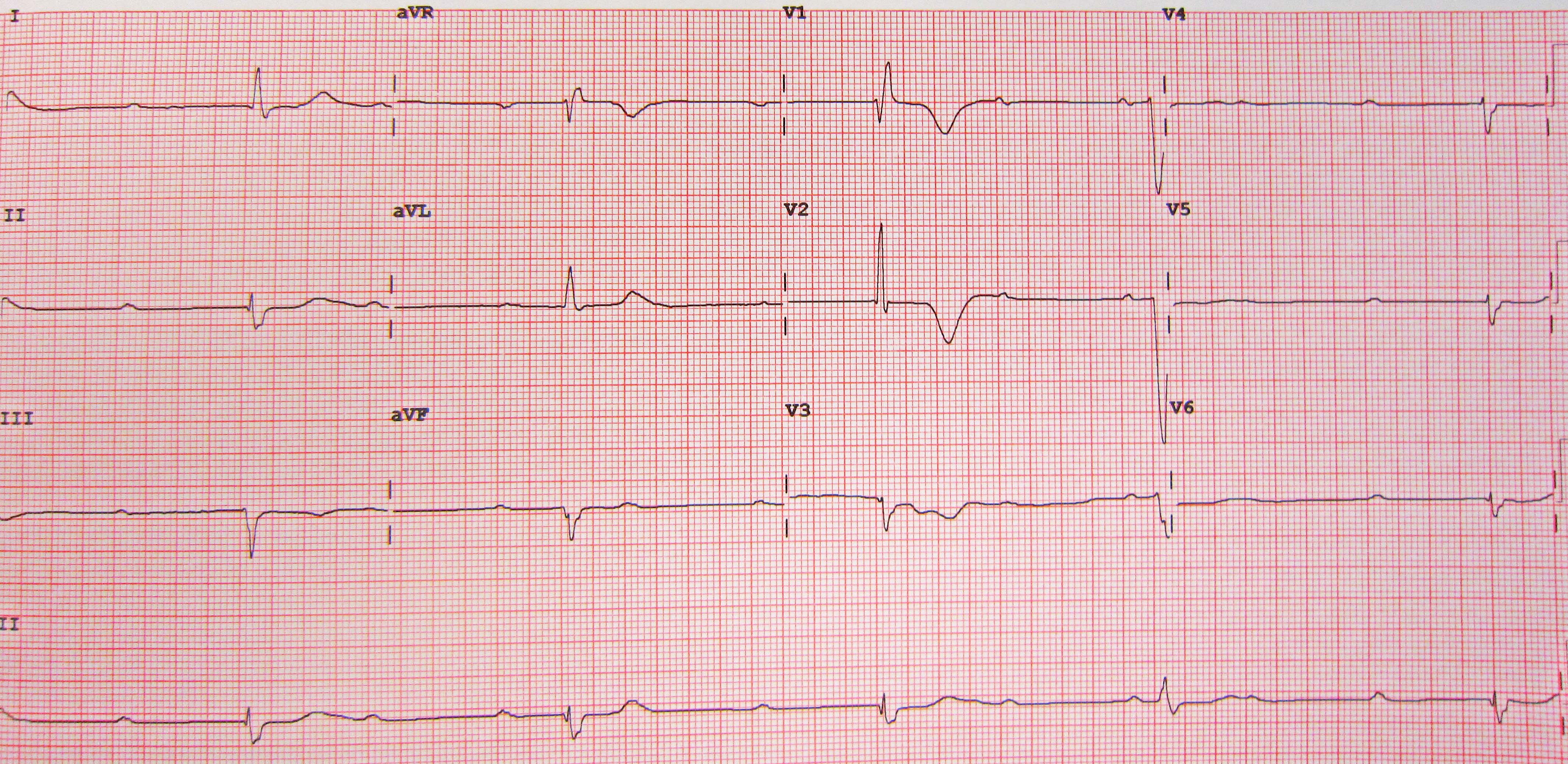
James Heilman, MD, enhanced by YeraC · CC BY-SA 3.0 · reproduced without further alteration.

Practice boundary: device selection, conduction-system pacing/CRT criteria, post-TAVR/postoperative pathways, neuromuscular/infiltrative disease, and EP escalation require specialist assessment.
A
Pattern recognition
clues, not proof| Pattern | Features | Useful next step |
|---|---|---|
| Reflex / vasovagal | Prolonged standing, heat, pain/emotion, prodrome of warmth/nausea/diaphoresis, pallor, rapid recovery | Education, individualized fluid/salt advice, and counter-pressure maneuvers when a prodrome gives time. Avoid routine salt/fluid loading in HF or supine hypertension. Tilt testing can help when the diagnosis remains uncertain. |
| Situational | Micturition, defecation, cough, swallow, instrumentation | Address trigger and injury risk. Consider cardioinhibitory monitoring when recurrent/severe |
| Orthostatic | Standing-related. Volume loss, autonomic disease, vasodilator/diuretic, postprandial. Delayed forms can be missed by a quick reading | Standardized supine/standing BP/HR, medication/volume review, autonomic evaluation when persistent |
| Arrhythmic | Sudden, little prodrome, exertional/supine, palpitations, abnormal ECG/heart disease/family history | Monitoring matched to frequency, echo, EP/inherited-disease evaluation as indicated |
| Structural / cardiopulmonary | Exertional with murmur/valve disease/HCM. PE, PH, tamponade, aortic disease. HF/shock clues | Urgent disease-specific imaging and management |
B
Common mimics
ask the witness- Seizure prolonged postictal confusion, lateral tongue bite, stereotyped aura. Brief myoclonic movements can also occur during syncope.
- Hypoglycemia / metabolic often slower recovery. Confirm glucose and context.
- Intoxication persistent altered state and exposure clues.
- Psychogenic pseudosyncope prolonged apparent unresponsiveness, frequent events, closed eyes and normal physiology during captured episode. Diagnose positively, not dismissively.
- Falls without recalled LOC in older adults may conceal syncope. Reconstruct injury and medications.
Measure the response to standing
- Rest supine 5–10 minutesRecord BP and heart rate.
- Stand with assistanceRecord symptoms and BP/heart rate at about 1 and 3 minutes.
- Interpret the changeA sustained fall ≥20 mmHg systolic or ≥10 mmHg diastolic within 3 minutes meets classic orthostatic hypotension criteria.
A normal brief test can miss initial or delayed orthostatic hypotension. Correlate the BP fall with symptoms and the event history.
C
Orthostatic medication pass
preserve indication- Review vasodilators, alpha blockers, nitrates, diuretics, psychoactive drugs, dopaminergic agents and alcohol. Do not stop HF/vascular therapy without defining the indication and a safer dose/timing alternative.
- Look for anemia/bleeding, dehydration, adrenal disease, neuropathy/Parkinsonism, deconditioning and postprandial effects.
- Document supine hypertension when treating neurogenic orthostasis. It constrains salt/fluid loading and pressor use. Compression is generally used while upright.
Mechanism statement: “Pattern favors ___ because ___. Competing dangerous cause ___ assessed by ___. Injury risk ___. Targeted test/intervention ___. Reassessment ___.”
A
Pre-implant questions
define the job| Question | Why it matters |
|---|---|
| What is the pacing indication? | Prognostic AV block, symptom-correlated sinus disease, pause-dependent syndrome, postoperative disease, or temporary bridge |
| What rhythm / atrial function? | Sinus rhythm, atrial arrhythmia burden and AV conduction determine atrial lead and tracking needs |
| Expected ventricular pacing burden? | High RV pacing burden can worsen synchrony in susceptible patients. LV function and HF risk affect physiologic pacing/CRT discussion |
| Access / infection / anatomy? | Dialysis access, congenital/surgical anatomy, bacteremia, prior leads, tricuspid disease, radiation and future MRI matter |
| What are the goals? | Symptoms, survival, HF prevention, procedural burden, future generator changes, and end-of-life preferences |
B
When the pacing question expands
EP / HF / imaging- LVEF 36%–50% with anticipated substantial ventricular pacing → cardiac physiologic pacing (CRT, His-bundle or left-bundle-branch-area pacing) is reasonable to reduce pacing-induced cardiomyopathy risk. The 2023 guideline uses approximately 20%–40% or more ventricular pacing to define substantial exposure. With normal LVEF, physiologic pacing may be considered when substantial pacing is expected. The 2025 device AUC also stratify selection by anticipated pacing burden and LV function.
- LBBB / HF symptoms → optimize GDMT and document QRS morphology/duration, rhythm, LVEF and timing before device eligibility decisions.
- Unexplained conduction disease → consider ischemic, infiltrative/inflammatory, genetic/neuromuscular or Lyme/infectious context when clinically appropriate.
- Post-TAVR / postoperative → use procedure-specific monitoring and implant timing. Early conduction injury may recover. A device pacing percentage also reflects programming and does not by itself prove pacing dependence.
C
After implant
capture the baseline| Before discharge | Longitudinal |
|---|---|
| Device type/model, indication, settings, underlying rhythm/dependence, lead parameters, wound/access, CXR when indicated, remote monitoring enrollment, emergency/device card and restrictions | Wound and device checks, battery/lead trends, atrial arrhythmia, pacing burden and symptoms. Periodic LVEF assessment with substantial RV pacing or chronic LBBB. MRI/procedure planning. |
D
Device red flags
urgent interrogation- Syncope/presyncope in a pacing-dependent patient, capture/sensing failure, unexpected bradycardia, diaphragmatic stimulation, lead alert or urgent generator replacement status.
- Fever/bacteremia, pocket drainage/erosion, rapidly expanding hematoma, pneumothorax symptoms, arm swelling/venous obstruction.
- Pocket erosion, purulence or a sinus tract implies device infection. Obtain blood cultures and urgent device/infectious-disease assessment. Definite infection generally requires complete system removal plus antibiotics. Superficial incisional infection without pocket involvement is different and needs close follow-up.
Practice boundary: verify current CRT/conduction-system pacing criteria, post-TAVR monitoring, device follow-up, MRI compatibility, and extraction/infection escalation with the device team.