Emergency
Actions
Actions
Arterial air detected
stop pump clamp both arterial & drainage cannulas · 100% FiO2 · perfusion STAT. Treat an air alarm as real until the circuit is checked.
Sudden low flow / chugging
Reduce RPM first to break suction. Look: kink · cannula · MAP. 3 D's: Drainage · Drugs · Device (preload · afterload · obstruction).
Right radial SpO2 ↓
North–South syndrome until proven otherwise. Optimize vent · ↑ ECMO flow (transient) · consider V-AV via IJ return. Right-radial gas screens upper-body oxygenation.
Limb ischemia · return side
Check pulses · Doppler · exam q1h. If absent / concerning exam → place / re-site DPC · page MCS limb team. (securement & DPC → P.9)
01
Indications & Selection
Refractory cardiogenic shockConsider VA-ECMO
Refractory cardiogenic shock with worsening hypoperfusion despite appropriate treatment · selected SCAI D–E or rapidly deteriorating C · post-cardiotomy shock · massive PE with RV failure · myocarditis · refractory VT/VF · bridge to recovery / decision / LVAD / Tx.
ECPR trigger (refractory arrest)
Selected refractory arrest · short no-flow interval · reversible cause · rapid cannulation-capable system. Aim for flow within 60 min. Shockable rhythm has the strongest trial evidence, but selected PEA may qualify.
Reasons to avoid or reconsider
Irreversible neurologic injury or no recovery/bridge option · uncontrolled bleeding. Aortic dissection and significant aortic regurgitation make peripheral VA support particularly hazardous and require a specialist alternative.
Pre-cannulation checklist
- Vascular bilateral femoral US (size, atheroma) · peripheral pulses · aorta if dissection risk.
- Echo assess aortic regurgitation, LV thrombus, tamponade, RV/LV size and aortic-valve opening before choosing support.
- Labs CBC · INR/PTT · fibrinogen · T&X · lactate · ABG.
- Access right-radial monitoring for femoral arterial return, plus reliable IV access. Sampling sites depend on the return configuration.
- Anticoag select cannulation and maintenance anticoagulation with the ECMO team, accounting for bleeding, prior heparin/HIT, renal function and the program assay (P.8).
Set destination early
ECMO is a bridge, not a destination. Frame at cannulation: recovery (myocarditis, post-MI stunning, post-cardiotomy) · decision (unclear neuro / oncologic context) · LVAD / Tx (chronic low EF). Revisit on every MDT round.
02
Cannulation & Limb
Peripheral VA — femoro-femoralCircuit anatomy
Drainage (venous, blue) tip near right atrium/caval region, according to cannula design → centrifugal pump → pre-membrane (Ppre) → oxygenator → post-membrane (Ppost) → arterial return (red) to femoral artery, retrograde aortic flow. The oxygenator has separate blood, sweep-gas and heat-exchanger connections.
Drainage cannula
- Size 21–25 Fr femoral vein, multistage examples. Position the tip and side holes for the selected cannula under imaging.
- Too shallow (iliac/IVC) → tip suction at small caliber → chugging.
- Too deep (across TV) → tricuspid impingement, arrhythmia.
- Confirm with echo/fluoroscopy at insertion and after suspected migration. Mark and document skin depth.
Arterial return + DPC
- Size 15–19 Fr femoral artery. Balance vessel caliber, limb perfusion and the pressure–flow curve.
- DPC 5–7 Fr in SFA antegrade — establish a distal-perfusion strategy at cannulation. Treat ischemia immediately.
- Limb monitoring q1h — pulses · Doppler bilateral. DPC patency.
- Limb ischemia absent pulses, mottling, ↑ CK/myoglobin → page MCS limb team. Fasciotomy if compartment syndrome.
Avoid post-cath manipulation
Do not manipulate a secured cannula blindly. Suspected migration requires the cannulating team, imaging and a controlled rescue-ready setting.
03
Four Levers — Initial Settings
Flow · FdO₂ · Sweep · Temp
1
Flow
Perfusion support
An initial flow around 60 mL/kg/min is a starting estimate. Titrate to perfusion, lactate trend, oxygen delivery and LV loading. RPM ≠ flow. Individualize alarms and MAP, often ≥65 mmHg.
2
FdO₂
Oxygenation
Start 1.0 during stabilization, then titrate using patient and post-oxygenator gases. Avoid hypoxemia and unnecessary hyperoxemia. A good post-oxygenator gas does not prove cerebral or coronary oxygenation.
3
Sweep
CO₂ removal
A gas:blood flow ratio near 1:1 is one initial approach. Titrate sweep to serial PaCO2. Avoid abrupt correction of hypercapnia, especially at initiation.
4
Temp
Demand
Prevent fever and monitor core temperature. After arrest, use a protocolized temperature-control strategy when unresponsive to commands (P.4). Cooling is not a routine treatment for inadequate flow.
LV unloading — when & how
- Why retrograde flow ↑ LV afterload → LV distension, pulmonary edema, stasis / thrombus, poor coronary perfusion. LV emptying + echo example → P.10
- Watch / trigger absent AV opening, rising PAD, ↑ pulm edema, low or declining pulse pressure.
- Options pharmacologic optimization, IABP, transaortic pump or direct LV/LA drainage. Rescue unloading differs from routine prophylactic unloading.
Routine ECMO is not a default for every infarct-related shock
ECLS-SHOCK found no reduction in 30-day mortality with routine early ECLS and more bleeding and vascular complications. Select rescue support by trajectory, reversibility and an exit plan.
T
First 2 hours after cannulation
Stabilize · sample · set alarmsFlow & pressures
Set patient-specific flow and alarms. Record measured pressures and ΔP with the sensor locations, then reassess after changes.
Gas + ABG
Obtain patient and post-oxygenator gases soon after initiation and after changes. Establish an upper-body oxygenation baseline.
Echo
TTE / TEE for cannula tip, AV opening, LV distension, RV, AI, tamponade. Mark depth at skin.
Limb
Verify DPC patency and bilateral limb perfusion repeatedly after cannulation. Record Doppler and examination findings.
Anticoag
Document agent, dose, assay, target and next sampling time. Trend bleeding, platelets, fibrinogen and hemolysis with the circuit examination (P.8).
Daily
levers
levers
Flow to patient need
Centrifugal output → O2 delivery + MAP. RPM ≠ flow · watch suction & afterload cut-off.
FdO₂ 1.0 → titrate
Check both patient and membrane gases. In femoral VA support, right-radial sampling screens for upper-body hypoxemia.
Sweep ~1:1 with flow
Clears CO2. Adjust using serial gases. Avoid a rapid fall in PaCO2 at initiation.
Temperature control
Check core temperature and heat-exchanger settings. Prevent fever. Follow the postarrest plan when applicable.
Anticoagulation plan
Agent-specific assay and target. Review hemolysis, platelets, fibrinogen, bleeding and circuit clot (P.8).
1
System-based assessment — eight quick passes
Head-to-toe · special considerations on ECMONeurologic
Assess sedation and neurologic examination daily when safe. Circuit sequestration and organ failure alter drug exposure unpredictably. Titrate to effect. Any acute change prompts evaluation for stroke, hemorrhage or seizure.
Cardiovascular
Assess pulsatility daily. Serial echo for LV/RV recovery + AV opening. Invasive monitoring can mislead — balance afterload, ECMO flow, & native output.
Pulmonary
Use lung-protective ventilation, e.g. VT 4–6 mL/kg predicted weight, with individualized PEEP/FiO₂ and limited driving pressure. Native lungs still affect upper-body oxygenation. Check right-radial gases with femoral return. Avoid oxygen extremes and rapid CO₂ correction. Extubate/mobilize when safe.
GI
Stress-ulcer prophylaxis when indicated. Start cautious enteral nutrition after hemodynamic stabilization and assess tolerance. Delay feeds during uncontrolled shock or suspected bowel ischemia. Examine for intolerance, bleeding and ischemia.
Renal
AKI is common & prognostic. Use CRRT for accepted indications, including refractory fluid overload. Avoid depletion that impairs drainage or perfusion. In-line CRRT may reduce additional vascular access. Use a circuit configuration approved by the treating program.
Hematologic
Review the agent-specific anticoagulation plan (P.8). Assess hemolysis with plasma-free hemoglobin plus supporting LDH, haptoglobin and urine findings. LDH alone is nonspecific. Consider HIT after heparin exposure. Use viscoelastic testing when useful for bleeding.
Vascular / MSK
Limb ischemia · DPC function q1h (pulses, Doppler, exam). Access-site bleeding or hematoma · pseudoaneurysm. Reposition q2h to prevent pressure injury.
Infectious
High burden of nosocomial infection. Culture and treat suspected infection by syndrome and severity, then de-escalate. ECMO alone does not mandate ongoing antibiotics. Review lines and source control daily.
2
Adequacy of support — three questions before any number changes
Numbers in physiologic context
Q1
Is the patient supported?
Perfusion endpoints- MAP often ≥65 mmHg, individualized to perfusion and comorbidity. Avoid unnecessary vasoconstriction that worsens LV afterload.
- Lactate trend toward clearance. A rise prompts reassessment of flow, oxygen delivery, bleeding, sepsis and regional ischemia.
- End organs LFT & Cr direction · urine output · stool · ileus on exam.
- Clinical skin perfusion · cap refill · mottling · mental status (sedation-adjusted).
- Venous saturation trend with Hb, flow and lactate. Specify the sampling site. Drainage-line saturation may not represent true mixed venous saturation.
Q2
Is ECMO flow appropriate?
Flow is not a vital sign alone- Too low reassess patient perfusion and localize the cause. Correct drainage, obstruction or excessive afterload before increasing speed.
- Too high → hemolysis, LV distension, circuit stress — ↓ RPM, optimize native function.
- Context body size · fever (↑ VO2) · residual native CO · RV–LV balance · vasopressor state.
- Don't titrate flow to "look right" — titrate to Q1 endpoints.
- Daily document flow, RPM, measured circuit pressures, ΔP, post-oxy ABG, hemolysis labs.
Q3
Native cardiac function optimized?
Pulsatility · AV opens · stasis- Pulsatility trend pulse pressure with the rhythm and echo. New loss prompts immediate assessment for absent ejection, LV distension or circuit/patient deterioration.
- AV opening track opening frequency, LV size, spontaneous echo contrast and congestion. A single pressure target does not establish adequate emptying.
- Inotropes minimize without losing pulsatility. Low-dose dobut / milrinone often enough.
- Stasis closed AV + low PP = LV thrombus risk. Re-assess venting strategy.
- Unload discuss a suitable strategy for persistent LV distension, pulmonary edema, stasis or inadequate ejection.
3
Patient complications — daily look-for list
Sepsis · bleeding · thrombus · limb · neuro- Sepsis high baseline risk. New fever / vasoplegia / leukocytosis → pan-culture, broaden empirically.
- Bleeding · coagulopathy access site, GI, intracranial, surgical drains. Trend fibrinogen, plt. Daily exam.
- Thromboembolism stroke, limb, mesenteric, pulmonary. Sub-therapeutic AC + circuit stasis = high risk.
- Differential hypoxia Right-radial saturation / ABG screens upper-body oxygenation during femoral VA support.
- Limb ischemia pulses · Doppler bilateral q1h. DPC patency.
- Renal failure CRRT for refractory overload, electrolyte/acid-base problems or other accepted indications.
- Neurologic injury stroke / ICH / hypoxic injury — low-threshold imaging.
4
Circuit complications — daily look-for list
Pump · oxy · cannulae · DPC- Access insufficiency drain P very negative · chugging · oscillating flow → reduce RPM first.
- Hemolysis rising LDH, falling haptoglobin, brown urine — pump head + oxygenator + suction.
- Bleeding cannula site, IV sites, mucosal — fix AC + correct underlying diathesis.
- Thrombosis pump head, oxygenator (rising ΔP), or DPC. Plan a controlled change-out before crisis. DPC can fail silently: it is a passive side circuit, so normal displayed ECMO flow does not prove distal-limb flow. Trace it by hand for kink/clot and verify bilateral pulses/Doppler, color, temperature, refill, and NIRS when used. Escalate a concerning limb exam urgently.
- Cannula migration / kink sudden flow drop or pressure change → inspect and image. Manipulation requires the cannulating team and rescue readiness.
- Cannula rupture profuse site bleed → clamp circuit + pump off. Never re-suture a leaking cannula.
5
Forward trajectory & exit — revisit destination on every change
Set at cannulation · reaffirm dailyRecovery
LV/RV improving · AV opens · lactate clears · pulsatility ↑ — turn-down trial per P.5.
Bridge to VAD
No recovery · Tx ineligible or long wait — early HF / surgery huddle.
Bridge to Tx
Acceptable candidate · early listing · UNOS status & center protocols.
Decision pending
Time-limited observation · neuro & end-organ recovery define path.
Palliation
Futile / no candidate — goals-of-care meeting · planned withdrawal pathway.
The circuit is part of the physical examination. Rounds note skeleton:
ECMO type · configuration · day of support · flow · RPM · measured circuit pressures · ΔP · post-oxy ABG · hemolysis labs · AC · cannula depth marks · DPC patency · limb exam · bleeding sources · transfusions · sedation · vent · nutrition · mobility · destination reassessed (recovery / decision / VAD / Tx / palliation).
4
Pressure Cheat Sheet
Localize in 10 seconds- Pdrain pre-pump, usually negative during active drainage. Reflects how hard pump is pulling. Set limits for the circuit and patient using its IFU.
- Ppre upstream of oxygenator — after pump.
- Ppost downstream of oxygenator — closest to patient on return side.
- ΔP = Ppre − Ppost trend reflects oxygenator resistance. Compare at similar blood flow. A rising gradient can suggest obstruction.
Increasingly negative drainage pressure + low flow
Reduce RPM to break suction, then assess volume, bleeding, obstruction and cannula position. Plasma-free hemoglobin supports hemolysis assessment. LDH alone does not establish suction injury.
High return pressure at comparable blood flow
Downstream obstruction — check cannula kink, patient position, clamps. Risk of tubing rupture / circuit decoupling rises with sustained high pressure.
Pattern recognition — 4 grids
A
Both ↑ together
↑Ppre
↑Ppost
B
Both ↓ together
↓Ppre
↓Ppost
C
ΔP rising at comparable flow
↑Ppre
→Ppost
D
Drain ↓↓ + chugging
↓↓Pdrain
~flow
5
Low-Flow Differential
Afterload · Preload · Obstruction
A
Afterload-limited
SVR / MAP too high- Clue circuit pressures rise · MAP very high · flow falls at given RPM. Common just after vasopressor up-titration.
- Mechanism centrifugal pump is afterload-sensitive — pump cut-off curve flattens flow as resistance rises.
- Fix reduce / hold pressors · vasodilate if appropriate (nicardipine, NTG) → then ↑ RPM. Individualize MAP to perfusion and LV loading.
B
Preload / drainage
Chugging — most common- Clue flow oscillates · audible chugging · drain pressure swings very negative · pressures drop when flow drops.
- Causes hypovolemia, bleeding, tamponade, pneumothorax, excessive RPM, cannula tip against wall / septum.
- First move ↓ RPM to stable flow before anything else.
- Then assess fluid responsiveness and bleeding, echo, patient position and cannula. Give volume when indicated, then re-titrate slowly.
C
Mechanical obstruction
Persistent, not oscillating- Clue low flow steady (not oscillating) · drain pressure stays very negative · no volume response.
- Causes kink, thrombus in circuit, tip migration, cannula too shallow (iliac/IVC), DVT propagation, abdominal compartment syndrome.
- Workup tubing walk-through · CXR / fluoro · echo (cannula tip) · bladder pressure if abdomen distended.
- Fix reposition · OR re-do if confirmed kink / thrombus · escalate to perfusion + CT surgery.
6
Oxygenator Function & When to Change
ΔP · gas exchange · hemolysisDaily checks
- Post-oxygenator saturation and PaO2 assess membrane output, not native lung or upper-body oxygenation.
- Post-oxygenator ABG: compare at similar blood flow, FdO2 and pre-membrane saturation. Sensor saturation alone can mislead.
- Gas source confirm blender / wall vs tank during transports — silent failure.
- Hemolysis / consumption plasma-free hemoglobin, LDH, platelets, fibrinogen and circuit findings. Test frequency follows stability.
"Sigh / burp" technique
Gas-side condensate can ↓ transfer. ECMO/perfusion: brief ↑ sweep only per oxygenator IFU, then restore prescribed sweep. Monitor gases and avoid hypocapnia. Repeat intermittently only if permitted. Recheck post-oxy gases at baseline settings.
When to plan a change-out
- Trending PaO2 falling on post-oxy ABG at comparable flow, FdO2 and inlet saturation.
- ΔP rising with clinical impact (hemolysis, oxygenation, alarms).
- Hemolysis rising LDH, falling haptoglobin, brown urine.
- DIC pattern fibrinogen falling, d-dimer rising, plt consumption.
- Visible clot in oxygenator on inspection (flashlight).
Plan, don't crash
Plan with perfusion + CT surgery for a controlled exchange. Use trends and patient impact to plan an exchange before catastrophic failure. No single age or PaO2 threshold mandates exchange.
Differential — gas exchange failure
- Gas source off / disconnected → check blender, tank pressure, line connection.
- CO2 clearance inadequate → assess gas supply and sweep. Increasing sweep chiefly changes CO2 removal.
- Oxygenator condensation → device-permitted sigh + reassess (P.7).
- Membrane clot / failure → gas exchange worsens, with or without a ΔP rise → assess for exchange.
- Recirculation / shunt on V-V or V-AV configurations — re-evaluate cannula tips.
- Differential hypoxia on peripheral VA — see P.4.
7
Air / Bubble Management
Prevent · detect · respondHigh-risk moments
- Opening a central line to atmosphere near drainage (IJ tip in SVC/RA).
- Loose connections during transport or line manipulation.
- Aggressive flushing or contrast near bubble sensor.
- Drainage cannula tip suctioning intermittently exposed to air (chugging + open lines = disaster).
Rule
No open-to-air manipulation of any line that drains into SVC/RA while on ECMO. Pause infusions, cap, then resume.
If air alarms
- 1. Look — pump head, oxygenator, tubing for visible air.
- 2. If air in arterial limb → Stop pump · clamp · 100% FiO2 · perfusion STAT.
- 3. Venous / pump air → ECMO/perfusion isolates and de-airs the affected segment using the circuit procedure. Do not push air forward.
- 4. Reset bubble detector only when circuit confirmed clear.
False positives
- Contrast microbubbles (Definity, Optison) can trigger bubble alarm / pump stop.
- If ultrasound contrast is needed, agree on a device-specific plan with perfusion before injection. Do not bypass an air safety response solely because contrast was given.
- Keep infusion and sampling connections closed and air-free. Follow the circuit-specific sampling procedure.
Stroke is the cost
Arterial air on VA → high stroke risk. Treat every bubble alarm as real until proven otherwise.
8
Differential Hypoxia — North–South Syndrome
Femoral VA + sick lungsOne-sentence mechanism
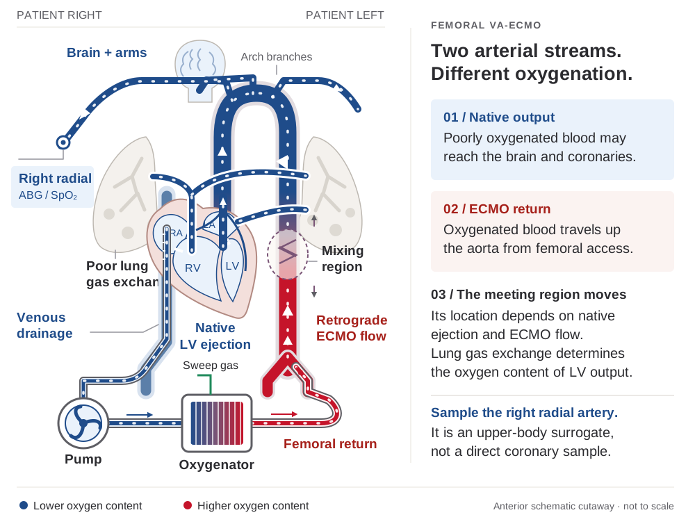
With poor lung gas exchange, native LV ejects desaturated blood toward the coronaries and brain. Oxygenated ECMO return travels retrograde. Their aortic mixing region shifts with native ejection, ECMO flow and lung function.
Monitor
Trend right-radial gases and perfusion. If unavailable, agree on alternative upper-body monitoring. Recheck after changes.
Detect
- Right radial SpO2 / ABG lower than expected despite good post-oxy sat. Right-radial sampling screens the upper body, but does not directly measure coronary oxygenation.
- Compare sites: upper-body hypoxemia may coexist with normal femoral or post-oxygenator gases. The pre/post membrane saturation difference does not locate the mixing zone.
- Reassess native ejection, lungs, cannulation and upper-body gases together. A right-radial sample cannot define the exact aortic mixing point.
Management ladder
Step 1
Optimize lungs — recruitment, PEEP, treat pulm edema, diurese, bronchospasm, infection. Best ROI first.
Step 2
↑ ECMO flow transiently — pushes mixing point proximally / shrinks native ejection effect. Watch suction and LV loading.
Step 3
Consider V-AV — add oxygenated venous return to improve blood reaching the native heart. Measure and balance both return limbs.
Step 4
Reassess unloading if LV distension or pulmonary edema is present. Impella moves LV blood into the aorta without oxygenating it. Effects depend on lung function and the balance of flows.
Step 5
Choose the configuration with the team: V-AV, axillary/central return, or VV if cardiac recovery permits. Measure both return flows in V-AV.
9
ECPR — Refractory Arrest Playbook
Perfusion first · cause secondActivation criteria
- Short no-flow interval with prompt CPR, usually a witnessed arrest. Special causes such as hypothermia require a distinct pathway.
- Rhythm and cause: shockable OHCA has the strongest trial evidence. Selected PEA with a reversible cause may be eligible under an established pathway.
- Activate early when conventional resuscitation is failing and candidacy is plausible. Continue high-quality CPR while the team evaluates.
- Low-flow < 60 min preferred (not an absolute exclusion).
- Age, comorbidities, baseline function permit recovery.
- Cannulation team available — perfusion · CT surgery · ECMO MD.
Candidacy limits
Established irreversible injury, no treatable cause or recovery/bridge option, and goals inconsistent with support. Uncertain downtime or major comorbidity requires rapid senior review.
Workflow
Early
Recognize refractory arrest — ACLS continues · activate ECPR team.
CPR
High-quality CPR continues. A trained team may use mechanical compression for transport/cannulation with minimal pauses.
Access
Rapid cannulation — ultrasound-guided access, imaging and a distal-perfusion plan. Avoid interrupting CPR.
Flow
Aim for ECMO flow within 60 min. Stop compressions only after effective circulation is confirmed. Follow pressure and perfusion.
Cause
Treat the cause promptly. Coronary angiography for suspected coronary occlusion. PE requires a reperfusion decision with the team.
ICU
Postarrest care — if unresponsive, temperature control 32–37.5 °C for ≥36 h. Multimodal prognosis after confounders clear, generally ≥72 h after normothermia.
"Why did they arrest?"
- Acute coronary occlusion → urgent cath / PCI. Most common reversible cause.
- Massive PE → thrombectomy or lysis pathway. RV support. CTA on table if feasible.
- Refractory VT/VF → cath ± ablation referral.
- 5 H's & 5 T's — work the full reversible-cause checklist:
HypoxiaTamponade (cardiac)HypovolemiaTension pneumothoraxH+ — acidosisToxins / drugsHypo- / hyperkalemia (& -Mg, -Ca, -glucose)Thrombosis — pulmonary (PE)HypothermiaThrombosis — coronary (ACS)
- Aortic catastrophe → if found, ECMO often contraindicated. CT scan early.
Post-flow targets
- MAP ≥65 as an initial goal · improving perfusion and lactate trend
- Right radial ABG q1–2h until stable
- After reliable measurement, titrate oxygen to avoid hypoxemia and hyperoxemia using the postarrest target and ECMO sampling configuration
- Family meeting within 24 h · destination framing
10
Running with no / minimal anticoagulation
temporary interruption · reassess oftenWhen to consider
- Major / active bleeding — post-cardiotomy, intracranial, trauma, pulmonary or GI hemorrhage — or bleeding-predominant coagulopathy. HIT usually requires a non-heparin anticoagulant, not simply no anticoagulation.
- Selected observational series report feasible temporary anticoagulation interruption. They do not prove equal thrombotic safety or support routine anticoagulation-free VA-ECMO.
Safety parameters
- Avoid stasis: maintain effective circuit flow and inspect low-flow segments. No universal flow rate makes anticoagulation interruption safe.
- Watch the LV if it isn't ejecting (no pulsatility, AV stays shut) → aortic-root / LV stasis → thrombus. Vent or preserve some ejection — a reason to anticoagulate.
- Surveil the circuit oxygenator ΔP & gas exchange, visual clot, fibrinogen / plt / D-dimer trends.
- Reassess frequently: new clot, embolism or gas-exchange failure requires urgent team review. Restart when hemostasis permits, balancing the competing risks.
VA caveat: the arterial limb means a circuit / LV clot can embolize systemically (stroke, limb, mesenteric) — higher stakes than VV. Keep AC-free periods as short as the bleeding requires, document the plan, and re-anticoagulate once bleeding is controlled.
Readiness — biochemical
Lactate clearing · end-organ recovery · adequate perfusion pressure on low vasoactive support.
Readiness — pulsatile
Aortic valve opens with sustained pulsatility and stable perfusion during support reduction.
Readiness — echo
Assess both ventricles, AV opening, LVOT VTI and filling pressures at reduced support. Thresholds below are cohort clues.
Do not wean
Active bleed · circuit issue · rising lactate · arrhythmia storm · refractory hypoxia · severe lung disease. Fix first.
14
Weaning & Turn-Down Trial
Staged · controlled · echo-drivenReadiness domains — assess together
- Etiology corrected — the cause of cardiac failure has been treated (PCI, ablation, anti-inflammatory, time).
- Pulsatile waveform — sustained ejection, interpreted with AV opening and ventricular loading.
- MAP holds — > 65 without high-dose inopressors.
- Metabolic resolved — lactate normal · pH normal · K corrected.
- Lungs / AC — lung function not severely impaired · tolerating anticoagulation.
Turn-down trial workflow
- Stabilize first — bleeding / circuit issues addressed. Timing follows the cause and evidence of recovery.
- Stepwise flow ↓ — use the team's controlled protocol with continuous pressure/echo assessment and serial perfusion checks. Stop deterioration promptly.
- Minimum flow trial — the minimum and duration depend on circuit and program technique. Confirm adequate anticoagulation and rescue readiness before low-flow testing.
- Echo focus — LV recovery (LVOT VTI, EF, AV opening) + RV function (TAPSE, S', size). RV often easier on TTE than TEE.
- Pass → plan controlled percutaneous or surgical decannulation with vascular closure and rescue support ready.
- Fail → back to support, reassess destination on next MDT round.
High vasoactive need argues against removal
Persistent high vasoactive requirements or deteriorating perfusion during turn-down argue against removal. Restore support and reassess.
Staged decannulation strategies
Standard
Pass turn-down → percutaneous or surgical decannulation in OR · vascular closure (Perclose / suture / cut-down) · daily limb exam.
Chronic ↓ EF
If LV dysfunction persists but RV and lung function permit, consider transition to isolated LV support. ECMO can worsen LV afterload, so good systemic perfusion does not establish LV recovery.
RV failure
Consider an appropriate RV-support configuration when isolated RV failure persists. Account for pulmonary vascular resistance, oxygenation and device suitability.
Bi-V failure
Begin LVAD/transplant evaluation early if recovery is unlikely. Timing follows etiology, neurologic and end-organ trajectory, candidacy and goals.
Non-recovery
Destination conversation: LVAD candidacy, transplant listing, or transition to comfort. Engage palliative early.
Limb & access after decann
Continue access-site and distal-limb surveillance after removal. Use the program ultrasound strategy to detect thrombosis, pseudoaneurysm or arterial injury. Reassess urgently for swelling, pain, ischemia or bleeding.
▸
Weaning framework — what each trial tests
V-A and V-V differV-A — wean the flow
- Support reduction tests native cardiac output, filling pressures and biventricular reserve. Assess systemic arterial gas exchange as flow falls.
- Echo clues LVOT VTI ≥10 cm, LVEF >20–25% and lateral mitral S′ ≥6 cm/s were associated with success in a small observational cohort, not validated stand-alone cutoffs.
- Technique flow reduction, a brief clamped trial or pump-controlled retrograde trial off require a trained team and a defined circuit protocol. Low-flow support can still mask RV failure.
- Stop for falling perfusion pressure, rising filling pressures, ventricular dilation, hypoxemia or intolerance. Restore support and reassess the cause.
V-V — keep flow, wean gas
- Ready native lungs can maintain acceptable oxygenation and ventilation on a lung-protective ventilator strategy.
- Wean gas reduce FdO₂ and sweep using the VV protocol, then assess a monitored sweep-off trial.
- Maintain blood flow sufficient to limit circuit stasis during the gas-off trial. Minimum flow follows the circuit and protocol.
Other temporary devices (IABP · Impella · TandemHeart) & detailed device-by-device decannulation technique → MCS Weaning & Decannulation card.
15
Exit pathways & destination
Set at cannulation · revisit dailyBridge to…
Recovery
Myocarditis · post-MI stunning · post-cardiotomy · refractory VT/VF. Turn-down → decannulate. Expect days, not hours.
VAD
Chronic ↓ EF. Impella 5.5 overlap → decannulate ECMO → HF ICU → LVAD eval. Tx-listed pts often go direct.
Transplant
Listed candidate or rapid eval feasible. Requires acceptable oncologic / functional candidacy + center pathway.
Palliation
No reversible cause + no destination — engage palliative early, name surrogate, document plan in EHR.
TBD
Trajectory unclear → daily MDT, family meeting in 24 h, set time-limited trial with explicit milestones and a reassessment date.
When to call who
Perfusion
Immediately for critical alarms, suspected air, loss of flow or oxygenator failure. Involve the team early for planned exchange.
CT surgery
Cannulation · decannulation · bleeding from access · suspected dissection · cannula migration / rupture.
MCS limb team
Limb ischemia · pseudoaneurysm · DPC placement / failure · compartment syndrome.
Advanced HF
LVAD / Tx evaluation · destination decisions · LV venting strategy.
Palliative
Family meetings · goals-of-care conflict · time-limited trial framing · re-cannulation discussions.
Three immediate circuit threats
(1) Air in the arterial limb. (2) Bleeding + over-anticoagulation. (3) Decannulation / cannula rupture by movement or migration. Hard-stop checks for each.
VitalFlow™
Centrifugal · touchscreen · MC3 / Medtronic
Illustrative control layout
1Oxygenator dock sits on top. Clip-on as single piece
2Status light bar green/yellow/red — read from across the room
3Tile screen all pressures, sats, temp on one glance
4Big numbers Flow (L/min) + RPM — the two you titrate
5Three hard keys silence · alarm settings · lock
6Physical knob RPM — confirm response and delivered flow
First touch
Confirm flow, alarms and lock state after every change. Use the exact console version's instructions.
Numbers on screen
Pin · PΔ · Pout · SO2 in/out · T · Flow · RPM all integrated. Confirm SaO2 with a real ABG daily — sensors drift.
If screen freezes
Confirm actual flow and activate the trained emergency support procedure. A separate emergency handcrank is available. Do not assume any control remains functional during a console fault.
Don't
Not for cardiotomy suction. Centrifugal pump — needs preload. Don't ↑ RPM to fix chugging.
Cardiohelp
Compact · HLS clip-on · Getinge / Maquet
Transport / legacy
AHLS Set — pump + oxygenator as one disposable, clips to right
BTile screen + tap-to-confirm hard keys ✓ ✕ alongside
CVenous probe values Hb · HCT · SvO2 · Temp on the line
DPump well HLS pump head clips directly into console
ERetaining bar verify secure attachment before transfer
FStatus LEDs on the bar — green / yellow / red, visible from across room
VitalFlow
One disposable
HLS Set combines pump head + oxygenator. Change-out is one swap, not two. Don't bump the cassette during transfers.
Venous probe
Live Hb · HCT · SvO2 · Temp on screen — available values depend on the installed sensors and console version. Values drift over days — confirm with lab daily.
Emergency
Know the emergency drive location and rehearse transfer with perfusion. Verify motor engagement and forward flow during recovery.
When you'll see it
US labeling revised January 2025 describes specified surgical bypass uses for <6 h. Prolonged ECMO is outside that label. Verify the current regional IFU and safety notices.
B
Console Buttonology — VitalFlow vs Cardiohelp
High-yield onlyMedtronic VitalFlow
Maquet Cardiohelp
Lock / unlock
Identify lock state and RPM control on the installed software. Confirm flow and re-lock after changes.
Use the installed version's unlock and confirmation sequence. Confirm displayed flow after changes.
Set low-flow alarm
Individualize the low-flow threshold and verify the alarm response.
Individualize the threshold and review after flow targets change.
Bubble / air action
Visual + audible alarm. Stop pump = arterial air action when configured.
Verify the active bubble response. Contrast requires a planned, supervised strategy rather than automatic safety bypass.
Pressures / sats
Pre + post + ΔP + pre/post sat all on the tile screen — trend in chart.
Pre + post + ΔP shown. Integrated probe adds Hb · HCT · SvO2 · Temp — confirm with ABG.
Emergency mode
Use the emergency support procedure for the fault. Locate and inspect the separate handcrank.
Know the emergency drive and transfer procedure. Confirm actual forward flow, not motor rotation alone.
Sweep / FdO₂
Blender on top of oxygenator. Verify gas source on transport.
Verify the separate gas supply, blender, sweep and backup cylinder. Console power does not prove gas delivery.
C
Cannula Quick Reference
Adult VA · verify with IFU + your team| Role | Common product | Sizes (Fr) | Notes · positioning |
|---|---|---|---|
| V Drainage — fem-RA | Bio-Medicus Life Support™ multi-stageMedtronic | 21 · 23 · 25 | Multi-stage with fenestrations distributed along shaft · tip at cavoatrial junction · TEE/fluoro confirm · mark depth at skin. |
| V-V — right-atrial dual-lumen | Crescent™ RA dual-lumenMedtronic | 13 · 15 · 19 | Pediatric-sized V-V catheter with both drainage and reinfusion lumens. Not an adult VA single-stage drainage cannula or a generic IJ return adjunct. |
| V-V — bicaval dual-lumen | Crescent™ jugular bicavalMedtronic | 24 · 26 · 28 · 30 · 32 | For long-term V-V (not VA). RIJ → bicaval. Confirm position by TEE / fluoro. Recirculation if malpositioned. |
| A Return — femoral artery | Bio-Medicus arterial / Opus™ access kitMedtronic | 15 · 17 · 19 | Match vessel caliber and pressure–flow needs. Larger cannulae may compromise limb perfusion. Plan and verify distal perfusion. |
| A Distal perfusion (DPC) | Antegrade SFA sheath (institutional) | 5 · 6 · 7 | Plan antegrade SFA perfusion at femoral VA cannulation. Use a secure, de-aired circuit connection and verify actual distal flow. |
S
Six scenarios — the ones the console will throw at you
VitalFlow sim · bedside
01
Low flow + chugging
Preload
Flow2.1
RPM3400
P drain−110
P pre120
SO₂ in72%
Audiblechug
Stop
↓ RPM first — break suction before anything else. Never ↑ RPM to "fix" low flow.
Think
Tip suction · hypovolemia · bleeding · tamponade · pneumothorax · cannula against wall.
Do
Check bleeding and fluid responsiveness, echo, position and cannula. Give volume only when indicated, then re-titrate.
Call
Call ECMO/perfusion early, immediately if perfusion is impaired. Involve the cannulating team for suspected migration.
02
Condensation: post-oxy sats fall
Gas side
SO₂ out↓
PaO₂ out↓
ΔPstable
Flowstable
Clot seennone
Exhaustwet
Check
Confirm post-oxy gases at unchanged flow / FdO₂. Check gas supply, connections and exhaust patency.
Think
Gas-side condensation can impair exchange. No visible clot or ΔP rise does not exclude thrombosis or membrane failure.
Sigh
ECMO/perfusion: brief gas-side ↑ sweep per oxygenator IFU, if gases permit → restore prescribed sweep. Monitor CO₂ and stop for hypocapnia. Repeat intermittently only if IFU / protocol permit.
Recheck
Recheck gases at baseline. Persistent decline or wet exhaust → urgent assessment for other causes, including plasma leak, and need for exchange.
03
Bubble alarm — arterial air
Air
BubbleDETECT
PumpSTOPPED
Flow0.0
RPM0
SpO₂ R rad88%
Ptdeteriorating
Stop
stop pump · clamp arterial · 100% FiO2.
Think
Treat an air alarm as real until the circuit is checked. Otherwise air source = open central line near drainage, loose connection, suction event with open line.
Do
De-air via designed ports — do not push air forward. Reset bubble detector only when circuit confirmed clear.
Call
Perfusion STAT. Document neuro exam. Treat stroke pathway if focal deficit.
04
North–South syndrome
Differential
Flow4.5
SO₂ out99%
R rad SpO₂82%
Fem SpO₂98%
PEEP8
AV opensyes
Stop
Don't be fooled by femoral sat. Right-radial sampling screens the upper body, but does not directly measure coronary oxygenation.
Think
Native LV ejecting desaturated blood to arch / coronaries / brain while ECMO returns to lower body.
Do
Optimize lungs and reassess flow. Discuss V-AV or alternate arterial return for persistent upper-body hypoxemia. Unload for LV distension or stasis, not as a fixed step for every case.
Call
ECMO MD + CT surgery early if V-AV / central conversion likely.
05
Arrest on VA ECMO
Pseudo-PEA
A-lineflat
RhythmVF / asys
Flow4.2
MAP62
Pulsatilitylost
SO₂ out99%
Stop
Assess effective perfusion immediately. If ECMO maintains adequate pressure and organ perfusion, loss of pulsatility alone is not an indication for compressions. If circulation is inadequate or uncertain, activate resuscitation and restore support.
Think
Circuit first: flow OK? Pump running? Decannulation / rupture? Air? Pump-head clot? If circuit fails → ALS until ECMO re-established.
Do
Treat the rhythm and reversible cause with the resuscitation/ECMO team. Use current defibrillator-specific ALS dosing when arrest care is needed. Set ventilation from lung function and gases, with the appropriate resuscitation or ECMO ventilation strategy.
Call
ICU consultant + ECMO coordinator immediately. Remember the pt may be awake — sedate/analgese before painful interventions.
06
Cannula rupture / decannulation
Exsanguination
Sitebleeding
Flow↓↓
MAP42
Hb↓
P drain−0 / +
Air alarmpossible
Stop
Clamp circuit proximal & distal to the breach · turn pump off. Direct pressure to the site. Never re-suture a leaking cannula — needle puncture ruptures it.
Think
Return (arterial) breach → rapid exsanguination + air risk. Drainage breach → air entrainment. Cause: suture needle, withdrawal, disconnection, cut-down site bleed.
Do
Massive transfusion · pressure · ALS if no flow. Recannulate (rewire if possible, otherwise new site) — OR or cut-down. Resume ECMO once circuit intact.
Call
CT surgery STAT · perfusion · ICU attending · blood bank — activate MTP. Secure airway if not already.
Always
comes
first
comes
first
1 · Patient
Look at the patient before the console. Pulse · perfusion · pupils · vent. The screen is data. The patient is the truth.
2 · Reduce RPM
For suction/chugging, reduce RPM to break suction. Other low-flow causes require rapid circuit and patient assessment before speed changes.
3 · Call early
Perfusion + ECMO MD. Two minds beat one at 3 am. "I'm not sure" is the right thing to say on the phone.
▶
VA-ECMO learning resources
For residents · fellows · attendingsConsole simulators
- VitalFlow™ training sim browser-based — walk the tile dashboard, alarms, and emergency mode.
infusemeddev.com/projects/medtronic/vitalflow_sim - Cardiohelp / HLS Getinge training portal — touch flow, HLS module orientation.
getinge.training
Complications & troubleshooting
- Low-flow troubleshooting intensiveblog.com/oh-no-low-flow
- Venous insufficiency / chugging intensiveblog.com/an-ecmo-earthquake
- Differential hypoxia (N–S) intensiveblog.com/different-hypoxia-va-ecmo
- Cardiac arrest on VA ECMO intensiveblog.com/cardiac-arrest-on-ecmo-does-it-still-count
- Cannula rupture / dislodgement intensiveblog.com/everything-ecmo-018
Curricula & guidelines
- ELSO General Guidelines for All ECLS Cases · Adult Cardiac Failure Guideline · ECMO Specialist curriculum. elso.org
- EuroELSO Lorusso/EACTS expert consensus on V-A ECMO (2021).
- INTENSIVE / Alfred ICU Everything ECMO series (Nickson, Hoffman, Diehl, Burrell) — case-based Q&A.
- CICU teaching structured CICU rounds curriculum (Gottula & Elliott).
A therapeutic assay does not guarantee a safe circuit. Interpret anticoagulation alongside bleeding, circuit clot, hemolysis and cardiac stasis. Use a validated program nomogram with a stated agent, assay, target and recheck interval. The exact aPTT window varies by reagent and patient.
16
Choose the agent for the patient
UFH or direct thrombin inhibitor| Agent | Why choose it? | What changes the plan? |
|---|---|---|
| Unfractionated heparin | Established ECMO agent, familiar assays and protamine reversal. Requires antithrombin. | Bleeding, HIT, antithrombin deficiency and assay discordance. |
| Bivalirudin | Directly inhibits thrombin without antithrombin. An option for HIT or a program DTI strategy. | Renal dysfunction, changing clearance and stagnant blood. No specific antidote. ECMO dosing is off-label and differs from PCI dosing. |
| Argatroban | Another non-heparin option, including when renal dysfunction favors it. | Hepatic dysfunction can markedly prolong effect. Use an agent-specific protocol. |
Evidence boundary: adult comparisons favoring bivalirudin are predominantly observational and cannot establish a survival advantage. A small pediatric pilot randomized trial does not establish superiority in adult VA-ECMO.
17
Match the measurement to the drug
trend and reconcile discordance| Measurement | Useful for | Common trap |
|---|---|---|
| Anti-Xa | UFH activity using the laboratory's heparin-calibrated method. | Not a measure of bivalirudin. Hemolysis, bilirubin, lipemia and assay design can alter results. |
| aPTT | Commonly used for UFH or DTI titration, with an agent-specific target. | Baseline coagulopathy, lupus anticoagulant and acute-phase changes can disconnect the value from drug effect. |
| ACT | A rapid whole-blood adjunct where included in a validated strategy. | Affected by platelets, temperature, anemia and factor levels. Do not equate it with a drug concentration. |
| Dilute thrombin time / ecarin | Drug-calibrated DTI assessment when available and aPTT is unreliable. | Availability and calibration differ. Consult the coagulation laboratory. |
Recheck after change
- Check after initiation, dose adjustment, bleeding, organ-function change or CRRT change. Stable monitoring frequency must be explicit.
- With heparin resistance, examine assay validity and antithrombin before escalating blindly.
Watch patient and circuit
- Trend CBC, fibrinogen, coagulation tests, visible clot, ΔP at comparable flow and membrane gas transfer.
- Use plasma-free hemoglobin when available for hemolysis. LDH and haptoglobin are supporting, nonspecific markers.
18
Bleeding, procedures and clearance
source control first- Major bleeding identify and control the source, reduce or interrupt anticoagulation with the ECMO team, and replace demonstrated deficits. Reassess when it is safe to restart.
- Bivalirudin label half-life is about 25 min with normal renal function and about 3.5 h in dialysis patients. These are pharmacokinetic observations, not guaranteed procedure-hold times.
- CRRT can change clearance in either clinical direction as renal function, filter and prescription change. Recheck the assay rather than automatically raising the dose.
- Transfusion individualize hemoglobin, platelet and fibrinogen goals to bleeding, procedures and oxygen delivery. Active bleeding generally calls for at least as much hemostatic support as the nonbleeding state.
- Acquired vWS shear-related loss of large multimers can contribute to mucosal/GI bleeding despite near-normal routine coagulation tests.
- HIT assess probability and test appropriately. Stop all heparin exposure when clinically indicated and use an appropriate non-heparin anticoagulant.
19
Viscoelastic testing complements the workup
interpret the complete tracing| Finding | Interpretation in a bleeding patient |
|---|---|
| Prolonged R / CT | Anticoagulant effect or factor deficiency. Drug-related prolongation alone is not an indication for plasma. |
| Low angle or clot strength | Use platelet count and fibrinogen-specific testing to distinguish the deficits before targeted replacement. |
| Excess clot lysis | Confirm hyperfibrinolysis and the clinical context. Antifibrinolytics require a deliberate benefit–thrombosis assessment. |
Stasis remains dangerous: a non-ejecting LV, aortic root and poorly flowing side branches can thrombose despite a therapeutic systemic assay. No minimum circuit-flow number guarantees safety during an anticoagulation hold. Check the chamber and the circuit, then reassess anticoagulation and unloading together.
ECMO team members STOP & complete the checklist together — shift change (RN–RN) · after cannulation · before & after mobilization · arrival post-travel (RN / Perfusion / Provider). Mobilize with a trained ECMO team and an agreed plan. This check targets decannulation, air entry and power/gas failure.
19
The STOP checklist
every item, out loudCannulas & connections
- ☐ Arterial + venous cannula location & site assessment
- ☐ Cannulas secured in two places. All connections checked
- ☐ Tubing without kinks / sharp turns. Pressure lines flushed
Power · gas · emergency-ready
- ☐ Individualized low-flow alert and safety actions verified · emergency power confirmed
- ☐ Trace oxygen/air supply by labels and connectors · full backup gas · emergency drive
- ☐ Backup equipment ready under the program plan · clamps and approved connectors · emergency clamp site identified
☐ Team contacts & action plan at the bedside. ☐ Backup readiness includes circuit, gas, power and emergency drive availability. Priming/storage limits follow the component IFU and program policy. For power/driver failure, call immediately and follow the practiced procedure to prevent backflow and restore forward flow.
▤
Cannula securement — the three peripheral configurations
schematic — not to scale20
Securement standards & site pearls
two points · marked depth · the limb is the risk| Site | Secure — every cannula: 2-point fixation + marked depth | Watch |
|---|---|---|
| All cannulas | Use device-compatible fixation at two independent points. Mark insertion depth, anchor tubing to prevent traction and maintain a clean visible dressing. | Recheck fixation and depth at every handoff and move. Stop movement if unsafe and obtain immediate team help. Sedation does not substitute for securement. |
| Femoral venous | Long multi-stage drainage cannula, tip at the cavo-atrial junction. | Drainage obstruction, malposition and air entrainment. Classic return-to-drain recirculation applies to venous return in VV/V-AV, not simple femoral VA proximity. |
| Femoral arterial | Antegrade distal-perfusion cannula (DPC). Suture introducer at hub + eyelet, loop tubing (no kinks), Tegaderm so site + loop visible. | Limb ischemia is the headline risk — distal Doppler / pulses. Harlequin. |
| Axillary (graft) | End-to-side 8 mm Dacron side-graft → antegrade flow & ambulation. | Arm hyperperfusion / compartment syndrome. |
21
Perfusion touchbase & the ECMO record
daily + q-hourlyTeam touchbase — every day and change
- Mode & cannulation? Flow steady? Any bleeding? Which anticoagulant, assay and target?
- Clots / ΔP across the oxygenator? SvO₂ & Hct last updated? Weaning or road trips today (perfusion must travel)?
What perfusion charts (q-hourly + per change)
- Flow/RPM/sweep/FiO₂ · ABP/CVP/PAP/Pven/pre-post-oxy pressures · SpO₂/SvO₂/Hct/temp/ACT.
- Record support times, configuration, cannula sites/sizes/depth, gas settings, anticoagulant, assays, alarms and the current destination.
The lifeline rule: two-point fixation, a marked & documented depth, and tubing anchored to the patient — verified every shift & before every move (proning, transport, CT). Chart depth · sutures/device · dressing · distal pulses · flows/pressures. Check for bleeding, infection, ischemia, thrombosis and migration / dislodgement.
The SAVE score (Survival After Veno-Arterial ECMO) estimates in-hospital survival for patients placed on VA-ECMO for refractory cardiogenic shock — a planning & family-counseling aid, not a sole gatekeeper. Derived in 3 846 ELSO-registry patients (Schmidt 2015). Score = (sum of points below) − 6.
A
Point assignment — variables lined up by points
points high → most negative| Pts | Age (yr) | Etiology | Weight | Intubation | Other — add each if present (pre-ECMO) |
|---|---|---|---|---|---|
| +7 | 18–38 | ||||
| +4 | 39–52 | ||||
| +3 | 53–62 | Myocarditis · post heart/lung transplant | DBP ≥ 40 mmHg · peak inspiratory pressure ≤ 20 cmH₂O | ||
| +2 | Refractory VT/VF | 65–89 kg | |||
| +1 | < 65 kg | ||||
| 0 | ≥ 63 | any other cause | > 89 kg | ≤ 10 h | |
| −2 | 11–29 h | pre-ECMO cardiac arrest · pulse pressure ≤ 20 mmHg | |||
| −3 | congenital heart disease | acute liver (bili ≥33 µmol/L or AST/ALT >70 U/L) · acute renal (Cr > 1.5) · CNS dysfunction (stroke · encephalopathy · seizure · neurotrauma) · HCO₃ ≤ 15 | |||
| −4 | ≥ 30 h | ||||
| −6 | chronic renal failure (GFR < 60, ≥ 3 mo) |
Use pre-ECMO variables and the original definitions. Choose one age, weight and intubation category. Add each applicable diagnosis and other factor. Use the worst pre-cannulation values as specified by the model. SAVE = (sum of points) − 6.
B
Interpretation & evidence
score → survival| SAVE score | Class | Survival |
|---|---|---|
| > 5 | I | 75% |
| 1 to 5 | II | 58% |
| −4 to 0 | III | 42% |
| −9 to −5 | IV | 30% |
| ≤ −10 | V | 18% |
- Derivation 3 846 ELSO VA-ECMO-for-shock pts. excludes ECPR-during-CPR, primary respiratory failure, non-first-run.
- Validation prospective n = 161, c = 0.90.
- Modified SAVE adds lactate for urgent ED VA-ECMO < 24 h (derivation AUC 0.84 for 90-day mortality, versus 0.73 for SAVE alone).
- Intended population: cardiogenic-shock VA-ECMO. It is a pre-ECMO model, not a serial treatment-response score.
Use the ELSO SAVE calculator for the original definitions. The higher the score, the higher the predicted survival to discharge.
C
Using SAVE in context
worked example · companionsWorked example
- 45 yo, myocarditis, 70 kg, DBP 45, pulse pressure >20, PIP >20, HCO₃ >15, no arrest, intubated 8 h, normal renal/liver/CNS.
- Age +4 · myocarditis +3 · weight +2 · DBP ≥40 +3 = +12. Minus 6 = SAVE 6.
- Score > 5 → Class I (~75% survival).
Companion tools & caveats
- Right tool, right patient SAVE = cardiogenic-shock VA-ECMO. For VV/respiratory ECMO use the RESP score.
- Post-arrest prognosis requires delayed multimodal assessment with confounders excluded. Neither SAVE nor CAHP alone establishes neurologic outcome.
- Reassess trajectory daily while retaining the original pre-ECMO score. Historical class survival is not an individual certainty.
Use it as an adjunct. The SAVE score informs expected survival & family discussions — it does not by itself qualify or disqualify a patient. Combine with trajectory, reversibility, neurologic status, and the multidisciplinary shock-team decision.
▤
Beyond the score — assess LV emptying
Serial echo · separate from SAVE