Consider vasculitis when an unusual cardiac or vascular event accompanies systemic inflammation, injury in several organs or ischemia in an unexpected distribution. A positive antibody or unusual angiogram cannot establish the diagnosis alone.
- RecognizeIschemia, inflammation or unexplained cardiac injury.
- MapVessels, organs, time course and potential mimics.
- ConfirmTargeted imaging, laboratory tests and useful tissue.
Vessel size organizes the differential
| Predominant group | Diseases and clues |
|---|---|
| Large vessel | Giant cell arteritis (GCA), Takayasu: aorta and major branches. Pulse/BP asymmetry, bruits, claudication, cranial ischemia or aortic disease. |
| Medium vessel | Polyarteritis nodosa (PAN), Kawasaki: muscular arteries. Ischemia, aneurysm and thrombosis. PAN often involves renal or mesenteric branches. Kawasaki has a distinct coronary pathway. |
| Small vessel | ANCA-associated GPA, MPA and EGPA. Immune-complex disease including IgA and cryoglobulinemic vasculitis. Glomerular, pulmonary-capillary and skin findings help locate disease. |
| Other categories | Behçet is variable-vessel disease. Single-organ and secondary vasculitis also occur. Vessel-size categories describe a tendency, with overlap. |
Build the phenotype before ordering a panel
Ask about ENT disease, asthma, rash, eye symptoms, neuropathy, renal injury, abdominal pain, fever and weight loss. Examine both arm pressures and pulses, skin and neurologic function. An abnormality outside the heart may provide the safest diagnostic target. Coronary disease without conventional risk factors is a prompt to investigate, not proof of arteritis.
Initial evaluation commonly includes CBC with differential, creatinine, urinalysis with sediment and protein assessment, and ESR/CRP. Order PR3/MPO-ANCA, complement, infection studies or other tests to answer specific hypotheses. ANCA and biopsy assist diagnosis. Interpret discordant results against the clinical pattern. Organ-threatening disease may require treatment while confirmation proceeds.
A focused referral should identify the threatened organ, leading phenotype and unresolved alternatives. Transfer relevant images and pathology, not just a list of positive tests. This makes the next diagnostic decision explicit.
Research classification scores assemble comparable study cohorts. They are not bedside diagnostic rules or safe exclusion tests. This guide emphasizes adult cardiovascular recognition and coordination. Treatment follows the named disease.
Large-vessel vasculitis creates both inflammation and its structural consequences. A persistent stenosis may reflect healed injury. A normal inflammatory marker does not settle whether the artery is active.
| Clinical question | GCA | Takayasu |
|---|---|---|
| Recognition | Usually age ≥50. New headache, jaw claudication, visual symptoms, PMR or unexplained systemic illness. | Usually younger adults. Limb claudication, pulse/BP asymmetry, hypertension and aortic-branch disease. |
| Imaging approach | EULAR: temporal plus axillary ultrasound first line where expertise is available. MRI or FDG-PET alternatives. | MRI is preferred. CT, FDG-PET or ultrasound can address selected vascular territories and questions. |
| Cardiology focus | Aortitis, aneurysm and later structural injury. Direct coronary involvement is uncommon. | Coronary ostial/proximal disease, aortic regurgitation, renovascular hypertension and pulmonary-artery involvement. |
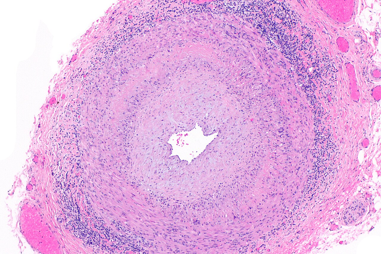

Do not make confirmation a treatment barrier
A normal eye examination or normal ESR cannot reassure away a convincing cranial-ischemic presentation. The 2026-published EULAR recommendations call for treatment without delay when GCA suspicion is strong. Initial oral prednisone-equivalent dosing for GCA or Takayasu is generally 40–60 mg/day. Threatened vision needs an urgent specialist-directed pathway rather than routine outpatient dosing.
ACR/VF 2021 conditionally favored temporal-artery biopsy in the US setting. EULAR prioritizes skilled ultrasound. Persistent suspicion after a negative test requires another diagnostic approach.
Treat inflammation. Track anatomy separately
Disease control
Selected GCA patients receive an IL-6 receptor inhibitor or upadacitinib with glucocorticoids. Methotrexate is an alternative. Upadacitinib carries boxed warnings for serious infection, mortality, malignancy, major cardiovascular events and thrombosis. Review individual risk before selection. All Takayasu patients should receive glucocorticoid-sparing therapy.
Structural surveillance
Compare symptoms, examination and prior imaging. CTA, MRA or ultrasound can monitor damage at previously inflamed sites. Imaging for suspected relapse is a different question. Persistent wall signal alone has uncertain significance in an otherwise quiescent patient.
Treat BP, ASCVD risk and drug toxicity. Statins and antiplatelets need their own cardiovascular indications.
PAN is necrotizing arteritis of medium or small muscular arteries without the glomerulonephritis or capillary/venular disease that defines many small-vessel syndromes. Renal dysfunction can arise from ischemia and hypertension.
- Kidney injuryMeasure BP, renal function, urine protein and sediment.
- Locate injuryArterial ischemia or glomerular inflammation?
- Target testsVascular imaging or tissue/serologic evaluation.
This is a diagnostic sorting aid, not a rule-out test. More than one mechanism can coexist. Glomerulonephritis should prompt reconsideration of an isolated PAN diagnosis.
Look beyond the coronary tree
| Territory | What changes the workup |
|---|---|
| Renal / visceral arteries | New hypertension, renal ischemia or unexplained abdominal pain may lead to abdominal vascular imaging. Aneurysms and stenoses support a medium-vessel process but do not name its cause. |
| Nerves, skin, muscle | Asymmetric painful neuropathy, weakness, nodules, livedo or other lesions may supply an accessible biopsy target. Coordinate the site and required depth rather than taking an uninformative superficial sample. |
| Heart | Coronary aneurysm, stenosis or thrombosis may occur. Establish whether symptoms arise from active arteritis, thrombus, fixed disease or another coronary mechanism. |
Obtain evidence from the involved territory
The ACR/VF guideline supports vascular imaging and tissue biopsy according to the presentation. In suspected abdominal PAN, noninvasive angiography can define involved beds. Catheter angiography may answer a remaining question when noninvasive studies are insufficient. A negative sample from an uninvolved site is weak evidence. Assess disease extent and the presence of threatened organ function before selecting induction.
Severity determines treatment intensity
For severe non-HBV PAN, cyclophosphamide plus glucocorticoids is the principal induction approach. Transition away from cyclophosphamide after remission limits toxicity. Less severe disease has different steroid-sparing options. Most PAN recommendations are conditional because comparative evidence is limited.
If pulmonary capillaritis, active glomerular sediment or a strong PR3/MPO-ANCA phenotype emerges, revisit GPA/MPA and other small-vessel processes. Antibody results should resolve a clinical question rather than override incompatible anatomy.
Kawasaki disease is a childhood inflammatory illness with a distinctive coronary risk. Adult follow-up depends on what happened to the arteries over time, not simply whether the current lumen looks smaller.
Carry the coronary history forward
| Required record | Why it matters |
|---|---|
| Largest documented abnormality | Record maximum dimensions, z scores, involved segments and the equation used. Historical severity remains relevant when an aneurysm regresses. |
| Current anatomy | Describe residual aneurysm, stenosis, calcification, thrombus, occlusion and collateral supply. Compare actual images when possible. |
| Events and procedures | Prior thrombosis, MI, ischemia, PCI or bypass alters surveillance and antithrombotic decisions. |
| Current treatment and access | Document each drug's indication, monitoring, adherence barriers and transition arrangements. |
Use a consistent z-score method during pediatric serial assessment. Measurement and equation changes can move a child across a risk category without a biological change. Coronary risk stratification incorporates both current and maximal involvement. A single descriptive label such as “ectatic” is inadequate.
Separate acute inflammation from late sequelae
Acute childhood disease
Prompt pediatric evaluation, IV immunoglobulin-based treatment and serial echocardiography aim to prevent coronary injury. Higher-risk presentations may warrant intensified initial therapy and more frequent imaging. Use the current pediatric algorithm rather than extrapolating an adult coronary strategy.
Adolescent and adult disease
Patients with coronary aneurysms need planned continuing cardiovascular care, including those whose aneurysms regressed. Establish an adult clinician, transfer original records and explain emergency symptoms, activity advice and medication responsibilities before transition.
Manage the complication that is present
Antiplatelet treatment, anticoagulation and ischemia testing depend on coronary risk, thrombus/event history and bleeding risk. Large or giant aneurysms require a specialist-directed thromboprophylaxis strategy. No single regimen applies to every dilated artery. Do not withdraw treatment solely because the measured lumen has normalized.
CCTA can characterize anatomy not fully seen on echo. Functional ischemia assessment and invasive studies are selected for the clinical question. New chest discomfort or unexplained exertional limitation deserves evaluation even at a young age. Prior Kawasaki disease changes the differential. It does not make conventional atherosclerotic prevention irrelevant.
GPA, MPA and EGPA share a small-vessel framework but have different clinical patterns. Cardiac disease in EGPA deserves deliberate assessment, including when the patient reports few cardiac symptoms.
| Disease | Pattern to recognize |
|---|---|
| GPA | Necrotizing granulomatous respiratory disease with small-vessel vasculitis. ENT, lung and kidney involvement often supply the diagnostic pattern. |
| MPA | Pauci-immune necrotizing small-vessel vasculitis, often with glomerulonephritis or pulmonary capillaritis. Granulomatous inflammation is not a defining feature. |
| EGPA | Asthma and eosinophilia with eosinophilic inflammation and vasculitis. Neuropathy and cardiac involvement can be decisive clues. |
A negative ANCA does not close the cardiac workup
PR3/MPO testing assists diagnosis in the appropriate setting. EGPA with cardiac involvement is frequently ANCA-negative, but negativity is not itself evidence that EGPA is present. Consider medication reactions, hypereosinophilic syndromes, infection and other causes of eosinophilia or myocarditis.
- At diagnosisSymptoms, examination, ECG and baseline echo.
- Resolve concernTroponin and CMR when cardiac injury is suspected.
- Define consequenceFunction, active injury, scar, thrombus and rhythm.
The ACR/VF guideline conditionally recommends an echocardiogram at EGPA diagnosis even without cardiac symptoms. An abnormal test changes disease-severity assessment. A normal EF cannot exclude every form of myocardial injury. Further evaluation should answer a remaining concern rather than become an unqualified schedule of repeated CMR.
In a retrospective specialist-clinic cohort, 45% of asymptomatic EGPA patients who underwent cardiovascular evaluation had an abnormal baseline assessment. This supports systematic attention to the heart, but is not a population-wide prevalence estimate: testing selection and the definition of an abnormality matter.
Organ-threatening disease needs an induction pathway
For life- or organ-threatening GPA/MPA, EULAR recommends glucocorticoids with rituximab or cyclophosphamide. For severe EGPA, EULAR favors glucocorticoids plus cyclophosphamide, with rituximab an alternative. ACR/VF favors cyclophosphamide for active cardiac EGPA because experience is greater. Mepolizumab does not replace severe-disease induction. Account for prior exposure, fertility, infection and toxicity.
Aneurysm, ectasia, beading and proximal narrowing describe coronary morphology. The clinical task is to determine whether inflammation, thrombus, atherosclerosis, dissection, prior intervention or external tissue explains the finding.
- LumenStenosis, dilation, flow and thrombus.
- Wall and tissueThickening, remodeling, dissection or encasement.
- Whole patientOther vessels, organs, exposures and disease activity.
Use imaging to narrow a differential
| Finding | Interpretation |
|---|---|
| Smooth concentric thickening | Raises the possibility of arteritis, especially with continuous or multifocal involvement. Not specific by itself. |
| Ostial / proximal stenosis | Consider aortic-root inflammation and Takayasu when the systemic distribution fits. Ordinary coronary disease remains possible. |
| Nodular or masslike tissue | Consider encasement from fibroinflammatory or neoplastic disease. IgG4-related disease may also produce smooth circumferential appearances. |
| Aneurysm / ectasia | Clarify reference-vessel disease, wall integrity, thrombus and distal flow. Dilation alone does not diagnose vasculitis. |
CCTA adds the wall and surrounding tissue to the angiographic lumen. Review prior imaging and other vascular beds. Targeted intravascular imaging may help a procedural team define anatomy and size a treatment. Slow flow and mural thrombus can complicate lumen assessment. Interpret measurements with the acquisition technique and comparator vessel in view.
Write two assessments before choosing treatment
Etiology and activity
What named disease best fits? Is the lesion currently inflamed, a remnant of prior injury, infected or noninflammatory? Which systemic finding or biopsy could distinguish the remaining possibilities?
Immediate coronary risk
Is there active ischemia, a culprit thrombus, impaired distal flow, important stenosis or structural progression? A difficult etiologic workup does not remove the need to address an acute coronary event.
These parallel questions prevent a common error: treating every aneurysm with immunosuppression, or assuming that a technically successful intervention treats the systemic disease. The workup and management need cardiology, imaging and disease-specialist agreement.
Related pathways: Chest Pain & ACS and Vascular Medicine.
Some patients have thrombosis because the vessel is inflamed. Others have thrombosis or arterial injury that imitates vasculitis. Establishing that distinction changes both immunosuppression and bleeding decisions.
Behçet connects inflammation, aneurysm and clot
Behçet is variable-vessel vasculitis involving arteries and veins. Recurrent mucosal ulceration, ocular disease and systemic features can accompany venous thrombosis, arterial aneurysm or intracardiac thrombus. The vascular presentation requires a disease-wide assessment.
- Define involvementVenous, intracardiac, arterial or pulmonary-artery disease.
- Control inflammationPrompt disease-specific immunosuppressive treatment.
- Assess anticoagulationBleeding risk and pulmonary artery aneurysms first.
The 2025 EULAR update, published in 2026, favors glucocorticoids plus immunosuppression for venous thrombosis, with monoclonal TNF inhibitors preferred in the stated pathway. Anticoagulants may be added if bleeding risk is low and coexisting pulmonary artery aneurysms have been excluded. Evidence is not sufficient to prescribe anticoagulation to everyone or to prohibit it universally. Optimal duration remains uncertain.
Actively test competing explanations
| Alternative | What helps distinguish it |
|---|---|
| Infection / secondary disease | Endocarditis and other infections can create inflammatory, renal and embolic findings, sometimes with positive ANCA. Use cultures and source-directed investigation when plausible. Antibody positivity alone does not justify immunosuppression. |
| Medication-associated disease | Review exposure timing, including hydralazine and other agents associated with ANCA disease. Attribute the syndrome using its clinical and laboratory pattern, not exposure alone. Suspected hydralazine-associated disease requires stopping the culprit drug and urgent specialist assessment of organ injury. |
| Noninflammatory arterial injury | Atherosclerosis, SCAD, intervention-related injury and other arteriopathies can resemble pieces of the coronary pattern. Review wall morphology and systemic distribution. |
| External tissue / systemic disease | IgG4-related disease is fibroinflammatory. Neoplastic tissue can encase coronaries. SLE/RA-associated vasculitis belongs under systemic-disease-associated vasculitis, not the variable-vessel category. |
Close the loop after the initial diagnosis
Record the evidence supporting the named disease, affected vascular beds, active manifestations and fixed damage. Assign ownership for drug monitoring, coronary/structural surveillance and new symptoms. A new vascular event should reopen activity, infection, thrombosis and treatment-toxicity questions rather than automatically be called relapse.
Chapter and selected source review: 17 October 2026. Original educational synthesis. Research criteria, observational frequencies and treatment recommendations have different purposes. The source accompanying each claim identifies its evidence base. Fibromuscular dysplasia is a noninflammatory mimic. Compare plaque, FMD and vasculitis, then see FMD diagnosis.