You cannot set a ventilator rationally without the mechanics. Every breath obeys a single equation of motion. pressure changes can reveal altered compliance, resistance, patient effort or circuit problems. Master these four ideas — the equation of motion, compliance, resistance, and the time constant — and the ventilator becomes a diagnostic instrument, not a black box.
A
The equation of motion
the master equationPapplied = V / C + (Flow × R) + PEEP
Elastic term distends the alveoli (revealed as Pplat on an inspiratory hold) · Resistive term drives gas through airways + ETT (Ppeak − Pplat) · PEEP is the baseline. In passive ventilation Papplied = airway pressure. a spontaneously breathing patient adds Pmuscle in parallel, so airway pressure understates true lung stress.
B
Compliance & resistance
read them every day| Property | Bedside formula | Typical values (variable) | Interpretation |
|---|---|---|---|
| Static compliance Crs | Vt / (Pplat − total PEEP) | 50–100 mL/cmH₂O | ↓ in ARDS (often reduced), edema, pneumothorax, ↑ intra-abdominal pressure |
| Dynamic compliance | Vt / (Ppeak − total PEEP) | — | A large peak–plateau gap suggests increased resistance. Compliance and resistance abnormalities can coexist |
| Resistance Rrs | (Ppeak − Pplat) / Flow | 5–15 cmH₂O/L/s | ↑ bronchospasm, secretions, kinked/small ETT, water in circuit |
| Lung vs chest wall | 1/Crs = 1/CL + 1/Ccw | — | Chest-wall stiffness can raise Paw without a proportional rise in lung stress. Esophageal pressure may help partition the load |
Poiseuille's law: For laminar flow, resistance ∝ 1/radius⁴. Halving radius predicts a 16-fold rise, but a 7.0-mm versus 8.5-mm tube predicts about 2.2-fold, not 16-fold. Real ETT flow is often turbulent. Measure resistance at constant inspiratory flow in a passive patient.
C
Time constants & regional heterogeneity
why lungs empty unevenly- τ = R × C ≈ 0.5 s (normal). Passive emptying (and filling) needs 3τ for 95%, 5τ for 99%.
- Heterogeneity fast units (low R·C) fill and empty first. slow units lag → pendelluft (gas sloshing between regions) and uneven distribution.
- Long τ (COPD, asthma): incomplete exhalation → dynamic hyperinflation → auto-PEEP — lengthen Te. → P.8
- Short τ (ARDS): empties fast, but low compliance → high pressures at modest volumes.
D
Advanced mechanics — what actually injures the lung
state of the art| Concept | Definition | Why it matters |
|---|---|---|
| Transpulmonary pressure PL | Palveolar − Ppleural | The true lung-distending pressure. Airway pressure misleads when the chest wall is stiff. Esophageal balloon estimates Ppl → EPVent-2 found no significant improvement in its death/ventilator-free-day composite |
| Driving pressure ΔP | Pplat − total PEEP = Vt / Crs | Associated with mortality in passive ARDS ventilation. A value around 15 cmH₂O is a warning signal, not a randomized-trial-validated treatment cutoff |
| Stress & strain | stress = PL. strain = ΔV / FRC | Regional lung stress varies with aerated volume and recruitability. Experimental strain thresholds are not universal clinical cutoffs |
| Mechanical power | energy transferred per breath × RR (J/min) | Depends on volume, pressure, flow, rate and mode. No single bedside threshold is a proven treatment target |
| P-SILI | patient self-inflicted lung injury | Vigorous spontaneous effort swings PL injuriously even on "lung-protective" settings — a reason to control drive |
Nunn's factoid — the oxygen cost of breathing: respiratory muscle oxygen demand rises during distress. Ventilation can unload this work, but induction and positive pressure can also precipitate circulatory collapse. Treat the respiratory and hemodynamic problem together.
Oxygenation and ventilation are separate problems with separate knobs. Oxygenation (PaO₂) responds to FiO₂ + mean airway pressure (PEEP, recruitment, prone). Ventilation (PaCO₂) responds to alveolar ventilation [RR × (Vt − dead-space volume)]. Treating a high CO₂ with more FiO₂ — or refractory hypoxemia with more rate — is the commonest bedside error.
A
Five mechanisms of hypoxemia
the differential in one table| Mechanism | A–a gradient | Corrects w/ 100% O₂? | Classic cause |
|---|---|---|---|
| ↓ Inspired PO₂ | Normal | Yes | Altitude. low-FiO₂ source / disconnection |
| Hypoventilation | Normal | Yes | Opioids, sedation, neuromuscular weakness |
| V/Q mismatch | ↑ | Largely yes | COPD, asthma, PE, pneumonia — most common |
| Shunt | ↑ | Limited response | ARDS, atelectasis, lobar pneumonia, intracardiac/PFO |
| Diffusion limitation | ↑ | Yes | ILD, emphysema, exercise |
Key: hypoxemia that barely responds to ↑ FiO₂ = shunt physiology → prompts evaluation of recruitability, PEEP and proning when appropriate (P.4–5). Intracardiac shunt may worsen with higher right-sided pressure.
B
Alveolar gas equation, A–a gradient & dead space
the gas-exchange mathPAO₂ = FiO₂ (Patm − PH₂O) − PaCO₂ / R ≈ FiO₂ × 713 − PaCO₂ / 0.8
Room air, sea level → PAO₂ ≈ 150 − 1.25·PaCO₂ ≈ 100 mmHg. A–a gradient = PAO₂ − PaO₂. approximately (age/4)+4 mmHg on room air. Pure hypoventilation or altitude does not widen it, but mixed disease can. P/F is a separate oxygenation index and varies with support.
- Dead space (VD/VT) ventilation without perfusion. normal ~0.3 (anatomic + alveolar). Rises with ARDS, PE, low cardiac output, high PEEP. Nuckton: a high dead-space fraction early in ARDS independently predicts death. ventilatory ratio is the bedside surrogate.
- Shunt (Qs/Qt) perfusion without ventilation. normal < 5% (bronchial/thebesian). A larger shunt reduces the response to oxygen. This is not specific for ARDS.
C
V/Q matching, West zones & hypoxic vasoconstriction
gravity shapes gas exchangeZone 1 · PA > Pa > Pv
Alveolar pressure limits perfusion → dead space. Overdistension and low blood pressure can enlarge this zone.
Alveolar pressure limits perfusion → dead space. Overdistension and low blood pressure can enlarge this zone.
Zone 2 · Pa > PA > Pv
Flow depends on arterial minus alveolar pressure → a vascular waterfall.
Flow depends on arterial minus alveolar pressure → a vascular waterfall.
Zone 3 · Pa > Pv > PA
Flow depends on arterial minus venous pressure. Common in dependent, well-perfused lung.
Flow depends on arterial minus venous pressure. Common in dependent, well-perfused lung.
PA = alveolar pressure, Pa = pulmonary arterial pressure, Pv = pulmonary venous pressure. These are pressure states, not fixed anatomical bands. Hypoxic vasoconstriction redirects flow away from poorly ventilated units.
D
Oxygen delivery & the hemoglobin dissociation curve
where PaO₂ stops matteringDO₂ = CO × CaO₂ × 10 · CaO₂ = (1.34 · Hb · SaO₂) + (0.003 · PaO₂)
~98% of arterial O₂ rides on hemoglobin. the dissolved term is trivial. Hb and cardiac output dominate delivery, not PaO₂ — raising an already high SpO₂ adds little content. Treat anemia and low output according to their indications, rather than transfusing to normalize calculated DO₂.
- Oxy-Hb curve P₅₀ ≈ 27 mmHg. Right shift (↑CO₂, ↑temp, ↑2,3-DPG, ↓pH = Bohr) unloads O₂ at the tissues. left shift (alkalosis, hypothermia, CO, ↓2,3-DPG, HbF) holds it. The sigmoid shape is why SpO₂ falls off a cliff below ~90%.
- SvO₂ / ScvO₂ reflects the DO₂ : VO₂ balance (normal SvO₂ ~65–75%). a falling value can signal reduced delivery or increased demand. ScvO₂ and SvO₂ are not interchangeable, and a normal/high value does not exclude impaired extraction.
- CO₂ transport most carried as bicarbonate, with additional carbamino and dissolved fractions. Haldane effect — deoxygenated Hb holds more CO₂, so oxygenating blood in the lung promotes CO₂ release.
Nunn's factoid — the oxygen cascade: At sea level on room air, inspired humidified PO₂ is about 150 mmHg and typical alveolar PO₂ about 100 mmHg. Arterial oxygen content, blood flow and tissue extraction then determine delivery. Tissue oxygen tension varies and is not a bedside oxygen target.
~21% of CICU patients require mechanical ventilation — respiratory failure is the top CICU admission diagnosis. Positive-pressure ventilation has profound cardiovascular effects: it reduces LV afterload (beneficial in HF) but increases RV afterload (dangerous in RV failure). Every vent setting is a hemodynamic decision.
A
Intubation indications in the CICU
when to intubate- Hypoxemia SpO₂ < 90% despite NIV/HFNC. P/F < 150 with accessory-muscle use.
- Ventilatory failure rising PaCO₂ with acidosis (pH < 7.25). respiratory fatigue.
- Airway protection Inability to protect the airway or manage secretions. GCS and post-arrest status inform, but do not replace, clinical assessment.
- Cardiogenic shock reduce work of breathing, redistribute blood flow to vital organs.
- Procedural TEE for MCS placement. cardiac surgery. prolonged hemodynamic procedures.
- Try NIV first in cardiogenic pulmonary edema, COPD — see P.7.
B
Initial ventilator settings
start here| Parameter | Setting | Rationale |
|---|---|---|
| Mode | AC-VCV (or AC-PCV) | Either mode can deliver lung protection. Check exhaled Vt, plateau pressure, effort and alarms rather than choosing by Ppeak alone |
| Tidal volume | 6–8 mL/kg PBW | Use predicted body weight (see box below). 6 mL/kg if ARDS |
| Resp. rate | 14–20 bpm | Adjust to pH > 7.25. allow permissive hypercapnia if needed |
| FiO₂ | 1.0 → titrate ↓ | Write a patient-specific oxygen target. Avoid unnecessary hyperoxia and titrate FiO₂ with PEEP (P.5) |
| PEEP | 5 cmH₂O (start) | ↑ per PEEP/FiO₂ table if ARDS (see P.4). watch hemodynamics |
| I:E ratio | 1:2 to 1:3 | Ensure complete exhalation. time constant ≈ 0.5 s |
| Pplat target | ≤ 30 cmH₂O | Check q4h or with any change. correlates with alveolar distension |
| ΔP (driving) | Trend ΔP | Use total PEEP during passive holds. Trend as a risk marker, not an isolated treatment mandate |
PBW calculation ♂ 50 + 2.3 × (height in inches − 60) | ♀ 45.5 + 2.3 × (height in inches − 60) | ♂ 50 + 0.91 × (height cm − 152.4) | ♀ 45.5 + 0.91 × (height cm − 152.4). Always use PBW, not actual weight — obese patients are routinely over-ventilated.
C
Common ventilator modes
what moves, what's set| Mode | Trigger | Control | Notes |
|---|---|---|---|
| AC-VCV | Patient / time | Volume | Set Vt, minimum RR and inspiratory flow/pattern. Pressure varies. Verify delivered ventilation when there is a leak or pressure limit |
| AC-PCV | Patient / time | Pressure | Set inspiratory pressure above PEEP, RR and Ti. Vt varies with mechanics and effort. High Ppeak alone is not an indication to switch modes |
| SIMV + PS | Patient / time | Vol or press | Mandatory + supported spontaneous breaths. Reducing the SIMV rate is not the preferred liberation strategy. Use a daily readiness screen and SBT |
| PSV | Patient only | Pressure | Patient controls RR & timing. set PS level + PEEP. Requires respiratory drive. weaning/SBT mode |
| APRV | Time-cycled | Pressure | Phigh (time long) / Plow (time short ~0.5 s). allows spontaneous breathing. may ↓ sedation needs |
Nunn's factoid: The time constant (τ = resistance × compliance) of the normal respiratory system is ~0.5 s. ≥3τ (1.5 s) required for 95% passive exhalation. At RR 30/min and I:E 1:2, Te is about 1.33 s. Obstructed units with long time constants may remain inflated. Read the expiratory flow trace.
A
Berlin oxygenation categories (2012)
classify severity| Severity | P/F ratio | Historical mortality | Management implications |
|---|---|---|---|
| Mild | > 200 to ≤ 300 PEEP/CPAP ≥ 5 | 27% | Low Vt. lower PEEP table often sufficient. monitor support failure closely |
| Moderate | > 100 to ≤ 200 PEEP ≥ 5 | 32% | Low Vt + higher PEEP table. consider prone if P/F < 150 |
| Severe | ≤ 100 PEEP ≥ 5 | 45% | Prone ≥ 16 h/d + low Vt + high PEEP. consider NMB, ECMO |
Plus: onset within 1 week, bilateral opacities and edema not primarily explained by cardiac failure or fluid overload. Cardiac disease can coexist. Mortality values above describe the Berlin derivation cohorts, not an individual prognosis.
Global definition (2024): also recognizes nonintubated ARDS on HFNO ≥30 L/min or NIV/CPAP ≥5 cmH₂O with P/F ≤300, or S/F ≤315 when SpO₂ ≤97%. Timing, imaging and edema-origin criteria still apply. Lung ultrasound and resource-limited criteria broaden access.
B
ARDSNet lung-protective protocol
the ARMA trial- Tidal volume 6 mL/kg PBW (range 4–8). ↓ to 4 if Pplat > 30.
- Plateau pressure ≤ 30 cmH₂O (check with inspiratory hold q4h).
- Driving pressure ΔP = Pplat − total PEEP in a passive patient. Reassess Vt and recruitability when high. Observational mediation analysis supports its prognostic value, not a proven ≤15 treatment cutoff.
- Oxygenation SpO₂ 88–95%. PaO₂ 55–80 mmHg.
- RR adjust to pH and expiratory flow, commonly up to 35/min in ARDS protocols. Accept hypercapnia when necessary for lung protection. There is no universal PaCO₂ ceiling of 60 mmHg.
- Permissive hypercapnia contraindicated with ↑ ICP. use with caution in severe RV failure (↑ PVR) and pulmonary HTN.
- Mortality benefit 31% vs 40% (p = 0.007). NNT ≈ 11. Non-negotiable standard of care.
C
ARDSNet PEEP/FiO₂ options
titrate togetherLower PEEP / higher FiO₂: example combinations from the 2008 protocol, grouped by FiO₂. These are options, not an automatic escalation sequence.
| FiO₂ | 0.3 | 0.4 | 0.5 | 0.6 | 0.7 | 0.8 | 0.9 | 1.0 |
|---|---|---|---|---|---|---|---|---|
| PEEP | 5 | 5, 8 | 8, 10 | 10 | 10, 12, 14 | 14 | 14, 16, 18 | 18–24 |
Higher PEEP table — moderate-severe ARDS (P/F ≤ 200). a conditional ATS 2024 option, without recruitment maneuvers. PEEP in cmH₂O.
| FiO₂ | 0.3 | 0.4 | 0.5 | 0.6 | 0.7 | 0.8 | 0.9 | 1.0 |
|---|---|---|---|---|---|---|---|---|
| PEEP | 5, 8, 10, 12, 14 | 14, 16 | 16, 18, 20 | 20 | 20 | 20, 22 | 22 | 22, 24 |
CICU caveat — PEEP & hemodynamics: PEEP may recruit lung or overdistend it. Recheck BP, cardiac output and RV function after changes. Do not automatically give fluid to a congested patient. Match PEEP to oxygenation, mechanics and circulation (→ P.6).
Nunn's factoid: The “baby lung” is the smaller aerated compartment, not a fixed fraction of total lung. A tidal volume that seems normal for body size may overdistend it. Respiratory-system compliance is not the same as isolated lung compliance.
A
Prone positioning
PROSEVA · mortality benefit in severe ARDS- PROSEVA entry P/F < 150 on FiO₂ ≥ 0.6 & PEEP ≥ 5, within 36 h of ARDS onset. This trial window does not prohibit later proning.
- Duration ≥ 16 h/day. continue until P/F > 150 on PEEP ≤ 10 & FiO₂ ≤ 0.6 in supine.
- Mortality 28-d: 16% vs 33% (p < 0.001). 90-d: 24% vs 41%. NNT ≈ 6.
- Mechanism ↑ V/Q matching. ↓ shunt. ↑ FRC. ↓ dorsal atelectasis. more homogeneous ventilation → ↓ VILI.
- RV benefit ↓ RV afterload → improves RV function. particularly valuable in CICU patients.
- Contraindications Unstable spine is a major contraindication. Open abdomen, unstable fractures, intracranial hypertension and recent surgery require an expert risk assessment. ECMO, chest tubes and RRT do not automatically preclude proning.
B
Neuromuscular blockade
ACURASYS vs ROSE| Trial | Population | Intervention | Outcome |
|---|---|---|---|
| ACURASYS 2010 | Early severe ARDS (P/F < 150) | Cisatracurium 48 h + deep sedation | Adj. 90-d mortality HR 0.68 (p = 0.04). ↑ vent-free days. no ↑ weakness |
| ROSE 2019 | Mod-severe ARDS (P/F < 150) | Cisatracurium 48 h vs usual care (lighter sedation) | Stopped futility. No mortality difference (42.5% vs 42.8%). ↑ CV events. lighter sedation may be sufficient |
Selective use: ATS 2024 conditionally supports NMB in early severe ARDS when needed for lung protection. In sepsis with moderate-severe ARDS, SSC 2026 suggests intermittent boluses over continuous infusion. Provide deep analgesia and amnesia before paralysis.
C
Recruitment maneuvers & PEEP titration
ART trial caution- ART trial (2017) aggressive recruitment + compliance-guided PEEP increased mortality (55% vs 49%, p = 0.04) with more barotrauma and cardiovascular events.
- Recommendation Avoid prolonged recruitment maneuvers. ATS 2024 favors higher PEEP without recruitment in moderate-severe ARDS. SSC 2026 recommends against an incremental PEEP titration strategy in sepsis-associated moderate-severe ARDS.
D
ECMO for refractory ARDS
EOLIA · rescue strategy- EOLIA (2018) VV-ECMO in very severe ARDS: 60-d mortality 35% vs 46% (p = 0.09). 28% crossover. The primary frequentist comparison was not statistically significant.
- Consider when P/F < 80 for >6 h despite optimized support. or P/F < 50 for >3 h. or pH < 7.25 + PaCO₂ ≥60 for >6 h despite optimized MV + prone. Refer early to a VV-ECMO center. VA support addresses a different circulatory indication. ATS 2024 conditionally supports VV-ECMO in selected severe ARDS.
E
Oxygen targets
avoid extremes- Avoid both extremes. ARDSNet used PaO₂ 55–80 or SpO₂ 88–95%. LOCO₂ raised safety concerns with a low-target strategy, but did not establish a universal PaO₂ floor of 70. HOT-ICU found no mortality difference between PaO₂ targets of 60 and 90.
- UK-ROX (2025): targeting SpO₂ 90% did not reduce 90-day mortality versus usual care in 16,500 ventilated adults. Individualize for ischemia, hypercapnia and measurement reliability. Recheck ABG when saturation is unreliable.
- Corticosteroids: ATS 2024 conditionally suggests them for ARDS. Regimen and duration are not standardized. Consider timing, infection and metabolic risk. This is separate from steroid treatment for septic shock.
F
RV-protective ventilation
critical in CICU- Acute cor pulmonale (ACP) occurs in ~25% of ARDS. associated with ΔP ≥ 18, PaCO₂ ≥ 48, P/F < 150.
- Strategy Limit distending pressure, avoid severe hypercapnia/acidosis and hypoxemia, and consider prone positioning. PaCO₂ 48 and ΔP 18 were observational risk markers, not universal therapeutic cutoffs. Titrate PEEP to recruitment and RV response. Inhaled vasodilators are a monitored rescue bridge.
●
How positive-pressure ventilation affects the heart
Venous return → RV preload
Higher pleural pressure can reduce the venous-return gradient. Hypovolemia and RV failure increase vulnerability.
Higher pleural pressure can reduce the venous-return gradient. Hypovolemia and RV failure increase vulnerability.
Lung volume → RV afterload
Recruitment may lower PVR. Overdistension compresses alveolar vessels and can raise PVR. More PEEP is not always more RV load.
Recruitment may lower PVR. Overdistension compresses alveolar vessels and can raise PVR. More PEEP is not always more RV load.
Pleural pressure → LV afterload
Higher pressure outside the LV reduces transmural systolic load. A congested, afterload-sensitive LV may eject more effectively.
Higher pressure outside the LV reduces transmural systolic load. A congested, afterload-sensitive LV may eject more effectively.
Net cardiac output → reassess
The balance depends on preload, ventricular function, recruitability and pressure transmission. Stiff lungs do not guarantee hemodynamic protection.
The balance depends on preload, ventricular function, recruitability and pressure transmission. Stiff lungs do not guarantee hemodynamic protection.
A
Hemodynamic effects of PEEP
titrate, don't ignore| Effect | Mechanism | Clinical implication |
|---|---|---|
| ↓ Venous return | ↑ intrathoracic pressure opposes venous inflow. abolishes "thoracic pump" of spontaneous inspiration | ↓ CO most common PPV side-effect. worse in hypovolemia. Give fluid only when likely responsive and tolerated |
| ↑ RV afterload | Overdistension raises PVR, while recruitment may reduce hypoxic vasoconstriction. Response is context dependent | Monitor for RV dilation (echo). limit PEEP in PH/RV failure. consider iNO |
| ↓ LV afterload | ↑ pleural pressure → ↓ LV transmural pressure. afterload reduction without drugs | Beneficial in LV failure. explains why HF patients tolerate PEEP well |
| ↓ Renal perfusion | ↓ CO → ↓ renal blood flow. ↑ venous pressure → ↓ renal perfusion gradient. ↑ ADH release | Assess perfusion and venous congestion. Oliguria alone is not an indication for fluid |
B
Spontaneous vs positive-pressure breathing
the mirror image- Positive-pressure inspiration ↑ ITP → ↓ venous return (↓ RV preload) & ↓ LV transmural afterload — helps the congested/failing LV.
- Spontaneous (negative-pressure) inspiration ↓ ITP → ↑ venous return (↑ RV preload) & ↑ LV afterload + ↑ work of breathing.
- Corollary the loading that makes NIV help acute cardiogenic edema is exactly what liberation reverses — extubation re-imposes the adverse loads.
- Two syndromes this drives weaning-induced pulmonary edema (→ P.9) and, at extremes of afterload, SCAPE (→ P.7).
C
Fluid responsiveness under PPV
pulse pressure & stroke volume variation- PPV / SVV pulse pressure variation > 13% or stroke volume variation > 12% predicts volume responsiveness (requires: sinus rhythm, Vt ≥ 8 mL/kg, no spontaneous breathing).
- Passive leg raise more reliable than static CVP/PCWP. ↑ CO ≥ 10% with PLR predicts fluid responsiveness regardless of rhythm or Vt.
- CICU principle if BP falls after PEEP increases, assess oxygenation, overdistension, RV function and fluid tolerance. Reverse an intolerable setting when appropriate. PLR needs a measured flow response. IVC size alone does not establish fluid responsiveness.
Nunn's factoid: The cardiovascular response to airway pressure varies with lung and chest-wall mechanics, blood volume and RV function. Do not predict a fixed percentage fall in cardiac output from the PEEP setting alone.
Cardiogenic pulmonary edema is a Starling-forces problem, not a lung-injury one: a rise in LV filling pressure raises pulmonary-capillary hydrostatic pressure until fluid floods the interstitium, then the alveoli. Here the ventilator is a hemodynamic device — positive pressure unloads the failing left ventricle.
A
Starling forces & cardiogenic vs high-permeability edema
two very different fluidsJv = Kf [ (Pc − Pi) − σ (πc − πi) ]
Filtration Jv rises when capillary hydrostatic pressure Pc climbs (cardiogenic) or when permeability Kf ↑ / reflection coefficient σ ↓ (ARDS). Hydrostatic and permeability edema can coexist. Treat congestion and the underlying lung insult when both are present.
| Feature | Cardiogenic (↑ Pc) | ARDS (↑ Kf) |
|---|---|---|
| PCWP / LAP | Often elevated | May also be elevated. PCWP ≤18 is not a current ARDS criterion |
| Echo / POCUS | ↓ EF or ↑ filling pressures. B-lines + effusions + plethoric IVC. congestion on VExUS | Cardiac dysfunction or congestion may coexist. B-lines identify interstitial involvement, not its cause |
| Edema fluid : plasma protein | Usually lower protein | Usually higher protein. Not a routine bedside diagnostic test |
| CXR | Central "bat-wing", cephalization, effusions, cardiomegaly | Peripheral, patchy. less cardiomegaly / effusion |
| Response | Rapid to diuresis / afterload reduction | Slow. treat the underlying insult |
B
Why positive pressure rescues the failing LV
afterload-sensitivity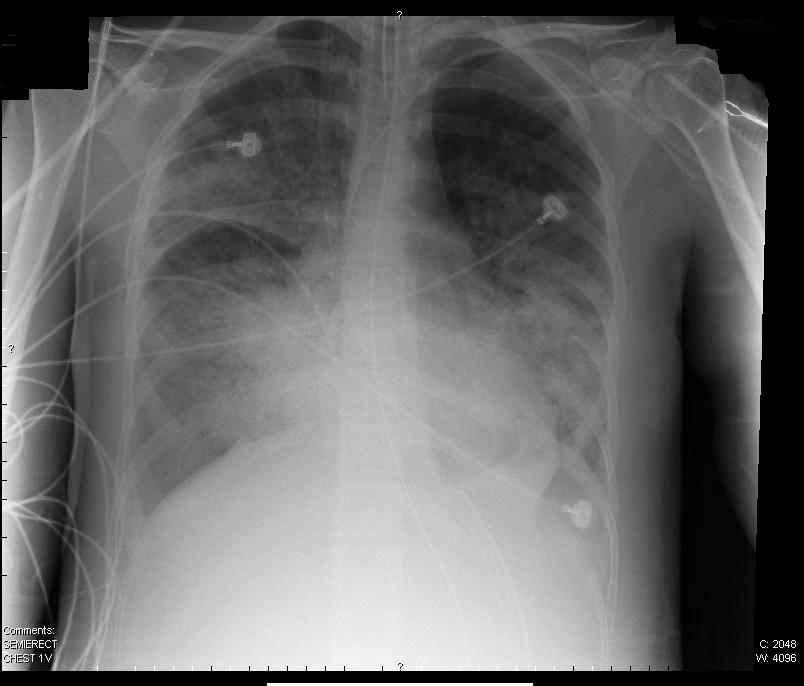

- Afterload reduction ↑ intrathoracic pressure → ↓ LV transmural pressure. An afterload-sensitive LV may increase stroke volume, provided the fall in venous return does not outweigh the benefit.
- ↓ Preload ↓ venous return → ↓ pulmonary congestion.
- ↑ FRC recruits flooded alveoli → ↓ shunt → ↑ PaO₂.
- ↓ Work of breathing ↓ myocardial O₂ demand and ↓ catecholamine surge.
C
NIV / HFNC in acute cardiogenic pulmonary edema
first-line — LV unloading- First-line CPAP 5–10 cmH₂O, titrated to distress, oxygenation and BP. Use BiPAP (IPAP 12–16 / EPAP 5–8) if hypercapnic, COPD overlap, or persistent work of breathing. Titrate FiO₂ to the prescribed saturation range, with a lower target when at risk of hypercapnic failure.
- HFNC may be useful when NIV is not tolerated, but delivers less predictable distending pressure. FLORALI studied nonhypercapnic hypoxemic failure and does not establish a mortality benefit in cardiogenic edema.
- Evidence NPPV ↓ intubation (RR ~0.52) and ↓ hospital mortality (RR ~0.66) vs standard O₂. 3CPO found symptom benefit without a mortality signal (crossover-limited).
- Avoid / caution shock needing intubation. depressed consciousness or can't protect airway. vomiting. Don't let NIV delay needed intubation or revascularization.
D
SCAPE — sympathetic crashing acute pulmonary edema
afterload crisis- Recognize abrupt severe hypertension (often SBP > 180–200) with flash edema. EF is frequently preserved. afterload and fluid redistribution may dominate. Total-body congestion can still coexist.
- Treat the afterload NIV + titrated IV nitroglycerin for severe hypertension when appropriate. High-dose/bolus regimens require an experienced team and close BP monitoring. Exclude recent PDE5 inhibitor use, riociguat and hypotension. Evidence for specific SCAPE regimens is limited. Give IV loop diuretic when congestion is present. Do not assume redistribution excludes volume overload.
- Airway avoid reflexive intubation (many respond to NIV within minutes). if intubation is required, anticipate post-induction hypotension and have vasopressors ready. Verify all doses against current labeling and pharmacy guidance.
Nunn's factoid — the edema safety factor: Lymphatic clearance and interstitial structure resist alveolar flooding. Chronic congestion can increase this reserve. Edema therefore depends on the speed of pressure rise and barrier permeability, not a universal wedge-pressure threshold.
A
High airway pressures — differential
peak vs plateau| Pattern | Causes | Action |
|---|---|---|
| ↑ Ppeak normal Pplat | ↑ Airway resistance: bronchospasm, mucus plug, kinked ETT, biting tube, small ETT, circuit obstruction | Suction. bronchodilator. check circuit/ETT position. consider larger ETT |
| ↑ Ppeak ↑ Pplat | ↓ Compliance: pneumothorax, pleural effusion, pulmonary edema, ARDS progression, abdominal distension, auto-PEEP, mainstem intubation | CXR. decompress if tension. ↓ Vt to keep Pplat ≤ 30. drain effusion. check ETT depth. rule out auto-PEEP |
| ↑ Ppeak cannot get Pplat | Patient-ventilator dyssynchrony. active expiration. pain/agitation | Assess sedation. check mode/trigger sensitivity. consider flow/rate changes. brief NMB for assessment |
B
Auto-PEEP (intrinsic PEEP)
trapped gas- Mechanism incomplete exhalation → dynamic hyperinflation → residual alveolar pressure at end-expiration. Common with high RR, high Vt, bronchospasm, short Te.
- Detect in a passive patient, end-expiratory hold: total PEEP − set PEEP = auto-PEEP. Active expiration or airway closure can invalidate or underestimate the measurement. Also: flow ≠ 0 at end-expiration on flow-time waveform.
- Consequences ↑ intrathoracic pressure → ↓ venous return → ↓ CO (acts as "a tourniquet to the right heart"). ↑ trigger threshold → missed breaths.
- Fix Reduce RR/Vt as tolerated and shorten inspiration to lengthen expiration. Treat obstruction. In expiratory flow limitation, carefully titrated external PEEP may ease triggering. Do not apply a fixed 80% rule to every patient. Monitor total PEEP, plateau pressure and BP.
C
Ventilator-induced lung injury (VILI)
four mechanisms| Mechanism | Pathophysiology | Prevention |
|---|---|---|
| Volutrauma | Alveolar overdistension from excessive Vt — the alveolar size, not the pressure per se, causes cellular injury | Vt 6 mL/kg PBW. Pplat ≤30. Track ΔP |
| Barotrauma | Sustained transmural pressure gradient → air leak → subcutaneous emphysema, pneumomediastinum, pneumothorax | Pplat ≤ 30 cmH₂O. minimize mean airway pressure |
| Atelectrauma | Cyclical collapse & reopening of small airways → shear stress → epithelial damage & surfactant dysfunction | Adequate PEEP to prevent end-expiratory collapse |
| Biotrauma | Mechanical forces transduced into inflammatory mediator release → local and systemic inflammation → MODS | Lung-protective ventilation. minimize mechanical power |
D
Patient-ventilator dyssynchrony
read the waveforms| Type | Finding | Fix |
|---|---|---|
| Trigger dyssynchrony | Missed triggers (auto-PEEP or low sensitivity). auto-triggering (leak, cardiac oscillations) | ↓ trigger threshold. add external PEEP for auto-PEEP. fix leak. switch flow → pressure trigger |
| Flow dyssynchrony | Patient demand exceeds set flow (VCV): "scooped" pressure waveform. patient pulling against ventilator | ↑ peak flow. switch to decelerating flow or PCV. consider PSV |
| Cycle dyssynchrony | Double-triggering (set Ti too short). premature termination. delayed cycling (expiratory effort during inspiration) | Adjust Ti or expiratory cycle-off %. preserve lung-protective Vt while correcting drive. match Ti to neural Ti |
| Breath stacking | Consecutive breaths without full exhalation → air trapping | ↓ RR. ↑ Te. check auto-PEEP. brief sedation/NMB if acute |
E
VAP prevention bundle
SHEA 2022- Core bundle HOB 30–45°. daily sedation awakening + SBT. early mobilization. subglottic secretion drainage. circuit changes when visibly soiled or malfunctioning. Provide toothbrushing without routine chlorhexidine.
- De-emphasized oral chlorhexidine (no clear benefit, possible harm). stress ulcer prophylaxis has separate indications.
Every ventilator day increases morbidity — VAP, deconditioning, delirium. Assess readiness daily. No single predictor is sufficient. use a systematic protocol (daily screen → SBT → extubation).
A
Readiness screen
daily · pass → SBT| Criterion | Threshold | Notes |
|---|---|---|
| Underlying cause | Improving or resolved | The precipitant that led to intubation is being treated |
| Oxygenation | P/F ≥ 150 (SpO₂ ≥ 90% on FiO₂ ≤ 0.4 & PEEP ≤ 8) | Adequate on minimal support |
| Hemodynamics | No/low-dose vasopressors. no active ischemia | Stable or decreasing support. No universal norepinephrine dose proves readiness |
| Neuro / drive | Follows commands (or baseline). spontaneous breaths, adequate cough | Pair the SAT with the SBT (awaken, then breathe) |
B
SBT & weaning predictors
30–120 min- Method SBT with or without low pressure support is acceptable. Use a standardized 30–120-minute assessment. Do not raise FiO₂ simply to pass the trial.
- Pass RR < 35. SpO₂ ≥ 90%. HR change < 20%. no distress/diaphoresis/accessory-muscle use.
- RSBI (f/Vt) may add context, but is not required to decide SBT readiness. A threshold of 105 must not be an automatic gate.
- NIF / cough MIP ≤ −20 to −30 cmH₂O. adequate cough clears secretions (poor cough → failure).
- Cuff leak test patients at high risk of stridor, not everyone. If failed but otherwise ready, give systemic steroid ≥4 h before extubation using an established regimen. Reassess the airway plan.
- Fail → retry resume support. address cause (volume, secretions, cardiac). retry in 24 h.
Weaning-induced pulmonary edema (WIPO) — the cardiac reason SBTs fail: switching from positive- to negative-pressure breathing raises venous return and LV afterload while the work of breathing drives an adrenergic surge — precipitating acute cardiogenic edema. In one selected cohort it accounted for 59% of failed SBTs. This is not a universal ICU rate. Suspect in HF, COPD, or obesity with a failing SBT showing rising BP, tachycardia, and distress. look for concordant SBT-related changes in echo, lung ultrasound, filling pressures and clinical congestion. No single E/e′ or BNP change confirms WIPO. Manage with negative fluid balance before the next SBT, afterload reduction / nitrates, and extubating high-risk patients directly onto NIV. → P.6 · P.7
C
Extubation & post-extubation
plan ahead- Before suction above & below cuff. HOB ≥ 30°. NIV/HFNC set up bedside. confirm reintubation plan.
- Prophylactic NIV/HFNC plan immediate prophylactic NIV for selected high-risk patients, particularly hypercapnic disease or HF. HFNC is an alternative/adjunct by phenotype. Preventive support is distinct from rescue NIV after failure.
- Post-extubation failure monitor closely for distress, airway obstruction, secretion failure and recurrent edema. Rescue NIV must not delay needed reintubation.
- Tracheostomy individualize for anticipated duration, airway protection, recovery and goals of care. No fixed day or failed-extubation count mandates it.
CICU liberation pearl: Historically, high mortality has accompanied invasive ventilation in CICU cohorts — MV is a marker of severity, not just a therapy. Optimize the cardiac cause of respiratory failure (diuresis, afterload reduction, revascularization, MCS) in parallel with the ventilator strategy. → Cardiogenic Shock · HF Inpatient cards