Don't
Miss
Miss
AL = oncologic emergency
Any abnormal monoclonal screen + suspected cardiac amyloid → urgent heme/amyloid referral. A monoclonal protein does not prove AL; obtain tissue and type the deposit. Do not wait for a bone scan.
Screen before you scan
Order serum free light chains + serum IFE + urine IFE in every suspected case. SPEP/UPEP alone are inadequate; a positive bone scan does not exclude AL.
HFpEF + LVH = think amyloid
Unexplained LV wall thickening, low-voltage ECG, BP-med intolerance, or discordant low strain → work it up. Suspicion is the rate-limiting step.
Treatable disease now
Tafamidis, acoramidis & vutrisiran improve ATTR-CM outcomes; dara-VCd is frontline AL therapy. Benefit is greatest when treatment starts early. ATTR AL
01
When to Suspect
Cardiac + extracardiac red flagsCardiac clues
- HFpEF with LV wall ≥ 12 mm not explained by HTN/AS, esp. age > 60.
- ECG–echo mismatch low / normal QRS voltage despite thick walls; pseudo-infarct (Q) pattern.
- Strain apical sparing ("cherry-on-top") on GLS; ↓ tissue Doppler velocities.
- Conduction AV block, need for PPM; AF with poorly tolerated rate control.
- Intolerance new hypotension / inability to tolerate prior antihypertensives or β-blocker.
- Hi-sensitivity troponin chronically elevated; NT-proBNP out of proportion.
Extracardiac (ATTR especially)
- Bilateral carpal tunnel (often precedes by years)
- Lumbar spinal stenosis
- Spontaneous biceps tendon rupture ("Popeye")
- Polyneuropathy (length-dependent)
- Autonomic dysfunction, orthostasis
- Vitreous opacities, glaucoma (variant)
AL-specific
- Periorbital purpura, macroglossia
- Nephrotic-range proteinuria
- Unexplained weight loss, fatigue
- Rapidly progressive HF (weeks)
Two diseases, one phenotype
AL light-chain (plasma-cell dyscrasia) — aggressive, urgent. ATTR transthyretin — wild-type (usually later-onset) or hereditary/variant (genotype-dependent, often + neuropathy). Management diverges completely after typing.
02
Diagnostic Algorithm
Suspicion → type → confirm
1 · Image
Echo ± cardiac MRI. Echo: thick walls, granular myocardium, biatrial enlargement, apical-sparing strain. CMR: diffuse subendocardial / transmural LGE, ↑ native T1 & ECV, abnormal nulling.
2 · Screen — DO THIS FIRST
Serum free light chains (κ/λ) + serum IFE + urine IFE. Run all 3 before interpreting any scan. CKD widens the FLC ratio; interpret with eGFR, assay-specific renal ranges, and both IFE results.
3 · Bone scintigraphy
⁹⁹ᵐTc-PYP / DPD / HMDP planar + SPECT. Grade myocardial uptake (Perugini 0–3); SPECT confirms myocardial (not blood-pool) signal. Nuclear: acquisition & grading.
4 · Interpret together
A nonbiopsy ATTR diagnosis requires a compatible phenotype, diffuse grade 2–3 myocardial uptake on SPECT, and a negative 3-part monoclonal screen. Any screen abnormality → tissue typing.
Grade 2–3 / MC − ATTR-CM nonbiopsy diagnosis → sequence TTR.
Any scan / MC + Clone possible; AL and ATTR+MGUS both occur → biopsy + mass spec.
Grade 0–1 / MC − If suspicion persists: CMR, expert review ± biopsy; do not use H/CL to rescue.
Workup negative Reconsider mimics; early ATTR and some variants may be scan-negative.
Biopsy when discordant
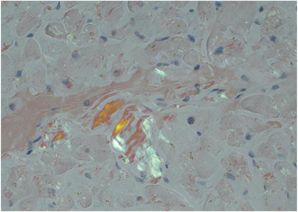
Sample fat pad + marrow or another involved organ when appropriate; a negative surrogate biopsy does not exclude cardiac amyloid. Endomyocardial biopsy has the highest cardiac yield. Congo red confirms amyloid; mass spectrometry types the protein.
03
Typing & Staging
Drives therapy & prognosisConfirm ATTR subtype
ATTRwt · wild-type
Cause: no pathogenic/likely pathogenic (P/LP) TTR variant; age-associated misfolding of normal TTR.
Pattern: usually later-onset and cardiac-predominant; bilateral CTS, lumbar stenosis, and biceps rupture are common clues. Marked autonomic, ocular, or renal disease is atypical; look for another/additional cause.
Family: not inherited; no cascade testing when sequencing finds no P/LP variant.
ATTRv · hereditary
Cause: germline P/LP TTR variant; autosomal dominant with age-/variant-dependent, incomplete penetrance.
Pattern: cardiac, neuropathic/autonomic, or mixed. Older age and a negative family history do not exclude ATTRv.
Family: each child has a 50% chance of inheriting the variant (not a 50% certainty of disease).
Variant clues, not destiny: p.Val142Ile (legacy V122I; ~3–4% with West African ancestry) is usually late cardiac-predominant; p.Val50Met (V30M) often causes neuropathy/autonomic disease ± CM, and early disease may be bone-scan negative. SCAN-MP: ATTR-CM in 17% of Black men >75 y with HF; ~53% of variant carriers with HF.
Act on the sequence, not assumptions
PYP cannot subtype ATTR. Sequence TTR in every confirmed ATTR with counseling: P/LP = ATTRv + targeted adult-relative testing; no P/LP = ATTRwt. A VUS does not establish ATTRv or direct treatment/cascade testing; genetics review + reclassification follow-up.
AL staging — Mayo 2012 (revised)
| Marker | Threshold |
|---|---|
| cTnT | ≥ 0.025 ng/mL |
| NT-proBNP | ≥ 1800 pg/mL |
| dFLC (involved − uninvolved) | ≥ 18 mg/dL |
Each marker = 1 point → Stage I–IV. Troponin cutoffs are assay-specific; use a validated local staging method.
ATTR staging — Gillmore / NAC
IBoth ✓
NT-proBNP ≤3k
eGFR ≥45
eGFR ≥45
IIEither
One cutoff
IIIBoth ✗
NT-proBNP >3k
eGFR <45
eGFR <45
Based on NT-proBNP + eGFR; predicts survival in wt & variant ATTR.
Baseline before therapy: NT-proBNP · hs-troponin · eGFR · 6-min walk/KCCQ · NYHA · ECG/echo.
▶
One-line pathophysiology
Misfolded protein → restrictive cardiomyopathyWhy the heart fails
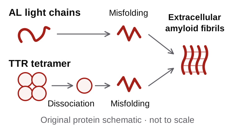
Misfolded monoclonal light chains (AL) or destabilized transthyretin tetramers (ATTR) deposit as insoluble β-sheet fibrils in the myocardial interstitium → progressive wall thickening with a stiff, non-compliant ventricle → restrictive physiology, HFpEF, low stroke volume, atrial dysfunction (AF + thrombus), and infiltration of the conduction system. In AL, circulating light chains are also directly cardiotoxic — explaining rapid decline and the urgency of suppressing the clone.
The
Rule
Rule
Non-biopsy ATTR (Gillmore)
Compatible phenotype + diffuse grade 2–3 myocardial uptake on SPECT + negative 3-part monoclonal screen → nonbiopsy ATTR-CM. Predictive value still depends on pretest probability.
SPECT is mandatory
Planar alone cannot separate myocardium from blood pool or bone. Always confirm with SPECT. Visual myocardial grading is primary; H/CL is optional.
Neither test alone types amyloid
AL can show uptake, occasionally grade 2–3. An abnormal monoclonal screen shows a possible clone—not AL; tissue typing resolves the disease.
01
Scintigraphy & Grading
⁹⁹ᵐTc-PYP / DPD / HMDPPerugini visual grade (vs rib/bone)
0None
No cardiac uptake; bone normal. Does not exclude amyloid.
1Equivocal
Uptake < bone. Indeterminate — see Col 2.
2Positive
Uptake = bone. Apply all ATTR gates.
3Strong
Uptake > bone, bone faint. Apply all ATTR gates.
Acquisition & interpretation
- Visual first assign the uptake grade, then use SPECT to confirm myocardial localization. H/CL is optional and must not override discordant images.
- Timing 2–3 h imaging is recommended; 1-h PYP is optional at experienced sites. If blood pool persists, repeat at 3 h.
- SPECT required confirms diffuse myocardial—not blood-pool, rib, or valve—retention.
Screen first, every time Order sFLC + serum & urine immunofixation before reading any scan. The scan only confirms ATTR once AL is excluded. CKD caveat: an abnormal free-light-chain ratio can occur in renal impairment without a true clonal process — use eGFR-adjusted FLC interpretation & immunofixation to avoid over-calling AL.
02
The Equivocal Scan
Grade 1 · focal · discordantGrade 1 / focal / discordant Obtain or review SPECT. Question: true diffuse myocardial retention vs blood pool, bone, valve, or focal injury?
SPECT = blood pool only Likely negative. If suspicion persists, re-image at 3 h or pursue CMR / biopsy.
SPECT = myocardial grade 1 ATTR remains possible, but grade 1 is not diagnostic. H/CL cannot rescue it; use phenotype, CMR, genetics, and biopsy as needed.
If monoclonal screen abnormal Do not assign AL or ATTR from scan → urgent hematology evaluation and tissue + mass-spec typing.
False positives / pitfalls
- Blood-pool contamination (planar-only, early imaging)
- AL amyloid uptake (occasionally grade 2–3)
- Recent or healing MI
- Rib fracture, valve/bone overlap, hydroxychloroquine toxicity
- Rare AApoAI & other amyloid types
Grade 1 is a decision, not a diagnosis
Treat every grade-1 / borderline result as indeterminate until SPECT localization and monoclonal status are both in hand.03
Echo & CMR Clues
Supportive, not confirmatoryEchocardiography
- Apical sparing "cherry-on-top" GLS supports CA, but accuracy varies by population and mimic; absence does not exclude amyloid.
- Other ↑ wall thickness, granular myocardium, biatrial dilation, ↓ TAPSE.
Cardiac MRI
- LGE diffuse subendocardial → transmural; abnormal myocardial nulling.
- Mapping ↑ native T1 & ECV (tracks amyloid burden). High sensitivity; cannot type.
Biopsy = reference standard
Endomyocardial biopsy has the highest cardiac yield. Congo red confirms amyloid; mass spectrometry types it. Use when monoclonal tests are abnormal or the nonbiopsy pathway is not met.AI echo screening — EchoGo® Amyloidosis
FDA-cleared adjunct (K240860): single noncontrast apical 4-chamber clip; intended for adults ≥65 y with HF undergoing echo. A flag prompts the standard amyloid workup—it does not diagnose or type CA.
| Cohort | Sens | Spec | Uncertain |
|---|---|---|---|
| FDA intended-use N=1,164 · ≥65 + HF | 84.5% 80.3–88.5 | 89.7% 87.0–92.4 | 14.0% |
| 18-site external N=2,719 | 85% | 93% | 13% |
| Tc-PYP referral subgroup | 77% | 86% | — |
FDA and external-cohort metrics excluded no-class/uncertain outputs; AUROC 0.93 overall and 0.86 in PYP referrals. A negative result can miss CA; continue evaluation when clinical suspicion is high. Predictive values vary with prevalence.
▶
Decision limits & failure modes
What the result can—and cannot—prove| Finding | What it supports | Failure mode | Next action |
|---|---|---|---|
| Diffuse grade 2–3 SPECT + phenotype + MC screen negative | Nonbiopsy ATTR-CM | Low pretest probability; poor localization | Sequence TTR; stage and treat |
| Grade 1, focal, or discordant | Indeterminate | H/CL falsely “rescues” blood pool or bone | Expert review; CMR/repeat/biopsy |
| Negative scan | ATTR less likely; AL not excluded | Early disease; low-uptake variant | Follow MC pathway; CMR/biopsy if high suspicion |
| sFLC + serum IFE + urine IFE | All negative: AL very unlikely; any abnormality: clone possible | CKD or MGUS misread as AL | Hematology + tissue typing when abnormal |
| Echo / CMR phenotype | Supports cardiac amyloid; cannot type | Hypertensive, HCM, storage mimics | Complete the amyloid typing pathway |
Do not let a ratio, planar image, or M-spike assign the disease. A negative scan does not exclude AL; an abnormal monoclonal screen does not prove it. Tissue & strain atlas · Scintigraphy atlas · Nuclear: acquisition & grading · Nuclear: diagnostic gates & reporting.
A
ATTR-CM — transthyretin
Stabilize the tetramer · silence the geneStabilizers (oral) — approved options
TafamidisVyndamax / Vyndaqel · Pfizer
61 mg PO daily
TrialATTR-ACT: ↓ all-cause mortality ~30% & CV hospitalizations over 30 mo.
EvidenceBest established in NYHA I–II; benefit is less certain in NYHA III; no trial evidence in NYHA IV.
NotesWell tolerated. Vyndamax 61 mg = Vyndaqel 80 mg exposure; formulations are not mg-for-mg interchangeable.
AcoramidisAttruby · BridgeBio
712 mg PO BID
TrialATTRibute-CM: win ratio 1.8; death/first CV hospitalization HR 0.64; recurrent CV hospitalization rate ↓ ~50%.
ClassNear-complete TTR stabilizer; outcomes were assessed over 30 mo.
NotesFDA approved 2024 (wt & variant). Diflunisal is an off-label NSAID stabilizer — avoid in HF/CKD.
Gene silencers — reduce TTR production
VutrisiranAmvuttra · Alnylam
25 mg SC q3 mo
TrialHELIOS-B: primary composite HR 0.72 overall; all-cause mortality HR 0.65 overall through 42 mo.
StatusFDA-approved for ATTR-CM (2025); also hATTR neuropathy.
PatisiranOnpattro · Alnylam
0.3 mg/kg IV q3 wk
TrialAPOLLO-B: preserved 6-MWT & KCCQ vs placebo at 12 mo.
NotesIV infusion; pre-meds. Approved for hATTR neuropathy; not FDA-approved for CM.
- Eplontersen ASO 45 mg SC monthly — hATTR-PN only; CARDIO-TTRansform missed its CM primary endpoint (Jul 2026).
- Inotersen ASO — older agent; monitor platelets & renal (thrombocytopenia, GN).
Choosing & combining in ATTR
Start as early as possible. Both ATTRwt-CM and ATTRv-CM are eligible for approved CM monotherapy; genotype does not establish a superior CM drug. In ATTRv, screen for sensorimotor/autonomic, ocular, and renal disease; prefer a silencer when clinically meaningful polyneuropathy coexists. Otherwise choose by route, comorbidity, access, and patient preference. Routine stabilizer + silencer combination is not recommended because incremental benefit is unproven.
B
AL amyloidosis — light chain
Suppress the plasma-cell clone — urgentlyFrontline regimen
- Dara-CyBorD daratumumab + cyclophosphamide + bortezomib + dexamethasone.
- Trial ANDROMEDA: adding daratumumab markedly ↑ hematologic CR & cardiac/renal organ response → now standard of care.
- Goal rapid, deep hematologic response (↓ dFLC) to stop light-chain cardiotoxicity.
Transplant & risk
- ASCT autologous stem-cell transplant for eligible (good performance, limited cardiac stage).
- High-risk cardiac AL Mayo stage III–IV poorly tolerate therapy — manage at amyloid center; consider dose-attenuation.
- Relapse specialist-directed: daratumumab-based, IMiDs, ixazomib; off-label venetoclax may be considered with t(11;14).
Monitor response
- Hematologic serum free light chains / dFLC, immunofixation each cycle.
- Cardiac NT-proBNP & troponin (organ response markers), echo strain.
- Caution NT-proBNP also changes with volume, renal function, infection, and some therapies (especially IMiDs) — interpret with clone status.
▶
Upcoming & investigational therapies
The next class — clearing deposits already there01
Stabilize
Approved. Hold the TTR tetramer — tafamidis, acoramidis.
02
Silence
Approved by indication. Cut TTR production — vutrisiran (CM/PN); patisiran (PN only).
03
Deplete new class
Investigational. Antibodies that clear existing amyloid via phagocytosis.
04
Gene-edit
Investigational. One-time in-vivo CRISPR knockdown of TTR.
| Agent | Class | Mechanism | Trial · status |
|---|---|---|---|
| Coramitug (NNC6019) · Novo Nordisk | ATTR depleter Ab | Targets misfolded TTR; promotes phagocytic clearance | Ph2: ↓NT-proBNP; 6-MWT NS. Ph3 CLEOPATTRA recruiting |
| ALXN2220 (NI006) · AstraZeneca | ATTR depleter Ab | Anti-fibril; Ph1 ↓ scan uptake, ECV, NT-proBNP / troponin | Ph3 DepleTTR-CM |
| Nex-z (NTLA-2001) · Intellia | ATTR CRISPR | One-time IV in-vivo gene editing → durable TTR knockdown | Ph3 MAGNITUDE resumed Mar 2026; enhanced safety monitoring |
| Eplontersen | ATTR ASO | Monthly SC antisense TTR knockdown | CARDIO-TTRansform missed primary (Jul 2026); PN only |
| Anselamimab (CAEL-101) · AstraZeneca | AL depleter Ab | Targets AL fibril deposits | CARES missed overall; nominal signal in prespecified κ-light-chain subgroup |
| Birtamimab | AL depleter Ab | Anti-AL amyloid antibody · Prothena | AFFIRM-AL negative (2025); development discontinued |
Investigational — not practice-changing. Signals from depleter and gene-editing studies require randomized outcome confirmation. Status checked 2026-07; verify before trial referral.
A
ATTR-CM landmark randomized trials
What each FDA approval rests onATTR-ACT
Tafamidis 20/80 mg PO
Maurer · NEJM 2018;379:1007
N = 441
DesignPhase 3 double-blind RCT; tafamidis (80 or 20 mg, pooled) vs placebo over 30 months; 2:1:2 randomization.
Pop'nATTR-CM (wild-type or hereditary), NYHA I/II/III = 8 / 60 / 32%; LV wall ≥12 mm; ~76% wt, ~24% hereditary.
PrimaryHierarchical (Finkelstein–Schoenfeld): all-cause mortality, then frequency of CV hospitalization at 30 mo.
EfficacyACM 29.5% vs 42.9% (HR 0.70, CI 0.51–0.96); CV hosp 0.48 vs 0.70/yr (RR 0.68). Less decline in 6-MWD & KCCQ. Survival curves separate ~month 18.
SafetyAEs & discontinuations similar to placebo — very well tolerated.
Bottom lineFirst therapy to improve survival in ATTR-CM. Benefit is best established in NYHA I–II; NYHA III evidence is less certain, and NYHA IV was not studied. → FDA approval 2019.
ATTRibute-CM
Acoramidis 712 mg PO BID
Gillmore · NEJM 2024;390:132
N = 632
DesignPhase 3 RCT, 2:1 acoramidis vs placebo over 30 months; near-complete TTR stabilizer.
Pop'nATTR-CM (wt or variant), ~90% wild-type, mostly NYHA II; eGFR ≥15 at entry. Primary efficacy analysis: 611 with eGFR ≥30. Tafamidis could start after 12 mo.
Primary4-step win ratio: ① ACM ② CV hospitalization ③ ΔNT-proBNP ④ Δ6-MWD.
EfficacyWin ratio 1.8 (CI 1.4–2.2). Death + 1st CV hosp HR 0.64 (↓36%); CV hosp ↓~50% (0.29 vs 0.55/yr); favorable NT-proBNP & 6-MWD. Serum TTR ↑~39%.
SafetyDiarrhea; early reversible creatinine rise/eGFR decline; overall AEs comparable to placebo.
Bottom lineOutcome-positive near-complete stabilizer. → FDA approval Nov 2024 (Attruby), wt & variant ATTR-CM.
HELIOS-B
Vutrisiran 25 mg SC q3mo
Fontana · NEJM 2024 (ESC 2024)
N = 655
DesignPhase 3 RCT, vutrisiran (siRNA) vs placebo up to 36 months; stratified by background tafamidis (~40%).
Pop'nATTR-CM (wt or hereditary), NYHA I–III; overall & tafamidis-naïve "monotherapy" co-primary populations.
PrimaryComposite of all-cause mortality + recurrent CV events.
EfficacyComposite HR 0.72 overall (↓28%) & 0.67 monotherapy. ACM HR 0.65 overall through 42 mo; less decline in KCCQ, 6-MWD, NYHA. ~40% used baseline tafamidis, but the trial did not prove combination superiority.
SafetyWell tolerated. Give vitamin A at the recommended daily allowance; assess new ocular symptoms. Do not megadose to normalize serum retinol.
Bottom lineFirst outcome-positive silencer in ATTR-CM. Routine stabilizer + silencer combination remains unproven. → FDA ATTR-CM approval March 2025.
APOLLO-B
Patisiran 0.3 mg/kg IV q3wk
Maurer · NEJM 2023;389:1553
N = 360
DesignPhase 3 RCT, patisiran (siRNA) vs placebo over 12 months.
Pop'nATTR-CM (wt or hereditary), NYHA I–III; ~⅓ on background tafamidis.
PrimaryChange in 6-minute walk distance at 12 months.
EfficacyPreserved 6-MWD (median diff ~14.7 m, p=0.02) & KCCQ. No significant CV-event benefit; tafamidis subgroup — no clear add-on.
SafetyInfusion reactions; otherwise well tolerated.
Bottom lineFDA declined the CM indication (Oct 2023) — functional, not outcome, benefit. Patisiran remains approved for hATTR-PN.
| Trial | Drug · class | N · follow-up | Primary result | Mortality signal | FDA-CM |
|---|---|---|---|---|---|
| ATTR-ACT | Tafamidis · stabilizer | 441 · 30 mo | ACM + CV-hosp hierarchy met | ACM HR 0.70 | Yes 2019 |
| ATTRibute-CM | Acoramidis · stabilizer | 632 · 30 mo | Win ratio 1.8; CV-hosp ↓~50% | Death+CV HR 0.64 | Yes 2024 |
| HELIOS-B | Vutrisiran · silencer | 655 · ≤36 mo | ACM + recurrent CV events ↓28% | ACM HR 0.65* | Yes 2025 |
| APOLLO-B | Patisiran · silencer | 360 · 12 mo | Preserved 6-MWD (~14.7 m) | Not powered | No |
*Overall population through 42 mo. Cross-trial caution: populations differ (ATTR-ACT had more NYHA III; ATTRibute-CM & HELIOS-B mostly NYHA II) — no valid head-to-head; no trial proves benefit in NYHA IV.
B
Applying the ATTR-CM evidence at the bedside
Translating trials into a treatment decisionEarlier = more benefit
- Evidence is strongest when therapy begins before advanced disease. Do not defer an indicated treatment in an eligible early-stage patient.
- Population-level curve separation does not define when an individual patient should expect benefit.
Stabilizer vs silencer
- No agent has proven head-to-head superiority. Choose an approved therapy by phenotype, route, comorbidity, interaction profile, and access.
- A silencer may be preferred when variant ATTR with polyneuropathy coexists; give vitamin A at the recommended daily allowance.
Combination & caveats
- ~40% of HELIOS-B were on baseline tafamidis, but there was no dedicated combo-vs-mono randomization. Routine combination is not recommended.
- APOLLO-B did not earn a CM indication. Evidence is insufficient to switch approved ATTR therapy solely for biomarker or functional progression.
AL frontline & emerging-agent trials → next page.
A
AL amyloidosis — frontline standard of care
Suppress the plasma-cell cloneANDROMEDA
Dara-VCd vs VCd
Kastritis · NEJM 2021;385:46
N = 388
DesignPhase 3 RCT; SC daratumumab + VCd (bortezomib/cyclophosphamide/dexamethasone) vs VCd, newly-diagnosed AL.
Pop'nUntreated AL with organ involvement; excluded NYHA IIIB / Mayo stage IIIB (advanced cardiac).
PrimaryHematologic complete response (CR).
EfficacyHemCR 53.3% vs 18.1% (OR 5.1). Cardiac response 41.5% vs 22.2%; renal 53% vs 23.9% @ 6 mo. Major-organ-deterioration PFS HR 0.58. Deeper & faster responses.
SafetyMore cytopenias, infections, pneumonia; low IRRs with SC dara.
Bottom lineEstablished Dara-VCd as standard frontline AL. → FDA accelerated approval 2021, converted to traditional (full) approval Nov 2025 (Darzalex Faspro) on longer-term MOD-PFS & survival. High-risk Mayo IIIB managed at amyloid centers, often dose-attenuated.
Hematologic response — the primary goal
| Depth | Definition (serum FLC) |
|---|---|
| CR | Negative serum & urine IFE + normal FLC ratio; interpret renal/discordant ratios with hematology |
| VGPR | dFLC < 40 mg/L (involved − uninvolved) |
| PR | dFLC ↓ > 50% |
| NR | Less than PR |
Aim for ≥ VGPR — deeper, faster response predicts organ recovery & survival. Interpret response using validated hematologic criteria, baseline dFLC and organ trajectory.
Cardiac & organ response
- Cardiac NT-proBNP ↓ ≥ 30% & ≥ 300 pg/mL (if baseline ≥ 650); interpret with volume and renal status.
- Renal proteinuria ↓ > 30% (or < 0.5 g/24 h) without eGFR drop > 25%.
- Lag organ response trails hematologic by months — don't judge cardiac benefit too early.
- Caveat NT-proBNP changes with volume, renal function, infection, and some therapies (especially IMiDs) — read alongside dFLC.
Transplant, risk & relapse
- ASCT for fit, low cardiac stage patients; daratumumab induction increasingly used first.
- Stage IIIb / NYHA IIIB high early-mortality — manage at an amyloid center, often dose-attenuated (e.g. reduced bortezomib/dex).
- t(11;14) may predict sensitivity to off-label venetoclax at relapse; specialist-directed.
- Relapse daratumumab-based (if naïve), IMiDs (lenalidomide/pomalidomide), ixazomib, ASCT.
B
Emerging trials — status 2026-07
Depleters · CRISPR · next silencersCARDIO-TTRansformATTR · Ph3
AgentEplontersen (ASO) 45 mg SC monthly
EndpointCV death + recurrent CV events; N≈1400
ResultMissed its primary endpoint (Jul 2026). Wainua remains approved for hATTR-PN, not CM.
DepleTTR-CMATTR · Ph3
AgentALXN2220 / NI006 anti-fibril mAb (depleter)
Ph1NEJM 2023: ↓ scan uptake, ECV, NT-proBNP & troponin @12 mo — signal of reversal
StatusPhase 3 enrolling · AstraZeneca
MAGNITUDEATTR · Ph3
AgentNex-z (NTLA-2001) in-vivo CRISPR, one-time IV
Ph1NEJM 2024: ~90% durable serum TTR knockdown after single dose
StatusPh3 resumed Mar 2026 (US clinical hold lifted) · Intellia / Regeneron
CLEOPATTRAATTR · Ph3
AgentCoramitug (NNC6019) depleter mAb
Ph2N=105: ↓ NT-proBNP @52 wk; missed 6-MWT
StatusPhase 3 recruiting · Novo Nordisk
CARESAL · Ph3
AgentAnselamimab (CAEL-101) AL depleter mAb
Pop'nMayo stage IIIa + IIIb AL (two trials)
ResultMissed overall primary (2026); nominal signal in the prespecified κ-light-chain subgroup; investigational.
AFFIRM-ALAL · Ph3
AgentBirtamimab AL depleter mAb
Pop'nMayo stage IV (advanced) AL
ResultDid not meet primary endpoint (2025); development discontinued by Prothena.
Investigational only: depleter and gene-editing signals require randomized outcome confirmation. Negative phase 3 results must not be repurposed from subgroup signals. Verify live status before referral.
C
hATTR polyneuropathy — approved silencers
Neuropathy indication ≠ cardiomyopathy indication| Agent (brand) | Class · dose | Pivotal trial | Key result and comparator |
|---|---|---|---|
| Patisiran Onpattro | siRNA · 0.3 mg/kg IV q3wk | APOLLO | Improved mNIS+7 & QoL; halted/reversed neuropathy progression |
| Vutrisiran Amvuttra | siRNA · 25 mg SC q3mo | HELIOS-A | mNIS+7 & QoL improved versus external APOLLO placebo, not concurrent placebo |
| Eplontersen Wainua | ASO · 45 mg SC monthly | NEURO-TTRansform | Improved mNIS+7 & QoL versus historical NEURO-TTR placebo. Self-administered auto-injector |
| Inotersen Tegsedi | ASO · 284 mg SC weekly | NEURO-TTR | Improved mNIS+7; boxed: thrombocytopenia & glomerulonephritis → weekly monitoring |
All silencers lower retinol-binding protein → give vitamin A at the recommended daily allowance only. Do not megadose or chase a serum retinol target; obtain ophthalmology review for ocular symptoms. Only vutrisiran also has an FDA ATTR-CM indication.
▶
Agents — indication, dose & benefit channel
Verify current label and plan| Drug (brand) | Class | FDA-approved indication | Dose / route | Access & critical note |
|---|---|---|---|---|
| Tafamidis Vyndamax 61mg · Vyndaqel 20mgPfizer |
ATTR stabilizer | Cardiomyopathy of wild-type or hereditary ATTR in adults — ↓ CV death & CV hospitalization. | 61 mg PO daily (or 80 mg = 4×20) | Pharmacy / Part D criteria vary; specialty distribution common. Not mg-for-mg interchangeable. |
| Acoramidis Attruby 356mgBridgeBio |
ATTR stabilizer | Cardiomyopathy of wild-type or variant ATTR in adults — ↓ CV death & CV hospitalization. | 712 mg PO BID (2 tabs) | Pharmacy / Part D criteria vary; specialty distribution common. Early reversible eGFR decline. |
| Vutrisiran Amvuttra · siRNAAlnylam |
ATTR silencer | ATTR-CM (wt/hereditary) — ↓ CV death, CV hosp & urgent HF visits; also hATTR polyneuropathy. | 25 mg SC q3 mo | Medical benefit HCP-administered. Vitamin A at RDA only; patient share varies. |
| Patisiran Onpattro · siRNAAlnylam |
ATTR silencer | hATTR polyneuropathy in adults. Not approved for CM (APOLLO-B declined). | 0.3 mg/kg IV q3 wk | Medical benefit IV + premeds; PN only; vitamin A at RDA. |
| Eplontersen Wainua · ASOIonis / AstraZeneca |
ATTR silencer | hATTR polyneuropathy in adults. Not approved for CM; CM phase 3 missed primary (2026). | 45 mg SC monthly (auto-inj) | Pharmacy benefit self-inject; PN only; vitamin A at RDA. |
| Inotersen Tegsedi · ASOIonis / Sobi |
ATTR silencer | hATTR polyneuropathy in adults. Boxed: thrombocytopenia, glomerulonephritis. | 284 mg SC weekly | Pharmacy benefit REMS; PN only; platelets & renal monitoring. |
| Daratumumab + VCd Darzalex Faspro + bort/cyclo/dexJanssen / J&J |
AL anti-CD38 | Newly-diagnosed AL amyloidosis — traditional approval Nov 2025 (accel. 2021). Not for NYHA IIIB/IV or Mayo IIIB outside trials. | Dara 1800 mg SC wkly→monthly + VCd | Medical / oncology notify blood bank before therapy; specialist-managed. |
| Diflunisal off-label · NSAIDgeneric |
ATTR stabilizer | Not FDA-approved for amyloidosis; off-label specialist option in selected patients. | 250 mg PO BID | Generic; avoid in HF / CKD / bleeding — monitor renal. |
▶
Access & affordability playbook
How these drugs actually reach the patientPrior authorization
Criteria vary. Submit the diagnostic packet: phenotype, SPECT report or typed biopsy, complete AL exclusion/typing, TTR genotype, stage, and prior therapy.
Specialty distribution
Oral and self-injected agents commonly use a plan-designated specialty pharmacy; confirm the required channel before prescribing.
Medical vs pharmacy
HCP-given SC/IV drugs usually bill the medical benefit; oral/self-injected drugs usually bill the pharmacy benefit. Patient share varies by plan/supplement.
Copay & PAP
Manufacturer copay cards generally exclude federal health programs. Screen Medicare/uninsured patients for independent foundations and patient-assistance programs.
Part D OOP threshold
For 2026, the standard annual out-of-pocket threshold is $2,100 for covered Part D drugs; verify plan-year design and what counts toward it.
Choosing therapy
No head-to-head superiority evidence. Do not let benefit-channel design masquerade as clinical superiority; individualize with the amyloid team.
Denials & appeals
Resubmit the complete diagnostic record, current label, and pivotal evidence; request a formulary exception or appeal when an alternative is inappropriate.
Vitamin A
Use the recommended daily allowance only. Do not megadose or chase serum retinol; ophthalmology for night blindness or other ocular symptoms.
Coverage, prices, and plan rules change. Verify the pharmacy or medical benefit, specialty channel, current label, and patient cost before prescribing.
▶
Class-specific monitoring & safety on therapy
What to check, and how often| Class | Agents | Baseline | On-therapy monitoring | Key safety / counseling |
|---|---|---|---|---|
| Stabilizer | Tafamidis, acoramidis | NT-proBNP, troponin, eGFR, ECG/echo, 6-MWT/KCCQ | Symptoms, volume, NT-proBNP & function periodically; acoramidis creatinine/eGFR at ~4 wk | Acoramidis: GI effects, early reversible eGFR decline; check labeled inducer/CYP2C9 interactions. Tafamidis: BCRP interactions. |
| siRNA / ASO silencer | Vutrisiran, patisiran, eplontersen | Cardiac/neurologic baseline; no serum vitamin A target; ocular history | Dosing adherence, injection/infusion reactions; ask about night blindness/ocular symptoms | Vitamin A at RDA only; do not megadose. Patisiran: IV premeds and infusion reactions. |
| ASO (inotersen) | Inotersen | Platelets, renal (UPCR), LFTs | Weekly platelet count & periodic renal (REMS) | Boxed: thrombocytopenia & glomerulonephritis. Hold for low platelets. |
| Anti-CD38 (AL) | Daratumumab + VCd | dFLC/IFE, cardiac markers, CBC, HBV; type & screen + notify blood bank | dFLC + IFE each cycle; CBC/infections; cardiac status; blood bank interference may persist 6 mo | Cytopenias/infection; antiviral prophylaxis. Advanced cardiac AL has high toxicity—amyloid-center management. |
Track ATTR clinically with symptoms, volume, biomarkers, and function; bone-scan uptake is not a response monitor. In AL, the depth and speed of hematologic response guide specialist regimen adjustment.
Volume = the lever
Loop diuretics (torsemide) ± MRA are the mainstay. Daily weights; individualize Na/fluid goals. Restrictive, preload-dependent — avoid over-diuresis.
Anticoagulate AF
Regardless of CHA₂DS₂-VASc — very high stroke / LAA-thrombus risk. TEE before cardioversion.
Fragile blood pressure
Fixed low stroke volume → rate-dependent CO. Standard HFrEF meds often not tolerated.
Treat the protein
Supportive care buys time — disease-modifying therapy (P.3) changes the trajectory.
01
Heart Failure — Use & Avoid
The drug list matters more hereDecongestion
- Loop diuretic mainstay; torsemide reliable absorption. Titrate to euvolemia.
- MRA spironolactone / eplerenone — useful adjunct; watch K⁺/renal.
- SGLT2 inhibitor reasonable add-on (dapa/empa) when BP/renal status permit; supportive ATTR-CM data are observational, not randomized.
- Over-diuresis drops preload → ↓ CO & pre-renal AKI. Find the narrow "dry-but-perfusing" window.
Drugs to use with caution / avoid
AVOIDCalcium-channel blockers
Non-DHP agents (verapamil/diltiazem) are generally avoided because negative inotropy and hypotension can be poorly tolerated. DHPs: individualize to BP/indication.
CAUTIONDigoxin
Not an absolute contraindication. If essential: low dose; follow renal function, K⁺/Mg²⁺, level, and clinical toxicity closely.
CAUTIONβ-blockers
Fixed SV makes CO rate-dependent — bradycardia ↓ output. Often poorly tolerated; deprescribe if hypotensive.
CAUTIONACEi / ARB / ARNI
Frequent hypotension (autonomic dysfunction). Low/slow; randomized amyloid-specific benefit is unproven. Individualize for EF, BP and other indications.
Most patients have HFpEF physiology — neurohormonal blockade has not shown the benefit seen in HFrEF and is limited by BP.
02
Rhythm · Conduction · Devices
Clots, blocks, and when to referAtrial fibrillation & thromboembolism
- Anticoagulate for AF irrespective of CHA₂DS₂-VASc — disproportionately high stroke risk; atrial mechanical dysfunction.
- Pre-cardioversion TEE to exclude LAA thrombus even if therapeutic — high thrombus prevalence.
- Rate vs rhythm rate control is often limited → amiodarone may be used in selected patients after toxicity/interaction review; consider ablation selectively.
Conduction disease & ICD
- Pacing AV block / sinus node disease common → PPM per standard indications; consider CRT if high RV-pacing burden.
- ICD no proven mortality benefit for primary prevention; PEA/brady deaths predominate. Individualize (secondary prevention, bridging).
Coexisting aortic stenosis
- ATTR + AS overlap common in elderly; suspect dual diagnosis in low-flow, low-gradient AS with LVH.
- Intervention TAVR reasonable for severe AS; treating AS can relieve symptoms even with amyloid present.
Advanced / end-stage
- Transplant select patients — heart transplant ± kidney; combined heart-liver only for selected hereditary phenotypes. In AL, require durable clone-control strategy. MCS is often anatomically difficult.
- Palliative integrate early in advanced stage III–IV; goals-of-care.
▶
Extracardiac care & follow-up
Multisystem disease — multidisciplinary clinicNeuropathy / autonomic
Orthostasis: compression/abdominal binder, midodrine or droxidopa. Fluid/salt changes must be individualized against congestion risk.
Musculoskeletal
Carpal tunnel release, spinal stenosis — flag for ATTR screening; tenosynovial biopsy can yield early Dx.
Renal / GI (AL)
Nephrotic proteinuria, malabsorption, weight loss → nutrition + nephrology co-management.
Family & genetics
ATTRv: autosomal dominant; counsel before targeted testing of adult relatives (each child 50% chance to inherit). ATTRwt: no cascade testing. A VUS is not a hereditary diagnosis.
Monitoring cadence
Symptoms/volume + biomarkers + ECG/echo + function at baseline and about every 6–12 mo, individualized to stage and change.
Team
Cardiology · heme-onc · neurology · genetics · imaging — manage in a dedicated amyloid program.
Vaccines / general
Standard HF self-care, Na/fluid education, cardiac rehab as tolerated.
Reassess goals
Re-stage; align disease-modifying therapy with prognosis & patient priorities each visit.
▶
ATTR-positive carpal-tunnel tissue
A wrist deposit ≠ ATTR cardiomyopathy1 · Confirm & phenotype now
- Confirm Congo red + LC-MS/MS ATTR. Congo red or TTR immunostain alone cannot direct therapy.
- Still run sFLC + serum IFE + urine IFE; MGUS and, rarely, a second amyloid type can coexist.
- Sequence TTR; do not assume wild-type by age. P/LP variant = ATTRv + genetics/adult cascade testing; VUS is not diagnostic.
- Baseline amyloid-center assessment: cardiac + neurologic/autonomic history, ECG, biomarkers, echo/strain, PYP-SPECT ± CMR.
2 · If PYP grade 0 + CMR negative
- With normal biomarkers/echo and no cardiac phenotype: document extracardiac ATTR without current ATTR-CM. Grade 1 PYP is indeterminate, not negative.
- Do not start tafamidis, acoramidis, or vutrisiran for the wrist deposit alone. Treat CTS locally; no repeat wrist biopsy is needed.
- Pathogenic variant + clinical polyneuropathy: refer neuromuscular amyloid clinic—PN-directed therapy may be indicated without CM.
- If cardiac suspicion remains high despite negative imaging, obtain expert image review and consider endomyocardial biopsy + mass spec only if the result will change management.
3 · Surveillance & triggers
- No evidence-based interval exists. Practical follow-up: annual symptoms/exam, ECG, biomarkers; echo based on risk.
- If normal and stable, repeat echo + PYP/CMR in about 3–5 years; sooner for dyspnea/edema, AF/AV block, biomarker rise, wall thickening, or GLS decline.
- TTR variant: genotype-specific annual cardiac + neurologic surveillance; PYP about every 3–5 y per expert hereditary-ATTR surveillance.
Why follow? In CACTuS II, 17% completing workup already had ATTR-CM; none without CM converted during mean 2.3-y follow-up. A positive wrist biopsy is a risk marker, not a treatment indication.
REMEMBER
Type before you treat, screen before you scan, and treat the protein early. The two diseases share a phenotype but diverge completely in therapy — AL is urgent clone suppression, ATTR is stabilize/silence. Supportive cardiac care manages volume and rhythm but does not alter the underlying deposition.
1 · See the deposit
2 · Recognize relative apical sparing
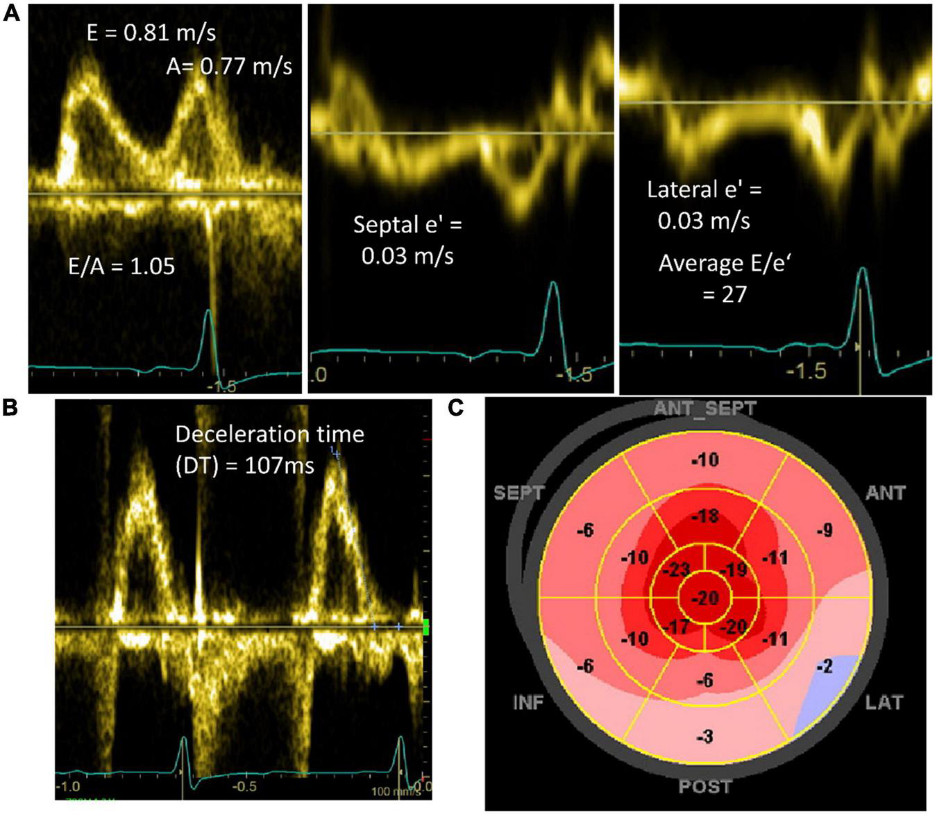
Select any illustration to inspect the full-size image. Source colors and annotations are preserved.
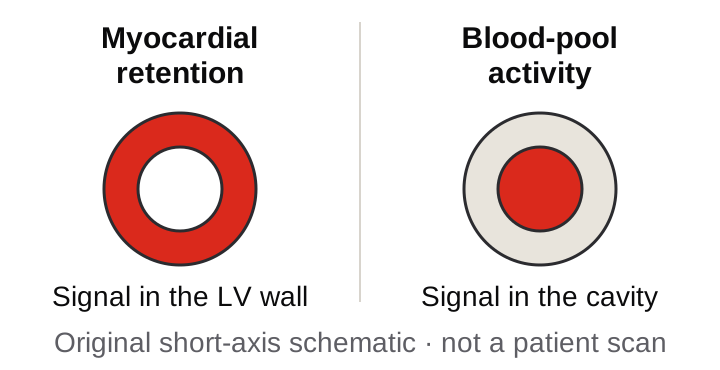
3 · Resolve apparent cardiac uptake
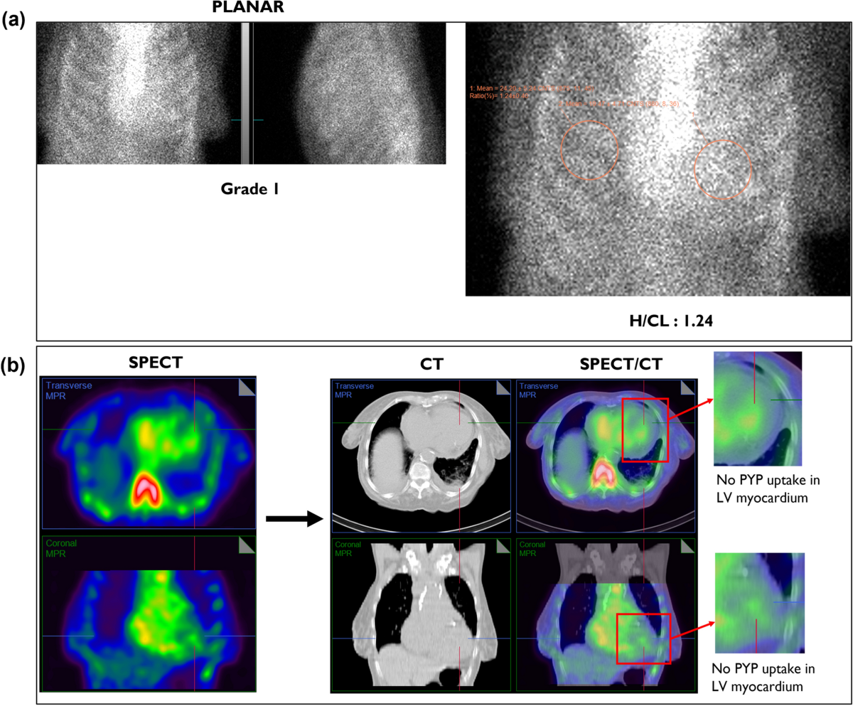
Apply the finding to the pathway
True myocardial uptake does not automatically mean ATTR. The nonbiopsy pathway also requires a compatible phenotype, grade 2–3 uptake and a negative complete monoclonal evaluation.
Complete the screen: serum free light chains, serum immunofixation and urine immunofixation. Interpret light chains with renal function and assay context. A possible clone requires further evaluation and tissue typing.
Do not rescue uncertainty with H/CL. Blood pool, focal uptake or true grade 1 uptake cannot establish nonbiopsy ATTR. A negative scan does not exclude AL, early ATTR or some variants.
Select a figure for full-size inspection. Diagnostic imaging colors are unchanged; the schematic is deliberately simplified.