Suction alarm with falling flow↓ P-level first to refill the inlet, then assess filling before fluids. Think hypovolemia · RV failure · malposition. Never ↑ P-level to chase a falling flow.
"Position Unknown / Wrong"Unknown may reflect low pulsatility or signal failure — look at the patient: MAP, A-line, flow before you touch it. Suspected migration → echo before adjustment (P.8).
Purge pressure low / "purge open"Risk of blood ingress and pump stop. Inspect purge circuit + cassette for leak/disconnect and follow the current alarm sequence urgently (P.7).
Cola-colored urine / ↑ LDHHemolysis — urgent imaging and plasma-free Hb. Assess suction, position and thrombus. Maintain perfusion at the lowest tolerable P-level. Reposition only if indicated (P.10).
01
What Impella Does
Trans-valvular axial unloading
Mechanism
A catheter-mounted microaxial pump sits across the aortic valve: it draws blood from an inlet in the LV and expels it from an outlet in the ascending aorta. It directly unloads the LV while providing systemic flow. Other support configurations can also unload the left heart.
Unloads ↓ LVEDP · ↓ LVEDV · ↓ wall stress → ↓ myocardial O2 demand, ↓ LAP, less pulmonary congestion, may improve myocardial oxygen balance. Routine infarct-size reduction in stable STEMI was not demonstrated (P.14).
Perfuses provides LV-to-aorta flow without rhythm synchronization. The perfusion response still depends on filling, pressure load and the rest of the circulation.
Preload-dependent output needs a filled LV and adequate pulmonary venous return. Inadequate filling promotes suction.
Unmasks the RV unloading changes venous return, pulmonary pressure and septal geometry. RV reserve may limit the response (P.6, P.10).
02
vs the other tMCS
load vs unload
IABP
Impella
VA-ECMO
Flow mechanism
Counterpulsation, no pump-flow lumen
LV → aorta, model-dependent
Venous drainage → arterial return
LV effect
mild unload
unloads
loads (↑ afterload)
Oxygenates
no
no
yes
Timing
Inflate in diastole, deflate before ejection
Continuous flow
Continuous flow
RV support
no
RP / RP Flex
yes
The unloading point: VA-ECMO returns blood to the aorta and raises LV afterload (distension, pulmonary edema). Impella does the opposite — it vents the LV forward. That's why an Impella is added to ECMO ("ECpella") to unload (P.10).
▦
The platform — three pumps, one console
full specs · P.11
Impella CP3.3–3.7 L/min mean
AccessPercutaneous femoral (14 Fr) retrograde across AoV · flexible pigtail
UseAMI / HF shock · high-risk PCI
Label≤4 days shock · ≤6 h PCI
Impella 5.5≤ 5.5 L/min
AccessSurgical axillary graft (or direct aortic) · no pigtail, soft tip
UseFull LV support · bridge · ambulatory
Labelup to 14 days
Impella RP / RP Flex> 4 L/min
AccessFemoral vein (RP) / IJ or femoral (RP Flex) → RA → PA
UseIsolated / predominant RV failure
Labelup to 14 days
Enlarge CP anatomy
Scroll horizontally to inspect the labels. Select again to return to the overview.
Enlarge RP anatomy
Scroll horizontally to inspect the labels. Select again to return to the overview.
Schematic cutaways, not to scale. Red = oxygenated blood, blue = venous blood, gold = device. Moving marks follow pump flow, not measured speed. The pump does not oxygenate blood. CP and RP shown, with model-specific variants discussed on P.5 and P.15. Anatomy informed by BodyParts3D and the Minnesota atlas.
Two separate routes on ECMO: peripheral VA return is retrograde in the aorta. Left Impella moves blood forward from LV to aorta to relieve distension. Neither route guarantees aortic-root washout. Follow valve opening, stasis and congestion (P.10).
1
Indications
shock · HR-PCI · RV failure · bridge
Cardiogenic shock (SCAI C–E)
Select by cause, LV/RV phenotype, organ injury, access risk and exit strategy. STEMI-shock benefit is trial-supported in selected patients (P.14). Pre-PCI timing associations are not randomized proof.
Protected high-risk PCI
Complex / last-patent-conduit or unprotected left-main PCI with severe LV dysfunction — elective hemodynamic support (CP / 2.5).
Isolated / predominant RV failure
Acute RV failure after LVAD, MI, transplant or open-heart surgery → consider RP / RP Flex within labeling (P.15). PE is not a labeled indication.
Bridge — name it at insertion
To recovery · decision · durable LVAD · transplant (5.5 supports up to 14 days by US label, mobilization possible).
2
Device choice — by phenotype and trajectory
shock-team decision
First define the shock phenotype. Integrate cause, trajectory, perfusion, echo and invasive hemodynamics when appropriate. CPO, PAPi and filling pressures support judgment, but no isolated value selects a device.
LV predominant
Poor LV output with adequate RV delivery and no major oxygenation failure.
Consider CP or 5.5 if expected benefit, access and labeling fit.
CP offers percutaneous support. 5.5 requires surgical access and can support an ambulatory bridge.
RV predominant
RV failure limits pulmonary flow and LV filling. Assess pulmonary vascular load and LV reserve.
Consider RP / RP Flex or another RVAD in an appropriate candidate.
RP pumps venous blood to the PA. The lungs must oxygenate it and the LV must accept it.
Both ventricles / gas exchange
Biventricular failure, severe hypoxemia or selected refractory arrest needs a broader support plan.
Consider VA-ECMO or tailored combined support through the shock team.
Assess LV distension on VA-ECMO and the need for unloading. Two pumps do not guarantee adequate support.
Before insertion → check reversibility, neurological prognosis, contraindications, vascular/valve anatomy and the exit strategy. After insertion → reassess perfusion, oxygenation, unloading and complications. Escalation follows the cause and trajectory.
3
High-risk PCI — support before you start?
heart-team assessment
Patient factors
LV reserve and active heart failure
Valve disease may increase risk or contraindicate Impella
Unstable rhythm despite therapy
Frailty, renal injury and bleeding/access risk
Procedure complexity
Multivessel PCI
PCI on the last patent conduit
CTO via retrograde through the last conduit
Large ischemic territory during planned occlusion
Hemodynamic findings
Elevated PCWP / LVEDP
Low output, perfusion and pressure reserve
Revascularization and surgical alternatives
No single factor mandates MCS. Balance procedural benefit against access and bleeding risk.
4
Contraindications — check before you cross
mostly anatomic
Left-sided (CP / 5.5)
LV thrombusabsolute — embolic stroke on crossing.
Mechanical AoVabsolute — can't cross the disc.
Significant AS (area ≤0.6 cm² by US labeling) — can't seat / valve injury.
≥ moderate AI regurgitation and recirculation can undermine forward support.
Access severe arterial disease. Also exclude septal defect, rupture and tamponade under the applicable label.
Right-sided (RP)
Mechanical TV/PV can't cross.
RA/caval thrombus or inaccessible venous route, including an obstructing caval filter.
Valve / PA anatomy severe TV/PV stenosis or regurgitation, or PA wall disease preventing safe position.
Pre-insertion echo is mandatory — rule out LV thrombus, define the aortic valve, document RV function. Significant RV or combined cardiorespiratory failure is excluded by the isolated-LV shock label.
1
Before you insert — checklist
access · purge · anticoag · imaging
AccessCP: femoral artery · RP: femoral vein · RP Flex: IJ or femoral vein · 5.5: axillary arterial graft. Use US/CTA for size & tortuosity
Echo no LV thrombus · define AoV · document RV
Anticoag verify procedural ACT target and heparin suitability with the current model IFU and concomitant therapy
Purge prime with heparinized D5W (25 U/mL) or NaHCO₃ 25 mEq/L in D5W
Prime follow the current on-screen and model-specific IFU sequence, confirm purge function and exclude air
Console white cable handed off & laid flat to the AIC (remove torque)
Wire 0.018″ × 260 cm placement guidewire in the LV
2
Access routes
arterial vs venous · percutaneous vs surgical
CP
Percutaneous femoral arterial
RP/RP Flex use venous routes
CP accessUS-guided femoral arterial micropuncture, single anterior-wall stick above the bifurcation. Angio to size. RP uses femoral venous access. RP Flex permits IJ or femoral venous access.
CP pre-close place a suture-mediated closure for the large-bore arteriotomy.
Cross backload over the 0.018″ wire. Retrograde across the AoV. Adjust orientation only under imaging by a trained operator. Avoid force and mitral/septal contact.
Seat inlet to depth (CP 3.5 cm). Slowly ramp the P-level.
5.5
Surgical axillary — 10 steps
graft conduit
1–3 expose & graftIsolate the axillary artery (vessel loops). Sew a 10 mm × 20 cm vascular graft end-to-side. Clamp & check hemostasis.
4–6 sheath & wireInsert introducer, secure with graft lock(s). Advance 0.018″ placement wire to LV. Confirm procedural anticoagulation and use the specified lubrication dilator.
7–8 place & seatBackload the catheter. Clamp the graft to avoid blood loss. Cross the AoV under fluoro. cannula bend at the annulus, inlet ~5 cm deep. Start support.
9–10 trim & secureShorten the graft. Peel the introducer. Slide & suture the blue suture hub into the graft.
3
Securement & first-hour targets
don't let it migrate
Fix3-point external fixation. forward suture on the repositioning unit. preserve the angle of entry. Touhy valve locked. Remove the yellow lock pin.
Securement use the approved fixation system and access-specific movement precautions. Recheck external depth after movement.
Chart the cm marking at the catheter lock unit daily. Confirm the suture hub/anchor each shift. No alcohol on the purge sidearm.
Position
Re-confirm by echo (TEE/TTE) on ICU arrival — inlet 5 cm (5.5) / 3.5 cm (CP) (P.5).
P-level
To goal MAP 65–75, CPO >0.6. don't run < P2.
Anticoag
ACT 160–180 s per IFU or an institutional anti-Xa/aPTT protocol. Reassess q4–6h (P.7).
Limb
Pulses · Doppler · NIRS on the access limb q1–2h.
4
Axillary graft conduit & post-insertion check
5.5 access · confirm before you leave
Route through the graft
Conduit a 10 mm × 20 cm vascular graft is sewn end-to-side onto the axillary artery.
Device route graft → axillary artery → aorta → across the aortic valve, with the inlet in the LV.
Securement the suture hub is secured to the graft. Follow the current 5.5 IFU for graft preparation, locking and fixation.
Before leaving the OR / lab
cannula bend at the annulus. Inlet ~5 cm (5.5) / 3.5 cm (CP) on echo
signals assessed against clinical state. Purge functioning
yellow lock pin removed. Touhy valve locked
3-point fixation + forward suture. Angle of entry preserved
peel-away sheath removed. Cm mark charted. Access-limb pulse documented
24/7 manufacturer clinical support for insertion, purge, anticoagulation, and alarm questions — use the current number on the device labeling or official support directory and call early, especially for heparin-delivery and positioning issues.
Read every round
P-level P0–P9
Pump speed → flow. Set to the hemodynamic goal, not a number. < P2 risks retrograde flow.
Flow max/min/mean
A calculated estimate, not a measured value — confirm perfusion (MAP, lactate, CPO).
Reflects the changing pressure load. A flat trace alone does not diagnose migration.
Purge mmHg · mL/h
Pressure & flow trend (P.7). Rising pressure suggests resistance. Falling pressure suggests leak or disconnection. Check the system.
1
The placement screen — waveforms inform position
aortic signal + motor current
Placement signal the pressure pattern sensed by the device. An aortic pattern supports expected position, while a ventricular pattern raises concern for deep placement. Confirm with imaging.
Motor current energy the motor draws across the cycle. Pulsatility can support a transvalvular pressure gradient. Flattening may reflect low native ejection, sensor problems or malposition.
Pattern change may reflect suction, loading, position or sensor error. Waveforms help frame the question, but do not identify the cause alone.
Low pulsatility ≠ migration in deep shock / full support / arrhythmia the signal flattens and the controller may alarm "Position Unknown" — confirm position rather than assuming migration or normal placement. Correlate with MAP, the A-line, flow and echo (P.8).
SmartAssist combines measured and derived parameters. Derived LV pressure and flow do not replace independent hemodynamics or imaging.
Flow is an estimate derived from current & speed — confirm perfusion clinically, especially at low P.
Controller check: interpret signals with perfusion and echo. RP/RP Flex sensor drift can corrupt flow, PA/CV signals and PAPi. Use independent hemodynamics and model-specific IFU ranges (P.15).
2
P-levels — the one knob you turn
P0 off → P9 max
P0
off
P1
min
P2
floor
P3
P4
P5
P6
P7
P8
support
P9
max
Higher = more flow each step raises speed. CP P9 mean flow is 3.3–3.7 L/min (systolic peak up to 4.3). Delivered flow varies with loading and position. Titrate to perfusion.
Don't go below P2 at very low speed blood can flow retrograde (aorta → LV) through the pump.
Going up for low MAP / rising lactate / CPO < 0.6 — but only if the LV is filled. An empty LV just suctions.
Coming down weaning is a stepwise P-level reduction with stability checks (P.9).
3
Suction physiology — the diastolic clue
why flow falls
A
Under-filled
Most common
Hypovolemia / bleeding, vasoplegia, or RV failure not delivering preload.
Signal diastolic suction can suggest inadequate filling, but is not specific.
Fix↓ P-level, assess filling and RV function, then treat the cause.
B
Malpositioned
Inlet on a wall
Inlet against septum / mitral apparatus, or device too deep.
Signal persistent suction may suggest wall contact. Confirm with imaging.
Fixecho + reposition. Don't just turn the pump up.
C
Over-pumped
P too high for filling
Demanding more than the LV can supply → repetitive suction + hemolysis.
Signal suction alarms cycle. PfHb / LDH climb (P.10).
Fix↓ P-level to a sustainable flow. Treat low preload.
The reflex: a suction alarm → reduce the P-level while assessing the cause to refill the inlet, then diagnose (volume · RV · position). Turning the pump up deepens suction and shreds red cells.
The placement signal can raise concern for migration. echo tells you where it is. Confirm at insertion, after every transfer, and whenever flow or the waveform changes — inlet in the LV, outlet above the valve, and measure the depth.
1
Verify proper placement — five checks
device IFU landmarks
Stable catheter in the mid-ventricular space, directed toward the apex.
Inlet free clear of the mitral subvalvular apparatus and the LV walls. Free-floating, surrounded by blood.
Depth annulus → mid-inlet ≈ 5 cm (Impella 5.5) · 3.5 cm (CP).
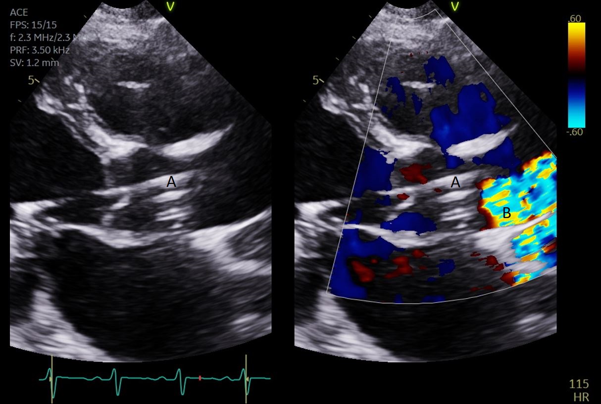
Outlet in the aorta well above the valve — confirm with color Doppler (outflow confined to the ascending aorta).
WindowsTTE: parasternal long-axis · TEE: mid-esophageal long-axis. Use off-axis cuts to follow the whole catheter.
Every echo assess LV unloading, RV function, new MR/AR, effusion and thrombus. Use TEE if TTE is inconclusive.
2
Echo landmarks — confirm in multiple views
TTE PLAX · TEE ME-LAX
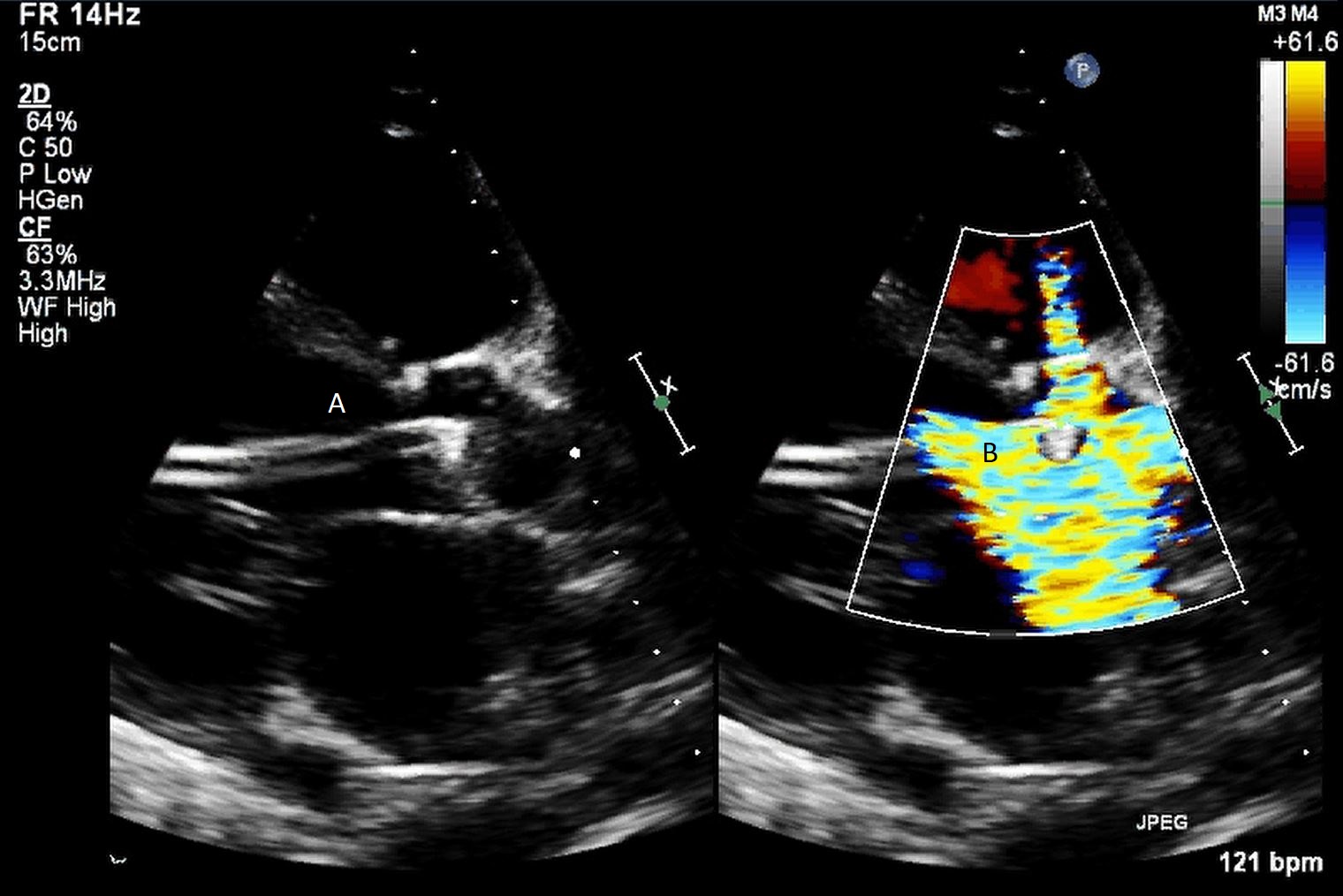
Identify the whole catheter: parallel cannula lines, the bend at the annulus, a free inlet in the LV, and outflow confined to the ascending aorta on color Doppler. Measure annulus to mid-inlet and verify against the current device IFU.
Outlet above the valve. In this published PLAX example, A marks the outlet and B its aortic color-flow signal.Outlet too low. A marks the catheter and B the outlet flow below the valve in the LVOT. Confirm in multiple views before adjustment.
Tracks two parallel echogenic lines = the cannula shaft.
Bend the cannula bends at the aortic annulus — a reliable landmark.
B
The inlet
teardrop
Teardrop the inlet area. The ends of the double lines mark it.
Anechoic can look black when the beam passes through inflowing blood.
Reverb artifact posterior to the distal cannula helps locate it.
C
Reposition
echo-guided
Techniquetrained operator, real-time imaging and device-specific instructions. No fixed-distance blind adjustment.
Outlet color jet confined to the ascending aorta. If at/below the valve → too deep.
Persistent hemolysis obtain additional views/TEE and assess suction, obstruction and thrombus (P.10).
4
Pitfalls & when to re-image
don't get fooled
Foreshortening an off-axis cut makes the inlet look too shallow — center the LV & AoV, follow the whole catheter.
Inlet at the MV a posteriorly-directed cannula bend points the inlet at the mitral apparatus → hemolysis, low/turbulent flow → reposition.
Anechoic inlet don't mistake the black inflow column for malposition — use the teardrop + reverberation.
Re-image whenever
on ICU arrival & after every transfer / proning.
flow falls or a waveform decouples / dampens.
suction alarms or a position alarm fires.
hemolysis (cola urine, ↑ LDH/pf-Hb) — obtain additional views if the first study is inconclusive.
before ambulation (5.5) — confirm the lock unit & inflow depth.
RP / RP Flex: confirm the outlet in the main PA and the inlet at the IVC/RA for femoral access or SVC/RA for IJ RP Flex access. The cannula crosses the tricuspid & pulmonic valves. Migration patterns & alarms → P.8.
1
Round every 6 h — three buckets
improving · mixed · worsening
↑
Improving
concordant
↑ CO & CPO, ↓ lactate at 12 & 24 h, ↑ UOP, pressors off, adequate ramp test.
Path recovery → wean (P.9), explant after a successful individualized reduction trial.
~
Mixed
discordant
Some params improve. Mixed CPO/lactate. Pressors lowered but not off. fails ramp.
Path continue support · assess escalation · if no recovery → durable VAD / Tx.
Path diagnose the cause, then consider device escalation matched to LV/RV and oxygenation needs.
2
Hemodynamic trends that inform escalation
CPO & PAPi
CPO = MAP × CO ÷ 451PAPi = (PASP − PADP) ÷ RAP
CPO ≤ 0.6 W is a high-risk signal in AMI shock. Reassess perfusion and support adequacy rather than using one number to assign a device.
Low PAPi (e.g. < 0.9 after AMI) raises concern for RV failure. Interpret with RAP/PCWP, echo, TR/PR and trajectory. A single threshold is not a universal RV-support gate.
Measurement use total CO from a documented method, not pump flow alone. Controller CO/CPO depends on an entered reference CO. Re-enter per IFU, including at 8 hours or when compliance changes, and verify independently.
Read the direction of travel
Serial pattern
Next assessment
CPO falls, lactate rises
Urgent shock-team reassessment of cause and support adequacy.
CPO improves, lactate remains high
Check ongoing ischemia, oxygen delivery, clearance and noncardiac causes.
CPO remains low, lactate clears
Verify CO measurement, vasoactive support and organ perfusion.
Both improve
Continue surveillance. Weaning still requires a supervised trial.
3
Targets & the daily bundle
individualize and trend
Target
Goal
MAP
65–75 mmHg
CPO
> 0.6 W
PAPi
Trend with RAP, PCWP and RV echo
Lactate
< 2, clearing
UOP
> 0.5 mL/kg/h
SvO₂
> 60%
Console P-level · flow (max/min) · waveform trend · purge P & flow · CO / CPO / LVEDP.
Position chart the cm mark at the lock unit daily. Suture/anchor secure each shift. Echo if any change.
Trend renal function, electrolytes, lactate and hemolysis to acuity. ABG/CXR for a clinical question. Echo for position or circulatory change.
Each shift chart the cm mark. Confirm suture/anchor. Access-limb pulse · Doppler · NIRS. Urine color.
Sidearm no alcohol on the purge sidearm. Keep the catheter straight, angle of entry preserved.
Sedation enough to protect groin precautions (femoral). 5.5 axillary → mobilize when stable.
Escalate, don't drift: reassess frequently and immediately with deterioration. Falling CPO despite increased support, low PAPi or rising lactate should prompt urgent shock-team reassessment of perfusion, device function and ventricular phenotype. Choose escalation for the cause and trajectory. No isolated threshold mandates RV support or ECMO.
Two separate tasks: purge pressure protects the motor from blood ingress. Systemic anticoagulation protects the patient and blood-contacting device. A heparin-free purge does not provide systemic anticoagulation.
1
Know the solution and the delivered dose
verify device IFU
Component
Practical approach
Heparin purge
SCAI recommends D5W with heparin 25 units/mL for ongoing support. Follow the current model-specific IFU and pharmacy preparation.
Bicarbonate purge
25 mEq sodium bicarbonate in 1 L D5W is the SCAI formulation when a heparin purge is unsuitable. The February 2026 FDA labeling change allows bicarbonate when heparin should not be used per physician judgment.
What not to add
No bivalirudin or argatroban in the purge. A systemic direct thrombin inhibitor can be used when indicated, with dose/assay adjustment for organ function.
Count purge heparin
Units/hour = units/mL × purge mL/hour. Example: 25 × 10 = 250 units/hour. Include this changing contribution when titrating IV UFH.
2
Choose one explicit anticoagulation protocol
targets are not interchangeable
During placement procedural ACT follows the IFU, procedure and concomitant antithrombotic therapy. Do not transfer a maintenance target to insertion.
During support IFU ACT targets commonly use 160–180 s. SCAI suggests anti-Xa 0.2–0.4 IU/mL or locally calibrated aPTT for ongoing UFH.
Sampling recheck after dose, purge or clinical changes according to the selected protocol. Track platelets, bleeding and hemolysis alongside the assay.
Discordance factor abnormalities can distort aPTT. Hemolysis, bilirubin and assay method can affect anti-Xa. Investigate unexpected results with the laboratory.
Apparent resistance check dose delivery, purge contribution, sampling and assay interference before escalating. Consider antithrombin assessment with specialist input.
No automatic escalation a high purge pressure or low anti-Xa alone is not a mandate for a higher systemic target, thrombolysis or pump exchange.
3
Bleeding and HIT are different problems
patient + pump
Clinically significant bleeding
Find and control the source, including retroperitoneal bleeding. SCAI's stepwise approach first stops IV heparin, then removes purge heparin and substitutes bicarbonate if bleeding persists. Severe bleeding requires urgent individualized decisions about all anticoagulants and antiplatelets.
Suspected / confirmed HIT
Assess clinical probability and test appropriately. When HIT requires heparin cessation, remove all heparin sources, including the purge. Select a nonheparin systemic anticoagulant with hematology/pharmacy and use a compatible heparin-free purge.
4
Purge fault → check the circuit before changing chemistry
Finding
First assessment
High pressure / low flow
Inspect kinks, clamps, tubing and cassette. Assess motor current, hemolysis and possible obstruction. If a higher-dextrose solution is used, lowering to D5W may reduce resistance. Do not increase dextrose to treat high pressure.
Low pressure / leak
Inspect bag, connections, cassette and sidearm urgently. Use the current alarm sequence and replace defective components. A persistent leak risks blood ingress and pump failure.
Disc not detected
Check the 2026 controller correction: reinsert the purge disc per IFU, then use a backup controller if the alarm persists. Plan the brief interruption of support.
2026 inventory check: Generation 1 purge cassettes and selected pump/controller units have specific field actions. Follow the applicable notice, including its continuity-of-care provisions (P.16).
1
On the console — correct vs the two migrations
echo detail → P.5
✓
Correct
Inlet at the labeled depth (≈3.5 cm CP · 5 cm 5.5), free in the LV cavity. Outlet above the valve. Waveforms can support this assessment but may be nonpulsatile during full support. Confirm & measure by echo (P.5).
↓
Too deep (in ventricle)
Outlet at/below the valve can recirculate blood into the LV. A ventricular signal or position alarm raises concern. Confirm by echo before imaging-guided correction (P.5).
↑
Too shallow (across)
Inlet near/in the aorta can cause ineffective LV unloading despite apparent pump flow. Confirm the inlet and outlet by imaging. Trained operator corrects position per IFU.
↓ P-level, image and assess filling/RV. Fluids only for demonstrated depletion.
Impella Position Unknown
Loss of pulsatility — the controller can't read a pulsatile signal (profound shock, low native output, full support, AF / arrhythmia). Does NOT mean it migrated into the aorta.
Look at the patient first — MAP, the A-line, flow. Review signals and echo. A supervised brief P-level reduction may reveal pulsatility if tolerated. Don't reflexively reposition.
Impella Position Wrong
Suspected malposition or unreliable positioning signals. Confirm the actual catheter position.
Verify with echo. Advance / reposition per SmartAssist (P.5).
Impella Position in Ventricle
Signal suggests deep position. Confirm outlet and inlet location by imaging.
Pull back to the labeled depth under echo (P.5).
Purge Pressure High
Kink, purge-path resistance, cassette problem or possible obstruction.
Inspect circuit, flow and solution. Follow the model IFU (P.7). Pressure alone does not diagnose thrombus.
Patient first. ↓ P-level if suctioning. Echo. Treat cause → tree below.
"Position Unknown" ≠ migrated into the aorta. Loss of pulsatility can prevent the positioning algorithm from resolving location. Migration and sensor failure still require assessment. Look at the patient: MAP, the arterial-line waveform, and flow. A supervised brief reduction may reveal pulsatility if tolerated. Obtain echo before adjusting catheter depth.
3
Low-flow / suction — work it down
don't reflexively turn it up
1 · Filling and perfusion
If suctioning, lower P-level as tolerated. Assess bleeding, LV filling and RV delivery.
Also look for tamponade. Elevated filling pressures do not prove adequate transmural filling. Give volume only for demonstrated depletion.
2 · Position and injury
Image the whole inlet-to-outlet route. Look for wall or mitral contact, valve injury and pericardial fluid.
Trained operator adjusts under imaging. A normal console does not exclude a mechanical problem.
3 · Load and device
Check pressure load, rhythm, purge circuit, motor current and hemolysis.
High purge pressure alone is not thrombus. Persistent low flow or deterioration needs urgent support-team reassessment.
4
Console & power emergencies
don't leave the pump stopped
Controller alarm check the white cable & connections (lay flat, no torque). If the AIC fails → swap to a charged backup controller — keep one at the bedside.
Power the AIC runs on internal battery (duration depends on controller and battery status) — keep it plugged in. On transport confirm charge + purge-bag volume + a compatible charged backup controller.
Pump stopped a stationary impeller risks thrombosis and retrograde flow (aorta → LV) — urgently follow the device-specific restart sequence. Suspected thrombus or obstruction requires expert assessment before repeated restart attempts.
Restart after any stop, re-confirm position by echo and the placement/motor-current waveforms before trusting the flow.
Readiness is physiologic. There is no universally validated Impella weaning schedule, mandatory 48-hour minimum, or single echo cutoff that proves readiness. A short PCI-support case and prolonged shock support require different plans.
1
Start with an exit strategy
recovery · bridge · goals
Recovery
Cause treated, adequate perfusion, improving organ function and manageable filling pressures.
Minimal or decreasing vasoactive support and a sustainable rhythm.
Bridge to therapy
If recovery is insufficient, assess durable LVAD, transplant or another temporary strategy early.
Reassess neurologic prognosis, RV reserve, organ injury and rehabilitation potential.
Goal-concordant limit
Clarify what additional support can achieve and the patient's goals.
A failing wean prompts reassessment, not indefinite escalation without a destination.
2
A supervised reduction trial
write the plan before changing speed
1 baselineRecord P-level, MAP, rhythm, vasoactive doses, lactate/urine trend, filling pressures and echo. Confirm the device is correctly positioned.
2 reduceReduce support in steps, with dwell time chosen by the shock team and response. Reassess before the next step. Avoid prolonged idle or stopped transvalvular support because of stasis and reverse flow.
3 challengeAt low support, assess whether native forward flow and perfusion remain adequate without rising filling pressures, pulmonary congestion or major vasoactive escalation.
4 decideIf intolerance develops, return promptly to the last tolerated level and investigate the cause. If stable, plan removal with a defined rescue strategy.
TIDE is an institutional framework, not a validated universal rule. It combines echo at full support, stepwise speed reduction with low-support reassessment, and selective stress echo/hemodynamics when recovery is uncertain.
3
Interpret three domains together
Domain
Evidence of tolerance
Reason to stop the trial
Perfusion
Stable mentation, lactate, urine output and skin perfusion on minimal vasoactive support.
Hypotension, rising lactate, oliguria, ischemia or new arrhythmia.
Hemodynamics
Adequate native/total output with acceptable filling pressures. Interpret CPO, CI and venous oxygen saturation as trends.
Falling output or rising RA/PCWP with congestion. A single CPO or PAPi threshold cannot certify success.
Echo
LV size and systolic function, LVOT VTI, aortic opening, MR/AR and RV function assessed at the same recorded support level.
LV distension, worsening MR, RV failure or a new mechanical complication. ECMO-derived numeric thresholds are not validated Impella explant criteria.
4
Removal is device- and access-specific
Configuration
Plan
Femoral CP
Operator-led withdrawal and stopping sequence per IFU, arterial closure strategy, anticoagulation plan and immediate vascular backup.
Axillary 5.5
Surgical team manages device extraction and graft ligation/repair. Consider thrombus, embolic risk and graft infection.
RP / RP Flex
Demonstrate RV tolerance of reduced support, then remove under the device-specific procedure with venous hemostasis and continued RV surveillance.
ECMO + Impella
If RV and pulmonary function recover, ECMO may be removed first while LV support continues. Severe respiratory or RV failure changes the sequence. Titrate unloading to echo and filling, not a fixed P2 rule.
After removal: watch for recurrent shock, access or retroperitoneal bleeding, limb ischemia, pseudoaneurysm and new valve dysfunction. Continue scheduled hemodynamic, access and hemoglobin reassessment. Link the next step to the MCS Weaning & Decannulation guide.
1
Hemolysis — the signature complication
look for a mechanical cause
Confirm hemolysis, then find its cause
Plasma-free Hb confirm and trend intravascular hemolysis. A value >40 mg/dL is used in some definitions, not a universal normal limit or an automatic exchange threshold.
Dark urine heme with few/no RBCs suggests pigment. Hematuria has urinary RBCs. Check microscopy and sample quality.
Supporting tests falling hemoglobin/haptoglobin and rising LDH/indirect bilirubin support the picture. LDH is also elevated by infarction and other tissue injury.
Mechanism suction, inlet/outlet obstruction, malposition or pump thrombus can produce shear. Check filling and the entire catheter route.
Treat the cause while maintaining perfusion
1 preloadAssess filling, bleeding and RV function. Replace volume only when depleted.
2 lower shearIf suctioning, reduce support as tolerated while maintaining perfusion. Correct the cause.
3 positionEcho/TEE: assess inlet, outlet, MR, RV and thrombus. Reposition only for an identified problem.
4 reassessTrend pf-Hb, hemoglobin and renal function after correction. Persistent severe hemolysis → urgent expert assessment for exchange/removal and alternative support.
2
Bleeding & the other complications
the harm signal is real
Access-site ooze — run the checklist
ACT in range (160–180)? heparin or bicarb in the purge?
peel-away sheath removed? forward suture used?
natural angle of entry preserved? leg immobilizer on?
arterial vs skin-tract ooze? catheter traction or movement?
Fixation / anticoagulation use approved securement and review all antithrombotic exposure. Control the bleeding source (P.7).
Bleeding the dominant driver of harm — balance against thrombosis.
Stroke embolic — never cross a known LV thrombus. Neuro checks.
Aortic valve / AI catheter sits across the valve — watch for new/worsening AI.
RV failure unmasked LV unloading exposes a failing RV (suction, ↑CVP, ↓PAPi) → cause-directed RV therapy and support reassessment (P.15).
Suspected thrombosis assess purge, motor current, imaging and hemolysis together. Expert team decides exchange/removal.
3
ECpella — unloading the LV on VA-ECMO
vent the loaded ventricle
Why VA-ECMO raises LV afterload → distension, pulmonary edema, stasis. An Impella actively vents the LV (alternative to IABP / surgical vent).
Wean order if RV and pulmonary function recover, ECMO may come off first while Impella continues. Follow perfusion, filling and echo during reduction (P.9).
Evidence mixed meta-analysis shows no clear early-mortality benefit overall (borderline in matched cohorts), with more bleeding, hemolysis, limb ischemia, RRT.
So reserve venting for a clear indication (distension / pulmonary edema / AV not opening), not reflexively.
4
Complication quick-reference
rate · watch · act
Complication
Risk context
Watch
Act
Hemolysis
definition-dependent
pf-Hb trend, hemoglobin, dark urine
assess filling · ↓P if suction · image · treat cause
Major bleeding
definition-dependent
Hgb, access & mucosal sites
source control and the stepwise anticoagulation plan (P.7)
Limb / vascular
access-dependent
pulses · Doppler · NIRS (large-bore arterial)
vascular STAT. Distal perfusion
Stroke
embolic
neuro exam
never cross LV thrombus. Imaging
AKI / RRT
common in shock
Cr, UOP, volume balance
renal support. RRT if needed
Device thrombosis
—
↑ purge pressure, ↓ flow, hemolysis
expert assessment and alternative support plan
Keep selection honest: the AMI-shock survival benefit came in a selected, LV-dominant population and was bought with more bleeding, limb ischemia, and RRT. registries link unselected use to higher complications & cost. Right patient, right pump, meticulous access & position.
◆
Side-by-side — verify against the current IFU
adult · current device family
Impella CP (SmartAssist)
Impella 5.5 (SmartAssist)
Impella RP / RP Flex
Supports
Left ventricle
Left ventricle
Right ventricle
Flow capacity
3.3–3.7 L/min mean at P9, peak up to 4.3
up to 5.5 L/min
> 4 L/min
Access
Percutaneous femoral (or axillary), retrograde across AoV
Surgical cutdown — axillary artery graft (or direct aortic)
Femoral vein (RP) / IJ or femoral (RP Flex) → RA → PA
Sheath / introducer
14 Fr peel-away
23 Fr × 6 cm via graft
~23 Fr (RP). Single-access (RP Flex)
Motor / catheter
14 Fr motor · 9 Fr shaft
19 Fr motor · 21 Fr cannula · 9 Fr shaft
22 Fr motor/cannula · 11 Fr shaft
Tip
Flexible pigtail
No pigtail — soft atraumatic tip
Distal pigtail in PA
Position sensor
Optical pressure (SmartAssist)
Optical pressure (no differential sensor)
Differential-pressure + fiber-optic (RP Flex)
Inlet depth (echo)
≈ 3.5 cm below annulus
≈ 5 cm below annulus
Outlet in main PA
Speed
up to ~46,000 rpm
0–33,000 rpm
—
US labeled duration
≤4 days shock · ≤6 h HR-PCI
up to 14 days
up to 14 days
Signature use
AMI/HF shock · high-risk PCI
Full LV support · bridge to recovery/LVAD/Tx
Acute RV failure within US labeling (P.15)
Dimensions refer to different parts (motor, cannula, shaft or introducer). Flow is load-dependent and is not guaranteed cardiac output. Verify the exact model and current device IFU.
P
P-level → flow · 5.5 accessory kit
console estimate · titrate
CP P9 mean flow 3.3–3.7 L/min, systolic peak up to 4.3. The mean and peak are different measurements.
5.5 P9 mean flow 5.0–5.5 L/min in the IFU table. Delivered flow depends on loading and position.
P0 / < P2 P0 = off. Below P2 blood may flow retrograde — use only in the model-specific controlled removal sequence.
Reminder displayed flow is a calculated estimate — verify with MAP, lactate, CPO.
Impella 5.5 axillary kit
Graft 10 mm × 20 cm vascular graft. One graft lock for this graft, plus a supplied spare.
Sheath23 Fr × 6 cm peel-away with hemostatic valve.
Dilator8 Fr silicone-coated lubrication dilator.
Wire0.018″ × 260 cm placement guidewire. Modular purge cassette.
↳
Picking the device — the 30-second version
detail → P.2
LV shock, percutaneous, fastImpella CP — femoral, no surgeon. Up to ~4 days.
LV shock, more flow / longerImpella 5.5 — surgical axillary, full support, ambulatory bridge.
RV-dominant failureImpella RP / RP Flex — RA → PA. RP Flex via IJ or femoral access.
Biventricular / respiratory failure select combined support. BiPella does not oxygenate. Significant hypoxemia may require an oxygenated circuit.
C
The Automated Impella Controller (AIC)
drives · monitors · purges
Drives the pump sets the P-level. Auto-regulates the purge. One console runs CP, 5.5, or RP.
Displays placement signal · motor current · flow (max/min/mean) · purge P & flow · SmartAssist CO / CPO / LVEDP / MAP.
Power AC + internal battery (duration depends on controller and battery status) — keep plugged in. Charge before transport.
Transport confirm battery + purge-bag volume + a backup controller. Lay the white cable flat (remove torque).
▸
The rest of the family
RP vs RP Flex · legacy
RP vs RP Flex
RP 23 Fr femoral vein. Effective but bed-bound.
RP Flex repositionable, IJ or femoral venous access. The IJ route can permit mobilization. Confirm inlet position for the chosen route.
Impella 5.0 / LD surgical predecessors of the 5.5. LD = direct-aortic.
Each Impella is a different operation — access, support level, position depth, the feel of anticoagulation, mobility, and removal all differ. Read down the column for the device you have. The generic workflow is on pp 3–10. This page is the per-device cheat-sheet.
◆
Management — side by side
read down your device's column
Impella CP
Impella 5.5
Impella RP / RP Flex
Role
LV · AMI/HF shock · high-risk PCI
LV · full support · bridge to recovery/LVAD/Tx
RV · eligible post-LVAD, MI, transplant, surgery (P.15)
Access
Percutaneous femoral (US-guided, pre-close) across AoV
Surgical axillary 10 mm graft (or direct aortic)
Femoral vein (RP) / IJ or femoral (RP Flex) → RA → PA
Flow capacity
3.3–3.7 L/min mean at P9, peak up to 4.3
up to 5.5 L/min
> 4 L/min
Support level
Titrate to perfusion, filling and suction
Titrate to unloading/perfusion, including on ECMO
Titrate to independent pressures, perfusion and echo (P.15)
Echo position
inlet 3.5 cm below annulus · pigtail in LV
inlet ~5 cm below annulus · no pigtail
outlet in main PA · femoral inlet IVC/RA or IJ inlet SVC/RA
Anticoagulation
Purge (D5W + heparin 25 U/mL, or bicarbonate when heparin is unsuitable) + systemic ACT 160–180 / institutional anti-Xa/aPTT target. Insertion per IFU (P.7)
Position confirm outlet in the main PA, inlet at IVC/RA (femoral) or SVC/RA (IJ). Cannula crosses TV & PV.
MobilityRP Flex via RIJ for ambulation. Dual sensors.
Wean with perfusion, filling pressures and RV echo at reduced support. No single TAPSE/PAPi value proves recovery.
Watch hemolysis if the cannula abuts the PA wall.
One-liner:CP = percutaneous LV support, hemolysis-/suction-prone, ≤ 4 days, bed-bound. 5.5 = surgical full LV support, ambulatory, the bridge — but it unmasks the RV. RP / RP Flex = the right-heart pump, RA → PA, RP Flex ambulatory via the RIJ.
▸
Device and surgical insertion
Impella 5.5 · verify current IFU
Impella 5.5: confirm the optical sensor, pump head, purge sidearm, graft route, and fixation steps against the current device IFU and surgical plan.
▤
The evidence base — read before you believe the flow number
Assess preload · lowest tolerable P · image before repositioning · trend pf-Hb.
04RV failure unmaskedRight heart
Flow2.4
CVP18
PAPi0.8
Suctioncycling
LVunloaded
MAP60
Stop
This pattern raises concern for poor RV delivery. Confirm by hemodynamics and echo.
Think
RV failure is possible with high CVP and low PAPi. Also exclude tamponade and malposition.
Do
Optimize RV loading/contractility and select support for the full phenotype (P.15).
Always comes first
1 · Patient
Look at the patient before the console — MAP · perfusion · rhythm · urine. Displayed flow is an estimate. The patient is the truth.
2 · P-level down
For suction, reduce P-level while treating its cause. Low flow without suction needs diagnosis.
3 · Echo & call
Confirm position by echo. Loop in shock team / perfusion / CT surgery early. Use the current manufacturer support route in the current device labeling or official directory.
1
DanGer Shock: a survival benefit with a substantial harm burden
Trial outcome
Impella CP + standard care
Standard care
Death at 180 days
45.8% (82/179)
58.5% (103/176)
Composite safety end point
24.0%
6.2%
Renal replacement therapy
41.9%
26.7%
Mortality HR 0.74 (95% CI 0.55–0.99). Absolute death reduction 12.7 percentage points, approximately 8 treated per death prevented. Safety events combined severe bleeding, limb ischemia, hemolysis, device failure and worsening aortic regurgitation. These end points overlap and cannot be subtracted to calculate a single “net benefit.”
The population that supports the result
360 randomized, 355 analyzed: STEMI with recent shock, hypotension/vasopressor need, hypoperfusion and LVEF <45%.
Important exclusions included severe RV failure, mechanical MI complications and persistent coma after out-of-hospital arrest.
CP was studied in experienced centers. This is not a randomized survival claim for 5.5, RP, every shock cause or all arrest patients.
What changed after the initial report
2025 US ACS guideline: microaxial support is reasonable for selected patients with STEMI and severe or refractory shock, Class 2a, B-R, with explicit attention to bleeding, ischemia and renal risk.
2025 long-term follow-up: the survival advantage persisted during follow-up extending to ten years. It remains follow-up of the same selected cohort, not evidence for broader indications.
2
Keep shock, stable STEMI and planned support separate
Clinical setting
What can be concluded
Anterior STEMI without shock
STEMI-DTU, 2026: 527 randomized patients. Unloading with a 30-minute delay before PCI did not reduce infarct size versus immediate PCI and increased bleeding. Do not delay reperfusion routinely to unload a stable STEMI.
High-risk PCI
Heart-team selection considers anatomy, ischemic territory, LV function, surgical options and access risk. The US CP/2.5 PCI label is temporary support ≤6 hours. DanGer Shock is not a high-risk PCI trial.
Preemptive cardiac surgery support
IMPACT, 2026: a 100-patient single-arm 5.5 study showed feasibility in selected surgical patients with poor LV function. Without a randomized comparator, favorable event rates do not prove a survival benefit.
HF shock / bridge
Physiology and observational experience inform 5.5 use. State the intended bridge, likelihood of recovery and duration-label boundary. Do not import a STEMI-specific treatment effect.
3
Before escalating support, answer four questions
What is failing? LV, RV, both ventricles, oxygenation, vasoplegia, or a mechanical lesion.
What is reversible? Revascularization, rhythm, valve/mechanical repair, myocarditis or another treatable cause.
Can this device be used safely? Valve, thrombus, vessel anatomy, bleeding risk and current labeling.
What is the exit? Recovery, durable LVAD, transplant, decision, or a defined goal-concordant limit.
Device choice follows the phenotype. A higher P-level cannot correct RV failure, tamponade, severe hypoxemia or vasoplegia. Escalating lactate despite apparently adequate pump flow requires a new diagnosis and shock-team reassessment.
1
Same purpose, different venous route
Feature
RP / RP with SmartAssist
RP Flex with SmartAssist
Access
Femoral venous route, inlet near IVC/RA
IJ or femoral venous route. IJ inlet: SVC/superior atrial junction or RA. Femoral inlet: IVC.
Flow path
Caval/right atrial drainage → pump across the right heart → pulmonary artery. Confirm outlet and inlet by imaging.
Oxygenation
None. The device increases pulmonary blood flow. It does not oxygenate blood or bypass pulmonary vascular obstruction.
Mobility
Femoral precautions limit mobilization
IJ access can support rehabilitation when stable and securely fixed. Femoral access requires femoral movement precautions
2
Use the current indication, not “any RV shock”
US labeling · checked Oct 17, 2026
Current US labeling covers temporary RV support up to 14 days, BSA ≥1.5 m², for acute right-heart failure/decompensation of <48 hours after LVAD implantation, MI, heart transplant or open-heart surgery, without profound shock, end-organ failure or acute neurologic injury.
Anatomic checks
Mechanical or severely dysfunctional tricuspid/pulmonic valve, PA wall disease, RA/caval thrombus, or an unsuitable insertion route may preclude use.
A caval filter is relevant to the intended route. Verify unobstructed access and the exact model IFU.
Massive PE and chronic pulmonary vascular disease are not equivalent to the labeled postoperative/MI population.
Evidence boundary
RECOVER RIGHT was a small single-arm study demonstrating hemodynamic improvement and short-term survival in selected patients.
It does not establish a randomized survival benefit in refractory arrest, prolonged RV shock, severe organ failure or PE.
If LV dysfunction or respiratory failure is substantial, reconsider the whole support configuration.
3
2026 sensor correction: do not treat a display in isolation
RP with SmartAssist and RP Flex: differential-pressure sensor drift can make displayed flow, PA/CV placement signals and PAPi inaccurate. The sensor fault itself does not prevent the pump from supporting circulation. Use independent hemodynamic monitoring and imaging before intervention.
Flow reference consult the model-specific IFU P-level ranges, which reflect controlled pressure gradients. Actual flow still depends on preload, afterload, position, suction and thrombus.
Abrupt change compare the trend with perfusion, invasive pressures and imaging. Do not increase speed or exchange the pump solely because an unreliable calculated flow/PAPi looks abnormal.
Pump interaction guidewires and other venous devices can contact the inlet. Plan insertion, adjustment and removal with the support team and imaging per the 2025 correction.
4
Reassess the circulation after each change
Pattern
Interpretation and next check
RA falls, systemic perfusion improves
Supports a useful RV bypass effect. Continue evaluating congestion, organ recovery and device complications.
Pulmonary edema or PCWP rises
Increased pulmonary return may exceed LV capacity. Reassess LV function, MR, afterload and need for left-sided support.
Low flow or suction
Assess drainage, volume losses, position, obstruction and the pulmonary pressure load. Treat the cause instead of chasing a display.
For configuration selection, oxygenated RVAD support and biventricular management, use the RVAD & BiVAD guide. Reassess native RV function during a supervised reduction trial rather than relying on a device-derived PAPi alone.
Checked October 17, 2026. A correction updates use instructions, while a removal targets inventory or servicing. Match the exact model, product code, serial/lot and current notice. This selected bedside summary is not a complete recall inventory.
1
Current actions to reconcile with the device at the bedside
FDA action
Problem
Action that changes care
Left-sided pumps 2024 correction
Catheter manipulation can cause ventricular perforation/free-wall injury.
Advance or torque only under imaging. Consider fragile infarcted tissue, small cavity or complex anatomy. New collapse → urgent echo for tamponade, device position and valve injury.
RP / RP Flex 2025 correction
Guidewire or other intravascular-device contact with the inlet can damage the pump/device.
Coordinate other line/device procedures with the support team and follow the current imaging and handling instructions.
RP / RP Flex April 3, 2026
Sensor drift can corrupt displayed flow and pressure-derived metrics.
Use independent hemodynamics, imaging and model-specific IFU flow ranges. See P.15.
Quarantine affected inventory and use Generation 2. The notice permits necessary Generation 1 use only when Generation 2 is unavailable, with increased purge monitoring.
Selected CP sets July 2, 2026
Specified units outside design specifications can develop low purge pressure.
Check the listed serial numbers. Do not use affected sets, quarantine and return per notice.
AIC purge flag September 24, 2026
Failed purge flag/retainer can prevent cassette recognition.
All AIC units are in the phased service/removal scope. Inventory may continue supporting care while awaiting service. For a disc alarm, reinsert per IFU, then use backup AIC if unresolved.
Separate AIC early alert September 23, 2026
Specified consoles need hardware, firmware and/or software updates for recognition, button or startup problems.
Check serial list and coordinate service. Notice permits continued inventory use while awaiting service. This is distinct from the purge-flag action.
2
A deteriorating patient needs two simultaneous assessments