1
Pre-LVAD: findings that raise risk
Labs · clinical state · echo01
Labs / end organs
Historical pre-LVAD cohort risk flags- Total bilirubin ≥2.0 mg/dL.
- Creatinine ≥2.3 mg/dL or renal replacement therapy.
- AST ≥80 U/L.
- BUN >39 mg/dL.
- Rising bilirubin / creatinine, oliguria, ascites and persistent edema add concern.
02
Clinical state
Less reserve before the operation- Vasopressor requirement or multiple inotropes.
- Preoperative mechanical ventilation.
- Shock, low venous saturation or worsening organ perfusion.
- Prior RV failure / RV infarction.
- Previous cardiac surgery.
03
Echo
A vulnerable RV- Severe RV systolic dysfunction.
- RV dilation, especially with poor contraction.
- Severe tricuspid regurgitation.
- TAPSE ≤1.7 cm or FAC ≤35% flags dysfunction. Read with global RV function.
- Pulmonary hypertension / high RV afterload.
2
Hemodynamic risk flags, with context
Cohort cutoffs · document support| Measure | Risk threshold | Use at the bedside |
|---|---|---|
| PAPi (PASP − PADP) / mean RAP | <1.85 before LVAD | Associated with early RV failure in a 132-patient cohort. Not a universal implant or explant threshold. |
| PAPi after optimization | ≤3.33 before LVAD | Residual-risk signal in a 315-patient cohort after PAC-guided optimization. Compare measurements under documented support. |
| PAPi in acute RV infarction | ≤0.9 | Severe RV dysfunction and risk of RV support / death in acute inferior MI. |
| RAP / CVP | >15 mmHg | Congestion, especially when paired with low CI and a dilated RV. |
| RAP / PCWP | >0.63 | Disproportionate right-sided congestion before LVAD. |
| Cardiac index | ≤2.2 L/min/m² | Low forward output. Concern increases when RAP stays high. |
| RV stroke work index | ≤250 mmHg·mL/beat/m² = ≤3.4 g·m/beat/m² | RVSWI = (mPAP − RAP) × SVI. SVI = CI × 1000 / HR (mL/beat/m²). Use CI in L/min/m² and HR in beats/min. Multiply RVSWI by 0.0136 for g·m units. |
Read the clusterLow PAPi + high RAP + low CI + rising bilirubin / creatinine + a poor RV should prompt a planned RV-support strategy. These cohort risk flags are not stand-alone pump triggers. Recheck after optimization. Record rhythm, ventilation and support when trending PAPi.
3
RV power output: equation and interpretation
Pressure in mmHg · CO in L/minRVPO = mPAP × CO / 451 WIndex: mPAP × CI / 451 W/m². Example: 30 × 4 / 451 = 0.27 W. Net hydraulic power uses (mPAP − RAP) × CO / 451. No established RVPO-alone cutoff predicts pre-LVAD RV failure. Falling flow / power with rising RAP is concerning. Higher power may reflect pulmonary load. Systemic CPO 0.6 W is not an RVPO cutoff.
4
Fick versus thermodilution in RV dysfunction
Before a CO / CI result drives treatment| Situation | Practical rule |
|---|---|
| Choose / name the method | Without shunt or circuit distortion, prefer thermodilution (TD) over estimated Fick. Direct Fick uses measured oxygen consumption. Estimated Fick assumes it. CI = CO/BSA. |
| TR / reliable TD | Severe TR does not automatically invalidate TD. Average 3 values within 10% with consistent injectate and respiratory phase. Repeat poor curves. |
| Low flow / disagreement | Decision-changing discrepancy: recheck Hb, paired arterial / PA samples and technique. Adjudicate with direct Fick. Do not average disagreeing methods. |
| Shunt or RVAD | Shunt: saturation run and shunt-specific Fick flows, not routine TD. RVAD: map drainage, return and sampling sites. See page 4. |
5
Before CABG or valve surgery
Items that raise concernA
Patient / substrate
Poor RV + greater pulmonary load- RV dysfunction, severe TR, pulmonary hypertension.
- sPAP >55 mmHg, LVEF <50%, AF were associated with postoperative RV failure.
- Renal/hepatic dysfunction, congestion and prior RV infarction.
B
Operation / perioperative stress
A smaller margin for error- Mitral or tricuspid surgery.
- Redo / complex operation, prolonged CPB or ischemic time.
- RV ischemia, hypoxemia, hypercapnia / acidosis, excessive intrathoracic pressure and loss of AV synchrony.
Choose
support
support
RV alonePoor RV delivery, with a left heart that can accept more flow → RVAD.
Both ventriclesInadequate LV output / unloading as well → BiV support.
Gas exchangeAdd an oxygenator to an external RVAD when needed. Assess lung and left-heart load.
Immediate collapseConsider VA-ECMO for rapid cardiopulmonary support. Assess LV unloading.
1
Recognize the support configuration
Device · access · blood-flow routeImpella RP Flex
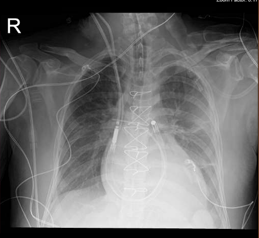
Jugular configuration shown. Inlet in the SVC / RA, outlet in the PA. Chest radiograph.
Agrawal et al. 2023 preprint, Fig. 3 · CC BY 4.0 · Unmodified.
Surgical CentriMag BiVAD
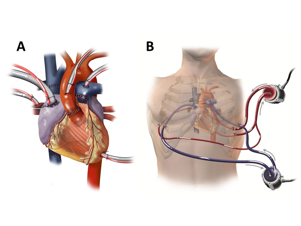
Right pump: RA → PA. Left pump in this case: LA + LV → aorta. Cannulas exit through the chest wall.
Kaczorowski et al. 2013, Fig. 1 · CC BY 2.0 · Unmodified.
Recognize the pump and the circuitImpella RP / RP Flex is a catheter pump. Surgical RVAD uses cannulas connected to an external pump such as CentriMag, with an oxygenator when needed. RVAD returns blood to the PA, then through the lungs to the LV. VA-ECMO returns to systemic arteries. Neither automatically unloads a failing LV.
2
Choose the device for the clinical job
Timing · lungs · access · rehabilitation| Device / configuration | Best fit | Why choose it? | Check before placement |
|---|---|---|---|
| Impella RP / RP Flex Intracorporeal pump | Acute, predominantly isolated RV failure after MI, LVAD, transplant or cardiac surgery. | Percutaneous RV support when native gas exchange is adequate and a short recovery course is expected. RP uses femoral access. RP Flex supports jugular or femoral placement. | Check venous / RA-to-PA access. Exclude right-heart thrombus, severe tricuspid/pulmonic stenosis or regurgitation, mechanical right-sided valves, PA-wall disease preventing safe placement/positioning, and caval obstruction/filter without adequate 22 Fr access. No oxygenator. |
| Surgical RVAD / CentriMag RA / venous drainage → PA | Failure to separate from CPB, severe post-LVAD RV failure, or planned support in a high-risk operation. | Direct control of cannula size/position, robust external support, optional oxygenator and integration with surgical LVAD / BiVAD. | Plan cannulation and explant together. Tunnelled grafts may permit closed-chest removal. Inability to anticoagulate is a contraindication. |
The Impella RP selection windowUS labeling: BSA ≥1.5 m², acute RHF lasting <48 hours after the qualifying event, support for up to 14 days, without profound shock, end-organ failure or acute neurologic injury. The <48-hour window refers to RHF duration, not simply time since the operation or MI.
RP Flex + PA or dialysis catheterAssess pre-existing catheter-associated thrombus and maintain the systemic anticoagulation plan. Additional right-sided catheters can increase thrombus risk.
CentriMag US labeling: up to 30 days for post-cardiotomy failure to separate from CPB (LV, RV or BiV support). The RVAS HDE covers acute RV-failure cardiogenic shock for up to 30 days. Effectiveness for the HDE indication has not been demonstrated.
3
Three practical selection calls
Choose the receiving circulation, then the pumpA
MI + isolated RV failure
Lungs and LV can handle the flow- Consider Impella RP / RP Flex when anatomy and the labeled clinical window fit.
- If gas exchange requires an oxygenator, choose an external RVAD circuit or VA-ECMO according to LV / systemic support needs.
B
Post-LVAD RV failure
LVAD already supports the left side- Consider Impella RP / RP Flex for percutaneous support when anatomy and labeling fit.
- Use surgical RVAD when already in the OR, an oxygenator is needed, or percutaneous support is unsuitable.
C
BiV / lung failure
Support the whole circulation- Add left support for LV failure. Add membrane gas exchange for lung failure.
- Use VA-ECMO when immediate systemic bypass is needed. Reassess LV distension and unloading.
Why an isolated RVAD can worsen pulmonary edemaThe pump increases PA delivery and pulmonary venous return. If the LV cannot clear the flow, left filling pressure rises. Check LV filling, LVAD unloading and outflow before turning up the RVAD.
CTICU
priorities
priorities
FillRelieve congestion. Give small, reassessed volume only for underfilling.
PerfuseSupport systemic pressure and forward output together.
UnloadLower pulmonary load and ensure adequate LV unloading.
SynchronizePreserve sinus rhythm / AV synchrony. Consider pacing when it improves output.
1
An individualized RV-protection plan
Pilot-derived targets · individualize to responseFilling
Aim for CVP about 8-14 mmHg with improving congestion and output.
Systemic perfusion
Aim for MAP about 70-90 mmHg. Follow lactate, urine output and venous saturation.
Left filling
Aim for PCWP <18 mmHg when measured. Check LVAD unloading and septum.
Rate / rhythm
Sinus rhythm + AV synchrony. The pilot used HR >100/min. Pace only when filling and output improve.
Lungs
SpO₂ >95% · PaCO₂ <50 mmHg
Keep plateau pressure <30 cm H₂O.
Keep plateau pressure <30 cm H₂O.
Geometry / oxygen delivery
Use echo-guided LVAD speed. Treat bleeding and anemia that limits oxygen delivery.
A
Underfilled?
Use a small, observed preload trial- Give 100–250 mL, then reassess flow, CVP and echo. Stop if pressure rises without improved output.
B
Congested?
Read MAP together with CVP- MAP 70 − CVP 25 leaves a perfusion gradient of only 45 mmHg. Support arterial pressure and relieve venous congestion.
2
Consider pacing before escalating catecholamines
Rate and AV timing are treatmentsA
Sinus bradycardia
Intact AV conduction- With reliable atrial wires, use atrial demand pacing to raise rate while preserving native ventricular activation.
- Test arterial waveform, CI/venous saturation, filling and echo. Keep the rate that improves output.
B
AV block / lost synchrony
Atrial + ventricular wires- Use AV sequential pacing when indicated. Optimize AV timing for filling and forward flow.
- With AF/flutter, avoid rapid atrial tracking. Correct triggers and restore a useful rhythm promptly.
C
Dependable pacing
Each shift and after a change- Confirm sensing, electrical capture and mechanical response. Record mode, rate, AV delay and thresholds.
- Keep backup available. New hypotension, arrhythmia or loss of capture needs immediate reassessment.
3
Vent and drips: give each intervention a purpose
Pressure · filling · flow01
Ventilator
Recruit without overdistension- Tidal volume 6-8 mL/kg predicted body weight. Check plateau pressure and auto-PEEP.
- Start with low PEEP, often 5 cm H₂O. Increase when recruitment / oxygenation improves and the RV tolerates it.
- Correct hypoxemia, hypercapnia, acidosis, pain and dyssynchrony. Aim for early extubation when perfusion and gas exchange permit.
Low and high lung volumes both raise pulmonary vascular load.
02
Drips
Treat the limiting physiology| Problem | Practical choice |
|---|---|
| Low output | Dobutamine 2.5–20 mcg/kg/min. Start low, titrate to perfusion. Watch tachyarrhythmia. |
| Low output + pulmonary load | Milrinone 0.125–0.75 mcg/kg/min. Start low. Watch hypotension and renal accumulation. Reduce for renal impairment. |
| Hypotension | Norepinephrine ± vasopressin. Support coronary/systemic perfusion while correcting the cause. |
| Pulmonary load | iNO 20 ppm or iEPO 50 ng/kg/min were used in INSPIRE-FLO. Ensure uninterrupted delivery, including transport / extubation. Wean stepwise to avoid rebound. |
| Congestion | IV loop diuretic. Escalate decongestion if venous pressure stays high. Consider renal support for refractory overload. |
4
Reassess after every important change
One team · one support planA
Trend perfusion and filling
Record the support state- RAP/CVP, PA/PCWP when available, CI, lactate, venous saturation, urine output and renal/hepatic trajectory.
- Pair these with RV/LV echo, rhythm, ventilator settings, vasoactive doses and LVAD speed.
B
Escalate early
Congestion + poor perfusion despite treatment- Exclude tamponade, bleeding, ischemia, PE, arrhythmia and device/outflow obstruction.
- Persistent RV failure needs an RV-support decision before progressive organ injury.
Why use a coordinated bundle?The RV Protection pilot randomized 20 patients, 10 per group. At 24 weeks, survival free of hemodynamic or hemocompatibility events was 80% vs 40%. Severe RV failure was 0/10 vs 3/10 (P=0.20). This supports further study, not a proven universal bundle.
On
RVAD
RVAD
IdentifyPump, cannulae, drainage/return sites, oxygenator and left support.
InspectTrace fixation, external marks, kinks, connections, gas and power.
InterpretRead flow with filling, echo, pressure and perfusion. Record RPM / P-level.
ActCorrect the limiting mechanism, then document the response.
1
Balance right delivery with left clearance
Especially RVAD + LVADOne circulation, two assisted routesRA → RVAD → PA supports lung delivery. LV apex → LVAD → aorta supports systemic delivery.
Too little right deliveryThe LV may underfill and the LVAD may suction.
Too little left clearancePulmonary venous pressure and congestion may rise.
Read the patientEcho, pressures and perfusion establish balance. Displayed pump flows need not match or sum to cardiac output.
Blue = venous blood. Red = oxygenated blood. Solid routes = assisted flow. Dashed routes = native flow. No oxygenator is shown. Motion illustrates direction, not measured flow.
A
LV underfilled / suction
Low delivery or excessive unloading- Check RVAD drainage, bleeding/volume loss, pulmonary resistance, cannula position and tamponade.
- Use LV size and septum to adjust left support. Correct the cause before escalating speed.
B
Pulmonary edema / high wedge
The LV cannot clear current delivery- Assess LV function, LVAD unloading/outflow, aortic regurgitation and volume.
- Correct left clearance and rebalance both pumps. An oxygenator adds gas exchange, not LV unloading.
2
Low flow: localize the problem
Patient → drainage → pump → return → receiving heart| Pattern | Think | First moves |
|---|---|---|
| Chatter / suction Variable low flow | Drainage limitation, underfilling, cannula-wall contact or excessive pump demand. | Echo + circuit inspection. Reduce excessive demand while treating the cause. Give fluid only when underfilled. |
| Flow down at same speed | More pulmonary/return resistance, kink/migration, thrombus or impaired LV acceptance. | Trace tubing, compare marks, check PA/left filling pressure and image cannula position. |
| Hemolysis / dark urine | Suction, malposition, obstruction, pump thrombosis or excessive shear. | Check plasma-free Hb/LDH trend and circuit/echo. Fix the mechanical cause. |
| RP display changes Patient appears unchanged | Position change or sensor error. | Use independent hemodynamics, imaging and the model-specific IFU P-level flow ranges, not AIC flow values. IFU ranges are bench ranges, not measured patient flow. The 2026 correction includes PAPi and PA/CV signals. |
| Patient suddenly worse | Bleeding, tamponade, arrhythmia, PE, circuit failure or a new LV problem. | Activate MCS/perfusion and surgery. Urgent echo and cause-directed resuscitation. |
3
Daily circuit and blood checklist
Make the rounds plan explicit01
Anticoagulation
One whole-circuit plan- Name drug, assay, target and who titrates. Include purge heparin in total exposure.
- RP: confirm purge solution / current IFU. Bicarbonate purge does not replace systemic anticoagulation.
- Bleeding: control source and coordinate interruption / restart. Check assay discordance before dose escalation.
02
Oxygenator
Blood flow ≠ gas flow- Verify gas source, sweep and delivered oxygen fraction. Trend pre/post-membrane gases and pressure drop at similar flow.
- Check gas supply and sampling before calling membrane failure.
- Oxygenated PA return alters mixed venous sampling and routine Fick assumptions. High PA saturation may reflect circuit return. Record the sample site.
03
Cannula + organ care
Each shift and around movement- Document marks/fixation. Inspect bleeding, infection, skin injury, migration and circuit slack.
- Follow perfusion, congestion, renal/hepatic recovery, nutrition and mobility.
- RA-to-PA bypass can distort PAC thermodilution. Pump flow is not native RV output. Validate CO for the circuit.
Pump stop, air or disconnectionCall MCS/perfusion immediately, assess the patient and follow the circuit emergency procedure. Keep matching backup equipment ready. Check current controller service notices and the Impella safety update.
4
Ready to test less support?
Recovery should survive a reduction in flow| Test separately | Change / observe |
|---|---|
| Lung recovery With an oxygenator | Step down oxygenator oxygen fraction and sweep, then perform a supervised off-sweep trial with blood flow unchanged. Follow arterial gases and work of breathing. |
| RV recovery | After the cause is treated and organs improve, reduce pump support stepwise. Follow filling pressure, perfusion and echo. Passing the lung test does not establish RV recovery. |
- Keep LVAD, rhythm, vent and vasoactive settings sufficiently stable to interpret the RVAD trial. Follow the device minimum-flow and anticoagulation plan.
- Watch for rising RAP, RV dilation, LV underfilling/suction, falling pressure or output, and worsening perfusion.
- If the trial fails, restore effective support and correct the limiting physiology. If recovery stalls, revisit the bridge to transplant or durable support.
1
Ready for a weaning trial?
Reassess dailyA
Cause / loading conditions
Remove reversible barriers- Ischemia, tamponade, bleeding and arrhythmia addressed.
- Decongested, adequate oxygenation, controlled pulmonary load and stable acid–base status.
B
Perfusion / support
Improving on less pharmacologic help- Low, stable inotrope / vasopressor doses.
- Lactate clearing, urine output adequate and renal / hepatic trajectory improving.
C
Echo / receiving LV
Enough reserve to test- RV size / contraction improving. LV fills and unloads appropriately.
- Exclude cannula obstruction, severe residual valve disease or a new mechanical problem.
Practical targets during reduced supportMAP ≥65 mmHg · CI ≥2.2 L/min/m² when the CO method is valid · RAP/CVP <15 mmHg with no progressive rise. These are practical trial targets, not validated stand-alone explant criteria. Require stable echo, perfusion and drug support.
2
Run a controlled reduction
Keep one interpretable support state| Step | What to do |
|---|---|
| Baseline / safety | Confirm anticoagulation and minimum-flow limits. Record flow / RPM or P-level, RAP, PA / PCWP, MAP, valid CI, sample site and echo. Hold optimized LVAD speed, rhythm, vent and vasoactive doses stable. |
| Reduce, pause, reassess | CentriMag example: decrease 0.5 L/min every 15–30 min toward 2 L/min, with echo and hemodynamic reassessment. Impella RP / RP Flex uses its device-specific P-level sequence. |
| Final low-support test | Final low-flow / interruption testing is brief and MCS / perfusion-led, following the device protocol. A pass at 2 L/min alone is not proof of independence. Keep effective support immediately available. |
3
What recovery should look like
Compare baseline with each flow step| Readout | Supports recovery | Stop / restore support |
|---|---|---|
| Filling pressures | RAP stays controlled as native RV work increases. | Progressive RAP rise, especially >15 mmHg, with poor LV filling. |
| RV echo | Global contraction holds. No progressive dilation, septal flattening or worsening TR. | RV enlarges, contraction deteriorates, septum shifts or TR worsens. |
| LV / LVAD | LV remains adequately filled and LVAD flow is sustained at the same speed. | LV underfills, suction develops or LVAD flow falls. |
| Systemic perfusion | MAP and valid CI hold, venous O₂ trend remains acceptable, no new drug escalation. | Hypotension, falling output / venous O₂, rising lactate, oliguria or new arrhythmia. |
Two high-yield interpretation rulesRAP >15 + PCWP <15 mmHg + low LVAD flow suggests inadequate right delivery. Restore support and assess RV failure, tamponade, pulmonary load and cannula obstruction. Judge RV size, septum, TR and perfusion together. TAPSE or FAC alone cannot decide explant.
4
Removal after a successful trial
Know the exit plan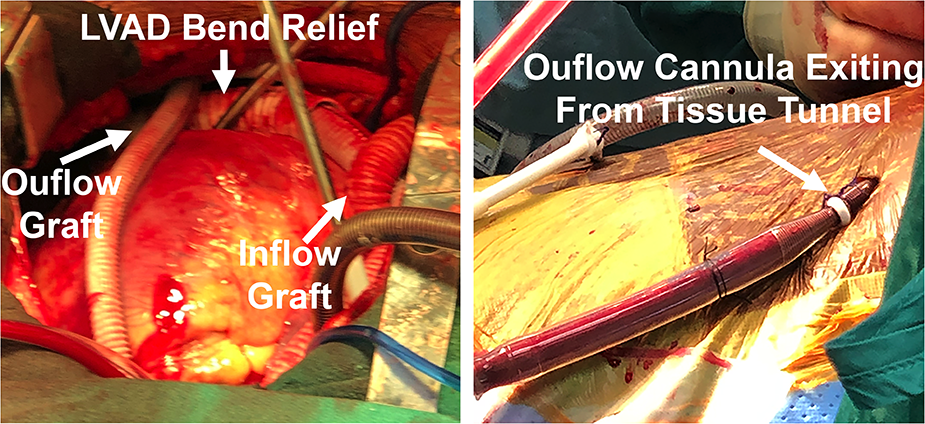
Magnified PA side-graft cutaway, not to scale. Gold = fabric graft. Blue = blood/cannula. After cannula removal, the free graft end is closed and buried. The PA attachment remains. This planned technique can avoid reopening the sternum.
A
Tunnelled RVAD: the essential point
Accessible graft allows closed-chest removal- In this planned technique, the team removes the return cannula, trims and closes the free graft end, and buries the remnant. The PA attachment remains.
- Remove percutaneous drainage separately. Direct RA entry may still need operative closure. Bedside versus OR depends on the actual cannulation / closure plan.
B
Confirm sustained recovery
Before and after decannulation- If an oxygenator is present, prove lung recovery separately using the gas-exchange trial on page 4.
- After removal, watch RAP, RV/LV echo, LVAD filling, perfusion and access-site bleeding.
- If repeated trials fail, revisit the cause and bridge to transplant or durable support.