Think
Infiltrative
Infiltrative
LVH + LOW voltage
Wall thickness up, QRS voltage low/normal — the classic discordance. This is chiefly an amyloid clue; Fabry and other storage phenotypes may have normal or high voltage, so do not generalize it to all storage disease.
Restrictive physiology
Biatrial enlargement, small/normal LV cavity, ↑ filling pressures, diastolic ≫ systolic dysfunction early. Differentiate from constriction.
AV block / VT young
Unexplained conduction disease or VT <60 yr → image for infiltration: sarcoid, Fabry, storage (PRKAG2/Danon), Chagas.
Multisystem clues
Neuropathy/CTS (amyloid·Fabry) · proteinuria (Fabry·amyloid) · eosinophilia (EMF) · transfusions (iron) · myopathy (Danon·Pompe).
01
The shared phenotype
why they look alike- Amyloid expands the interstitium. Fabry and glycogen disorders store material inside cells and can cause true myocyte hypertrophy. Low voltage is not a shared requirement.
- Cavity stays small / non-dilated until late; biatrial enlargement from chronic ↑ filling pressures.
- Diastolic dysfunction first (restrictive filling); systolic failure later as infiltration/scar advances.
Restrictive vs constrictive
- Restrictive (myocardial) no respirophasic septal shift; ↑ both atria; medial e′ low; ↑ BNP.
- Constrictive (pericardial) septal bounce, respiratory variation, pericardial thickening/calcification.
02
Workup ladder
cheap → targeted → tissue1 · ECG + Echo voltage-mass discordance, GLS apical sparing (amyloid), wall thickness, restrictive filling, atrial size.
2 · CMR mapping the pivotal step — LGE + mapping narrow the differential. Confirm with targeted tests.
3 · Targeted PYP (ATTR) · serum FLC + serum/urine IFE (AL) · ferritin/TSAT · α-Gal A/GLA · eosinophils · FDG-PET (sarcoid).
4 · Genetics / biopsy gene panel (familial); EMB when imaging + labs inconclusive or AL / active inflammation suspected.
03
Signature test
test combinations, not single-test labels- Amyloid ATTR Compatible phenotype + grade 2–3 myocardial uptake on PYP-SPECT + negative complete monoclonal screen.
- Amyloid AL Serum FLC + serum IFE + urine IFE. A clone needs urgent evaluation and typed tissue.
- Sarcoid FDG-PET (active) + LGE; extracardiac biopsy.
- Iron cardiac MRI T2* (<20 ms) — not ferritin.
- Fabry low native T1 + α-Gal A (♂) / GLA (♀).
- Storage LVH + WPW/block (PRKAG2), myopathy (Danon), infantile (Pompe).
- EMF / eosinophilic apical thrombus/obliteration. Eosinophilia may be absent in chronic EMF.
▦
Master comparison — red flag · key test · disease-modifying Rx
amyloid & sarcoid have their own cards ↓| Entity | Red flag | Key test | Disease-modifying Rx |
|---|---|---|---|
| Amyloid ATTR/AL | LVH + low voltage, apical sparing, bilateral CTS | PYP-SPECT + complete monoclonal screen | Tafamidis, acoramidis, vutrisiran. AL → chemo → card |
| Sarcoid | AV block <60, VT, patchy non-coronary WMA | FDG-PET + LGE; extracardiac biopsy | Steroids + steroid-sparing; device → card |
| Iron overload P.2 | Transfusions/thalassemia or HFE; restrictive→dilated | Cardiac MRI T2* <20 ms | Phlebotomy (primary) · chelation (secondary) |
| Fabry P.3 | Concentric LVH, neuropathic pain, cornea verticillata | Low native T1; α-Gal A / GLA | ERT (agalsidase) · oral migalastat |
| Storage P.4 | LVH + WPW/block; myopathy/ID; infantile | Gene panel; GAA enzyme (Pompe) | ERT for Pompe; transplant for Danon |
| EMF / eosinophilic P.5 | Apical obliteration/thrombus ± eosinophilia | Eosinophil count, echo/CMR | Steroids, anticoagulation, surgery |
◰
CMR parametric mapping — narrow the differential
native T1 / T2* / ECV narrow the differential| Deposit | Native T1 | ECV | T2* / T2 | LGE pattern & pearl |
|---|---|---|---|---|
| Amyloid | HIGH | HIGH (↑↑ interstitium) | — | Global subendocardial → transmural LGE; abnormal gadolinium kinetics (can't null myocardium) |
| Fabry | LOW | normal | — | Inferolateral mid-wall LGE; low T1 abnormal in ~40% before LVH |
| Iron | low | normal | T2* <20 ms (sev <10) | Usually no LGE; T2* is the load-bearing measure |
| Sarcoid | ↑ (active) | ↑ focal | ↑ T2 (edema, active) | Patchy non-coronary mid/epicardial LGE (basal septum, RV insertion); FDG-PET for activity |
| Eosinophilic / EMF | ↑ (acute) | ↑ | ↑ T2 (acute) | Endocardial LGE + overlying low-signal thrombus; apical obliteration |
01
Cause & phenotype
where the iron comes fromTwo routes to loading
- Primary / hereditary HFE C282Y homozygote (commonest); juvenile (HJV/HAMP) more cardiac. GI over-absorption.
- Secondary repeated transfusions — β-thalassemia major, MDS, sickle cell; ineffective erythropoiesis.
Cardiac course
- Iron in cardiomyocytes → oxidative injury.
- Early restrictive + diastolic dysfunction, AF & conduction disease.
- Late dilated / systolic failure can still improve with intensive chelation. Severe disease needs urgent specialist treatment.
02
Diagnosis
ferritin is not cardiac ironScreen
- Labs transferrin saturation >45% + ferritin; HFE genotype if primary suspected.
- ECG/echo low voltage, arrhythmia; restrictive then dilated pattern.
Cardiac MRI T2* — gold standard
- T2* < 20 ms on validated 1.5-T methods = cardiac iron overload; < 10 ms = severe, high arrhythmia/HF risk.
- Cardiac T2* is not reliably predicted by serum ferritin or liver iron — measure cardiac iron directly.
<20T2* ms abnormal
<10T2* ms severe
~25%cardiac iron in TM
03
Treatment
deplete the ironPrimary disease
- Phlebotomy ferritin target about 50 µg/L for induction, then 50–100 µg/L maintenance. Do not drive iron deficiency to normalize TSAT. Adjust for hemoglobin and tolerance.
Secondary — chelate (can't bleed an anemic patient)
- Treat-to-T2*; intensive chelation recovers EF.
- Cardiac iron is the leading cause of death in transfusion-dependent thalassemia — yet reversible.
℞
Iron chelators
match agent to severity & tolerance| Agent | Route / schedule | Niche & notes |
|---|---|---|
| Deferoxamine (DFO) | SC / IV (parenteral) | Continuous IV for severe cardiac loading / very low T2*; cumbersome but potent rescue. |
| Deferasirox (DFX) | oral, once daily | Convenient first-line for chronic transfusional overload; monitor renal/hepatic. |
| Deferiprone (DFP) | oral, formulation-dependent schedule | Effective myocardial iron clearance; combine with DFO for low T2*; agranulocytosis requires label-directed ANC monitoring. |
◷
Monitoring & when to refer
titrate to cardiac T2*Monitor
- Cardiac T2* stable >20 ms: about every 2 y. 10–19 ms: yearly. <10 ms: about every 6 mo, individualized to response.
- Function LVEF + diastology on echo/CMR; Holter for arrhythmia.
- Iron burden ferritin / TSAT trend + liver iron (tracked separately from cardiac).
Refer / escalate when
- T2* < 10 ms, falling LVEF, or arrhythmia → cardiology + intensify chelation (combine DFO + deferiprone).
- Advanced HF despite chelation → advanced-HF / transplant evaluation.
- Coordinate with hematology (transfusion & chelation program).
KEY
Ferritin cannot replace cardiac T2* MRI. Iron cardiomyopathy is reversible with intensive chelation (deferiprone ± deferoxamine) including some patients with systolic failure; phlebotomy is first-line for primary HFE disease.
◷
Cardiac T2* severity bands → action
treat-to-T2*; cardiac iron clears slower than liver| Cardiac T2* | Iron burden | Risk | Action |
|---|---|---|---|
| >20 ms | none / acceptable | low | Maintain regimen. Stable T2*: reassess about every 2 y |
| 10–19 ms | mild–moderate | ↑ — subclinical dysfunction | Optimize chelation. Usually yearly, sooner if clinical deterioration |
| <10 ms | severe | high — HF & arrhythmia; main cause of death in TM | Intensive combination (DFO + deferiprone). T2* about every 6 mo + cardiology |
⚑
Hemochromatosis beyond the heart & mimics on the differential
multisystem iron + who else gives restrictive LVHSystemic iron clues (primary HFE)
- Endocrine diabetes ("bronze diabetes"), hypogonadism, hypothyroidism, hypopituitarism.
- Liver hepatomegaly → cirrhosis → HCC risk.
- Joints / skin arthropathy (2nd/3rd MCP), slate-grey hyperpigmentation.
- Pattern juvenile (HJV/HAMP) & transfusional load are the most cardiac.
Don't mistake it for…
- Amyloid — also low voltage + restriction, but high T1/ECV & apical sparing (not low T2*).
- Fabry / storage — concentric LVH but normal T2*; check native T1, enzymes.
- Other siderosis — verify with cardiac T2*, not ferritin, before committing to chelation.
- Pitfall normal liver iron / ferritin does not exclude cardiac iron.
01
Biology & heart
GLA → Gb3 storage- X-linked GLA mutation → α-galactosidase A deficiency → globotriaosylceramide (Gb3) in myocytes, podocytes, endothelium, neurons.
- Males hemizygous, classic/severe. Females heterozygous — variable, can be fully affected.
- Later-onset "cardiac variant" GLA mutations can be heart-limited.
- Concentric LVH (mimics HCM/HTN/amyloid); short PR then conduction disease, AF; papillary muscle hypertrophy; inferolateral LGE.
02
Diagnosis
CMR + enzyme/geneCMR signature — LOW native T1
- Low T1 supports storage but is scanner/sequence-dependent. Fibrosis can pseudonormalize T1. Normal T1 does not exclude Fabry.
- Abnormal in ~40% before LVH (early marker); ECV normal (storage, not interstitial).
Confirm
- Males plasma/leukocyte α-Gal A activity (low).
- Females GLA sequencing (enzyme can be normal) ± plasma lyso-Gb3.
03
Therapy
treatable storage diseaseEnzyme replacement (ERT)
- Agalsidase beta or pegunigalsidase alfa are US-approved ERT options. Agalsidase alfa is used in other jurisdictions. Choose with a Fabry specialist.
Oral chaperone
- Migalastat for amenable GLA missense mutations — phase 3 placebo-controlled (FACETS); comparable renal outcomes over 18 mo (ATTRACT).
- Treat early — before extensive LGE/fibrosis.
⚑
Extracardiac red flags & who to screen
Fabry is multisystem — the clues are outside the heartMultisystem features → suspect
- Neuro childhood acroparesthesias (burning hands/feet), early stroke/TIA.
- Skin / eye angiokeratomas, cornea verticillata, hypohidrosis.
- Renal proteinuria → progressive CKD/ESRD.
- Other GI cramping/diarrhea, hearing loss, fatigue.
Screen these populations
- Unexplained concentric LVH — especially with low native T1 or a short PR.
- Cryptogenic stroke in the young; unexplained proteinuria / ESRD.
- Any family member of a known proband (X-linked cascade).
KEY
Concentric LVH + low native T1 + multisystem clues = test for Fabry — X-linked and treatable (ERT or oral migalastat for amenable mutations).
℞
Choosing therapy & monitoring
ERT vs oral chaperone · what to track| Feature | ERT — check regional label | Migalastat (oral chaperone) |
|---|---|---|
| Route | IV infusion every 2 weeks | 123 mg every other day, fasting 2 h before/after |
| Eligibility | Confirmed Fabry disease. Product age/indication limits apply | Adults with pathogenic GLA variants listed as amenable. Not recommended if eGFR <30 |
| Evidence | multi-organ benefit (heart · kidney · nervous system) | FACETS placebo-controlled; ATTRACT: comparable renal-function outcomes at 18 mo |
| Caveats | infusion reactions; anti-drug antibodies | nonsense / large variants → use ERT instead |
Monitor on therapy plasma lyso-Gb3 trend · eGFR + urine protein · echo/CMR (LV mass, native T1, LGE) · ECG / Holter for conduction disease & arrhythmia. Start before irreversible fibrosis (LGE) develops.
⊕
Differential of concentric LVH — how Fabry stands out
low native T1 + ECG & multisystem flags split the field| Cause | Native T1 | Discriminator | Confirm |
|---|---|---|---|
| Fabry | LOW | Acroparesthesias, cornea verticillata, short PR, inferolateral LGE | α-Gal A (♂) · GLA + lyso-Gb3 (♀) |
| Amyloid | HIGH | Low voltage, apical sparing, bilateral CTS | PYP scan · serum FLC + serum/urine IFE |
| Sarcomeric HCM | normal / ↑ | Asymmetric septum, SAM/LVOT gradient, FHx SCD | Sarcomere gene panel |
| Hypertensive / AS | normal / ↑ | BP history; valve gradient; regresses with load relief | Echo + clinical context |
| PRKAG2 / Danon | normal | Pre-excitation (WPW), conduction block, myopathy | Gene panel (see P.4) |
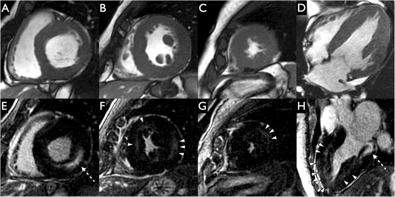
Do not stop at the imaging clue
Confirm with enzyme testing in males and GLA sequencing in both sexes. Normal enzyme activity in a female does not exclude Fabry.
Offer genetic counseling and targeted family testing after a pathogenic variant is established. Follow cardiac, renal and neurologic involvement.
Low T1 and inferolateral LGE are clues. Neither substitutes for biochemical/genetic confirmation.
These mimic HCM but are metabolic storage diseases — myocytes are vacuolated and glycogen-filled, with no sarcomeric disarray. Clues it isn't sarcomeric HCM: ventricular pre-excitation (WPW), conduction block, skeletal myopathy / ↑CK, X-linked pattern, intellectual disability, retinopathy, very high LV mass.
P
Pompe
GAA deficiency · AR- Defect acid α-glucosidase (GAA) deficiency → lysosomal glycogen.
- Infantile massive HCM, hypotonia, respiratory failure, death <1 yr untreated.
- Late-onset limb-girdle and respiratory weakness. Significant cardiomyopathy is uncommon.
- Dx GAA enzyme (dried blood spot) + GAA gene.
- Rx — ERT alglucosidase alfa q2 wk: 99% ↓ death risk, 92% ↓ death-or-ventilation vs historical (infantile).
G
PRKAG2
AMPK γ2 · AD- Triad LVH + ventricular pre-excitation (WPW) + progressive conduction block.
- Histology glycogen vacuoles, no myofibrillar disarray.
- Course Penetrance and pacing need vary by family/variant. Serial ECG and rhythm assessment remain essential.
- Rx supportive; ablation for pre-excitation; pacemaker for block.
D
Danon
LAMP2 · X-linked- Defect LAMP2 → impaired autophagy, glycogen vacuoles.
- Triad HCM + skeletal myopathy + intellectual disability (± retinopathy); males severe & early.
- Course rapidly progressive — advanced HF / arrhythmia by young adulthood.
- Rx supportive → early transplant referral; LAMP2 gene therapy emerging.
▲
Storage vs sarcomeric HCM — how to tell
when LVH is "too much" or has the wrong company| Feature | Points to storage / metabolic | Typical sarcomeric HCM |
|---|---|---|
| Pre-excitation (WPW) | PRKAG2, Danon, Pompe | absent |
| Conduction block | PRKAG2, Danon (also LMNA/Fabry) | uncommon early |
| Skeletal myopathy / ↑CK | Danon, Pompe (late-onset) | absent |
| Inheritance / extra clues | X-linked (Danon/Fabry); retinopathy, ID | autosomal dominant, isolated |
| LV mass | often extreme | asymmetric septal, variable |
⊕
Other metabolic & storage mimics
send a metabolic panel when LVH keeps odd company- Friedreich ataxia (frataxin / FXN) — concentric HCM + ataxia, diabetes, scoliosis; autosomal recessive.
- Mucopolysaccharidoses (Hurler / Hunter) — LVH + valve thickening / regurgitation; coarse features.
- Mitochondrial (e.g., MELAS) — HCM/DCM + deafness, lactic acidosis, myopathy; maternal inheritance.
- Others oxalosis & rare deposits — pursue when the phenotype + family pattern don't fit sarcomeric HCM.
KEY
LVH + WPW/block (PRKAG2), + myopathy/ID (Danon), or infantile HCM + hypotonia (Pompe) → glycogen storage, not sarcomeric HCM. Pompe is treatable (ERT); refer Danon early for transplant.
⌗
Gene · inheritance · diagnostic test · therapy at a glance
one panel for the metabolic / storage mimics| Disease | Gene · MOI | Heart & clue | Test | Therapy |
|---|---|---|---|---|
| Pompe | GAA · AR | Infantile massive HCM + hypotonia; late-onset limb-girdle | GAA enzyme (DBS) + gene | ERT alglucosidase alfa q2 wk |
| PRKAG2 | PRKAG2 · AD | LVH + WPW + progressive block | Gene panel (glycogen, no disarray) | Supportive; ablation; pacemaker |
| Danon | LAMP2 · XL | HCM + myopathy + ID ± retinopathy; males early/severe | LAMP2 gene; muscle biopsy | Supportive → early transplant; gene therapy emerging |
| Fabry | GLA · XL | Concentric LVH, low T1 (see P.3) | α-Gal A / GLA + lyso-Gb3 | ERT · migalastat |
| Friedreich | FXN · AR | Concentric HCM + ataxia, DM, scoliosis | FXN GAA-repeat expansion | Supportive; HF therapy |
| MPS (Hurler/Hunter) | IDUA / IDS · AR / XL | LVH + valve thickening/regurgitation; coarse features | Urine GAGs + enzyme/gene | ERT ± HSCT (type-specific) |
RX
Genotype changes management. Confirm the gene before labeling "HCM": it determines treatability (Pompe/Fabry/MPS have enzyme therapy), device need (PRKAG2/Danon block), transplant timing (Danon), and cascade screening of relatives. A negative sarcomere panel with extreme LVH + extracardiac clues should trigger a metabolic/storage work-up.
01
Spectrum & stages
eosinophil-driven endocardial diseaseRelated phenotypes, different etiologies
- Löffler endocarditis temperate; hypereosinophilic syndrome (clonal / reactive / idiopathic) — granule proteins are endocardial toxins.
- Tropical EMF endemic tropics; a major endemic restrictive cardiomyopathy. Eosinophilia is not required, and its pathogenesis is incompletely defined.
The three stages
1 · Necrotic acute eosinophilic myocarditis — myocardial necrosis, troponin rise.
2 · Thrombotic mural thrombus over damaged endocardium → embolism.
3 · Fibrotic endocardial fibrosis, apical obliteration, AV-valve tethering (MR/TR), restrictive physiology.
Eosinophil count is dissociated from cardiac progression — keep cardiac follow-up even after the blood eosinophilia is controlled.
02
Diagnose & treat
cause · clot · fibrosisDiagnose
- Labs eosinophil count/trend; evaluate cause (FIP1L1-PDGFRA myeloid, reactive, idiopathic HES).
- Echo apical obliteration, mural thrombus, restrictive filling, AV-valve involvement.
- CMR endocardial LGE + thrombus; EMB in the acute phase.
Treat
- Cause corticosteroids (reactive/idiopathic); imatinib if FIP1L1-PDGFRA; hydroxyurea.
- Clot anticoagulation for mural thrombus.
- Fibrotic stage HF therapy; endocardial resection ± valve repair/replacement — symptom-mitigating, not curative; poor prognosis untreated.
EMFgeographically variable
19.8%one Mozambique community, 2008
3stages
+
Complications & the restrictive-CM differential
what EMF causes · what else mimics itComplications to anticipate
- Thromboembolism apical / mural thrombus → stroke & systemic emboli — anticoagulate.
- AV-valve regurgitation chordal & leaflet tethering by fibrosis → MR / TR.
- Restrictive HF ascites, edema, low output; atrial arrhythmias (AF).
- Apical obliteration cavity loss → low stroke volume.
Other restrictive CMs to exclude
- Amyloid (PYP-SPECT + complete monoclonal screen), sarcoid (FDG-PET), iron (T2*), Fabry (low T1) — see the other pages.
- Radiation heart disease; carcinoid (right-sided); idiopathic RCM.
- Constrictive pericarditis — the key non-myocardial mimic (septal bounce, pericardial thickening).
◑
Acute vs chronic · Löffler vs tropical
tempo changes the priorityAcute (necrotic) — an emergency
- Fulminant eosinophilic myocarditis → high-dose corticosteroids urgently; treat the eosinophilia driver.
- Cardiogenic shock → supportive care ± temporary MCS; EMB confirms the diagnosis.
Löffler vs tropical EMF
- Löffler temperate; overt hypereosinophilia; often biventricular; steroid / imatinib-responsive early.
- Tropical EMF endemic; eosinophilia may have waned by presentation; chronic fibrotic disease — surgery for advanced cases.
℞
Hypereosinophilia: find the cause, target the driver
cause-directed therapy + endocardial protection| Driver | Clue | Targeted therapy |
|---|---|---|
| Myeloid (clonal) | FIP1L1-PDGFRA fusion; ↑ B12/tryptase | Imatinib (dramatically responsive) |
| Lymphocytic / idiopathic HES | aberrant T-cell clone or no cause found | Corticosteroids first-line; mepolizumab (anti-IL-5) steroid-sparing |
| Reactive / secondary | parasites, drug, allergy, EGPA, malignancy | Treat the cause. Urgent steroids for organ-threatening disease require attention to Strongyloides exposure and infection risk |
Endocardial protection: anticoagulate established mural thrombus; CMR (endocardial LGE + low-signal thrombus) tracks damage better than the eosinophil count, which is dissociated from cardiac progression — keep cardiac surveillance after the blood count normalizes.
KEY
Eosinophilia + restrictive heart + apical thrombus = eosinophilic / EMF. Treat active eosinophilic disease and its cause, anticoagulate thrombus, assess surgery for advanced fibrosis — and follow the heart even after the eosinophils normalize.