Match the indication, risk estimate and recovery plan to the actual operation.
A
Define the operation
benefit before testing- Indication Lesion/syndrome, symptoms, expected benefit, urgency and harm of delay.
- Procedure Repair/replacement, graft targets, added procedures and alternatives.
- Reserve LV/RV function, hemodynamics, rhythm and residual lesions.
- Patient risk Organ reserve, frailty and operation-specific STS mortality/morbidity.
- Modifiable risk Congestion, anemia, infection, metabolic state, lung disease and nutrition.
- Execution Last doses, blood/conduits, anesthesia/ICU needs and postoperative antithrombotics.
B
Choose the readiness branch
conditions + responsible clinician1 · Proceed Indication and operative plan established, relevant risk addressed.
2 · Optimize / reassess Name treatment, required result and remaining time. Requested tests and planned treatments remain pending.
3 · Urgent despite risk / reconsider Explicitly weigh residual risk, treatment alternatives and consequences of delay.
C
Use the correct risk and evidence
no interchangeable operations- STS model Isolated CABG, AVR, mitral repair/replacement and combined surgery need their corresponding model. Record date, inputs and missing data.
- Related decisions Coronary benefit/cirrhosis · transplant candidacy · drug details. Cardiac-surgery holds here apply to the planned operation.
Confirm the lesion, clinical consequence and benefit before completing the workup.
A
Indication and urgency
what does treatment accomplish?- Stable CAD Symptoms/prognosis, anatomy, LV function and medical/PCI/CABG alternatives.
- ACS / unstable ischemia Instability, threatened myocardium, feasible revascularization and safe waiting interval.
- Chronic valve disease Confirmed severity, symptoms, ventricular response, trajectory and surgical/transcatheter options.
- Acute valve failure / IE Hemodynamic compromise, infection/embolism and surgical time window.
B
Selected valve triggers
confirm severity first| Lesion / guideline | Trigger for intervention assessment |
|---|---|
| Severe AS | Symptoms or LV systolic dysfunction. Resolve discordant gradient, area and flow. Selected asymptomatic patients also merit Heart Team assessment for earlier intervention under ESC/EACTS 2025 |
| Severe primary MR · ESC/EACTS 2025 | Asymptomatic dysfunction: LVEF ≤60%, LVESD ≥40 mm or LVESDi ≥20 mm/m² |
| Severe AR · ACC/AHA 2020 | Asymptomatic dysfunction: LVEF ≤55% |
| Severe AR · ESC/EACTS 2025 | Principal asymptomatic indications: LVEF ≤50%, LVESD >50 mm or indexed LVESD >25 mm/m². Selected low-risk patients may merit earlier intervention |
C
Let instability shorten the workup
not a complete valve algorithm- Elective Optimize within the available interval.
- Unstable Refractory ischemia, shock, destructive valve disease or uncontrolled infection → abbreviated evaluation and coordinated surgery. Routine screening must not delay necessary surgery.
Severity, symptoms and ventricular response establish indication. Mechanism and anatomy determine repair options.
A
Imaging that changes the operation
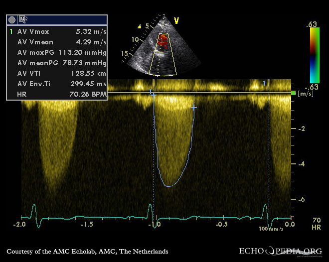
review images, not only the report- AS Vmax/mean gradient, area, flow/stroke volume, EF, morphology and root/aorta. Resolve low-flow or discordant data.
- AR Cusp versus root, integrated severity, LV volumes/EF, LVESD/indexed size and relevant aorta.
- Primary MR Etiology, leaflet/scallop and chordal lesion, calcification, severity and durable-repair probability.
- Secondary MR Ventricular versus atrial mechanism, geometry/tethering, ischemia, GDMT/CRT and persistence under appropriate loading.
- MS Rheumatic versus degenerative/MAC, area, gradient at recorded HR, MR, LA thrombus and balloon/surgical suitability.
- TR Primary/secondary/device mechanism, annulus, RV size/function, congestion and pulmonary vasculature.
B
Reconcile awake and operative physiology
BP + rhythm + loading state- Whole heart Other valves, LV/RV reserve, regional function, pulmonary pressure, rhythm and prior prostheses.
- TTE → TEE TTE establishes usual physiology. TEE defines repair anatomy and guides surgery. Sedation/unloading may reduce apparent MR, especially secondary MR. A smaller jet under anesthesia does not prove that the original lesion resolved.
- Before changing surgery Reconcile intraoperative MR with the awake study.
- Additional testing CMR/CT/stress imaging/hemodynamics only for an actionable question: regurgitant burden, low-gradient AS, calcium/aorta or discrepant pulmonary pressure.
C
Agree on repair and backup
before consent- Plan Repair, replacement backup and associated procedures.
- Consequences Conversion probability, prosthesis, anticoagulation and effects on future procedures/transplantation.

CABG planning requires usable targets and conduits, not a vessel count.
A
Map supply and technical risk
dated angiogram- Targets Left main, LAD/other territories, lesion location/severity, distal quality, diffuse disease, occlusions and competitive flow.
- Prior treatment Stent dates/locations, graft origins/course/patency, ACS and antithrombotics.
- Myocardium Regional/global LV function, actionable scar/viability question, aneurysm, MR and threatened territory.
- Conduits Arterial/venous quality, prior harvest, limb disease and access history.
- Technical constraints Aortic disease, redo entry, grafts near sternum, cannulation/access and pump strategy.
- Benefit horizon Symptoms, longevity, medical alternatives and time to prognostic benefit.
B
Conduit selection
2021 coronary guidelineLAD
Internal mammary
Prefer left when indicated.
Next important non-LAD
Radial over vein
Significantly stenosed target and suitable conduit.
- Radial suitability Prior radial catheterization, hand collaterals, kidney trajectory and potential dialysis-access need.
- Vein mapping When conduit availability or quality is uncertain.
C
Completeness and benefit
avoid reflex viability testing- Viability Test only if actionable. STICHES and REVIVED address different CABG/PCI questions. Neither mandates universal viability testing or immediate surgical-risk reduction.
- Final map Name likely grafts and untreated territories. Weigh completeness against procedural risk. Trial evidence.
Use the combined-operation risk estimate. Added lesion treatment also adds operative complexity.
A
Coronary disease during valve surgery
source-specific thresholdsESC/EACTS 2025
≥70%
Concomitant CABG recommended. 50–70%: consider.
Left main
≥50%
Assess separately under coronary guidance.
- Heart Team Review targets and physiology. Important valve disease can alter coronary physiology.
B
CTSN: moderate ischemic MR
301 randomized · 2 years| Outcome | CABG alone | CABG + repair |
|---|---|---|
| Mortality | 10.6% | 10.0% |
| Moderate/severe residual MR | 32.3% | 11.2% |
| LV reverse remodeling | No significant between-group difference | No significant between-group difference |
| Neurologic / SV arrhythmia events | Less frequent | More frequent |
- Result Combined-procedure mortality HR 0.90, 95% CI 0.45–1.83. Better MR correction without demonstrated survival/remodeling benefit.
- Limit Nonsignificance is not equivalence. Moderate ischemic MR, not severe primary MR or every contemporary repair decision.
C
Justify each added procedure
one final consent- Inventory Valve, coronary territory, aortic segment and AF/LAA procedure → indication, technical plan and incremental benefit.
- MR / TR Mechanism, annular/chamber findings and progression/residual-disease risk.
- LAA exclusion May add stroke protection in appropriate AF patients. Does not automatically replace anticoagulation.
- Consent Final operation, including added procedures and repair-to-replacement conversion.
Repeat a test for clinical change, poor quality or an unanswered operative question.
A
Core assessment
verify dated results- TTE / ECG LV/RV, valves, pulmonary pressure, rhythm/conduction, ischemia and postoperative comparison.
- CBC / chemistry / renal Anemia/platelets, electrolytes, kidney trajectory and drug clearance.
- Liver / albumin / coagulation Hepatic reserve, nutrition and anticoagulant exposure, especially with liver/bleeding concerns.
- Blood bank Antibodies, crossmatch, anticipated blood use and acceptable products.
- Coronary assessment CABG targets or concomitant revascularization during valve surgery.
B
Selective studies
question → test| Question | Study |
|---|---|
| Lung/pleura, previous surgery, aorta or access | Chest imaging |
| Lung disease / uncertain respiratory reserve affecting care | PFTs ± ABG |
| Redo entry, aortic calcification/disease, clamp/cannulation/access | CT chest/aorta |
| Usable conduit / harvest or access constraint | Conduit imaging |
| Exclude obstructive CAD in suitable low/moderate-risk valve candidate | CCTA. A nondiagnostic calcified scan cannot exclude disease |
C
Selection rules worth remembering
not routine for every operation- Carotid duplex ESVS 2023: consider before CABG with age >70, TIA/stroke, bruit or left-main disease. Abnormal results need individualized vascular/cardiac planning.
- No blanket carotid screening Routine duplex before every open-heart operation is not recommended.
- RHC Equivocal echo/hemodynamics. ESC/EACTS 2025 also recommends for all severe-TR treatment candidates. Record flow, filling pressures and PVR.
- Results Assign an abnormal-result reviewer.
Current organ reserve determines tolerance. A historical EF, creatinine or cirrhosis label is insufficient.
A
Find the limiting reserve
current trajectory- HF Congestion/perfusion, LV/RV, valve physiology and desired volume state.
- Kidney AKI versus CKD, electrolytes, dialysis schedule/access and nephrotoxic/contrast exposure.
- Cirrhosis Child-Pugh points, dated MELD/version, ascites/encephalopathy, portal bleeding, kidney reserve, nutrition and hepatology review.
- Lungs Baseline oxygen/ventilation, COPD/OSA, infection/effusion and expected postoperative support.
- Frailty / nutrition Mobility, independence, cognition, weight loss/intake and rehabilitation resources.
- Diabetes A1C, actual drugs, hypoglycemia, fasting insulin and SGLT2 exposure.
B
Cirrhosis: identify the applicable cohort
Jenkins · selected operated patients30-day mortality
4.1%
220 cirrhotic patients undergoing cardiac surgery with cardiopulmonary bypass.
90-day mortality
12.6%
Median MELD 10. Only four class C patients.
- Selection limit Declined patients absent. These rates do not describe all advanced cirrhosis.
- Advanced disease Assess isolated versus combined procedures and effect on transplantation. Compare cohorts, endpoints and complications.
C
Optimize within the surgical window
treatment → reassessment- Time Separate days, weeks and fixed risk. Prehabilitation must fit stability/urgency, without delaying necessary surgery solely for exercise.
- Readiness Diuresis, anemia or pulmonary treatment needs an outcome, reassessment time and responsible clinician.
Treat anemia before surgery. During bleeding, evaluate surgical source and hemostasis together.
A
Blood plan
before incision- History Bleeding/transfusions, antibodies/reactions, acceptable products and preferences.
- HIT Identify previous HIT and planned bypass anticoagulant.
- Anemia Cause, iron status and blood loss. Treat correctable causes when time allows. Avoid arbitrary normalization by transfusion.
- Hemostasis Drug clearance, platelets, liver disease and indicated hematology input. PT/INR/aPTT incompletely predict bleeding.
- Availability Type/screen or crossmatch, antibodies, rare products and alternatives.
- Conservation Antifibrinolytic, cell salvage, perfusion and laboratory/viscoelastic-guided pathway.
B
TRICS III
5,243 moderate/high-risk patients| Comparison / outcome | Restrictive | Liberal |
|---|---|---|
| Hb threshold | <7.5 g/dL | <9.5 g/dL OR/ICU, <8.5 g/dL ward |
| Death / MI / stroke / new dialysis | 11.4% | 12.5% |
| RBC transfusion | 52.3% | 72.6% |
- Endpoint Discharge or day 28, whichever first. Restrictive strategy met noninferiority in this population.
C
Thresholds do not replace resuscitation
source control + oxygen delivery- Hemorrhage / ischemia / shock Assess loss rate, perfusion, oxygen delivery and need for surgical source control.
- Escalation Surgeon, anesthesia, perfusion and blood bank share the plan. Empiric products must not delay evaluation of a correctable surgical source.
Use cardiac-surgery intervals, actual last doses, clearance and operation time.
A
Elective open-heart / CABG holds
exceptions require a surgical/anesthesia plan| Drug | Anchor |
|---|---|
| Aspirin for CABG | Usually continue. Review exceptional bleeding risk |
| Ticagrelor / clopidogrel / prasugrel | At least 3 / 5 / 7 days, respectively |
| DOAC | EACTS 48–96 hours, drug/CrCl dependent. Renal impairment prolongs clearance. No routine heparin bridge |
| VKA | EACTS at least 4 days. Surgery-day INR ≤1.5. Verify INR/bridging need |
| UFH infusion | EACTS 2–4 hours, then aPTT |
| LMWH bridge | EACTS 12–24 hours, preparation dependent. Therapeutic dosing/renal plans generally need the longer interval |
B
Urgency and thrombotic risk
drug offset is not the only clock- Urgent CABG ACC/AHA/SCAI 2021: ≥24 hours off clopidogrel/ticagrelor when feasible. Emergency surgery may precede ideal clearance.
- Timing rationale Record bleeding contingency. Reconcile ACS/PCI urgency with the chosen P2Y12 interval.
- VKA bridge Mechanical valve type/position, rheumatic-valve AF, recent thrombosis/LV thrombus, prior events and bleeding risk.
- Residual drug Platelet-function/drug-specific assay only if timing changes. Routine PT/aPTT cannot reliably exclude significant DOAC effect.
C
Plan beyond the hold
reversal + restart- Reversal / bypass Anesthesia/perfusion review. Urgent reversal concerns.
- Restart indication Graft patency, ACS, AF, prosthesis or other thrombosis. Name conditions and owner.
Preserve indicated therapy when tolerated. Name restart conditions and the clinician responsible.
A
Cardiac-surgery medication decisions
physiology + indication| Therapy | Before → reassess after |
|---|---|
| Beta blocker | Continue if tolerated → bradycardia, conduction, shock and POAF prevention |
| ACE inhibitor / ARB | EACTS: consider continuation, individualized by half-life/patient factors → BP, perfusion, kidney function, potassium |
| ARNI / MRA / diuretic | HF/anesthesia plan for congestion, BP and renal/electrolyte risk → current physiology |
| Statin | Preserve ASCVD indication, avoid reflex loading → secondary prevention/tolerance |
| SGLT2 inhibitor | Hold ≥3 days, 4 for ertugliflozin → resume after illness/ketosis risk resolves and intake is reliable |
| Insulin / other diabetes drugs | Fasting regimen, basal coverage, monitoring → nutrition, glucose and insulin transition |
B
STICS: loading is not secondary prevention
1,922 elective cardiac-surgery patientsRosuvastatin
20 mg
Versus placebo. No POAF or myocardial-injury reduction.
Postoperative AKI
+5.4 percentage points
Does not remove long-term lipid indication after CABG.
C
Avoid predictable medication errors
recheck perfusion, intake and laboratories- ACEi / ARB No universal noncardiac-surgery hold. ARNI needs its own decision and switching precautions.
- ICU glucose Usually 140–180 mg/dL. Selected cardiac-surgery patients may safely use a tighter individualized target.
- Type 1 diabetes Basal insulin despite fasting. SGLT2 exposure plus unexplained acidosis → consider euglycemic ketoacidosis.
- Other exposures Review GLP-1, gastric symptoms/aspiration, QT/rate interactions, steroids and immunosuppression with anesthesia/pharmacy.
An infection label needs a source-control and surgery-timing decision.
A
Define infection and destructive anatomy
before elective prosthetic-valve surgery- Sources Active infection, skin/wounds and dental concerns. Use cardiac-surgery screening/decolonization.
- Cultures Before antibiotics when feasible without unsafe delay. Record organism, susceptibility, doses and response.
- TTE / TEE ± advanced imaging Vegetation, abscess, prosthetic involvement and destructive complications.
- Antimicrobials Intraoperative treatment, tissue cultures/pathology and postoperative ID plan.
B
Identify the operative indication
Endocarditis Team- HF / acute valve failure Perfusion, congestion, mechanism and time available.
- Uncontrolled infection Culture trajectory, sepsis, perivalvular extension and extracardiac sources.
- Embolic risk Vegetation location/mobility, prior embolism and neurologic findings.
- Anatomy Annular/aortic destruction, other valves, prostheses and device infection.
C
ESC 2023 timing
indication and current physiologyEmergency
Within 24 hours
Timing follows the surgical indication.
Urgent
Within 3–5 days
Nonurgent: same admission.
- Ischemic stroke Indicated surgery often proceeds without routine delay when neurologic prognosis permits.
- Intracranial hemorrhage Individualize with neurology/neurosurgery and Endocarditis Team. No blanket delay for every neurologic event.
- Antibiotics Do not require course completion before indicated surgery.
- Pending decisions Name infection/surgical leads, outstanding microbiology/source actions and cost of waiting.
Before induction: preload, afterload, rhythm and support plans for LV failure, RV/PH, acute regurgitation or fixed outflow obstruction.
A
TEE findings that change the operation
record loading state- Valve / CABG Verify intervention result and complications. After CABG, review global/regional function and new ischemia.
- Mitral repair Residual MR, gradient and SAM/LVOT obstruction.
- Prosthesis Seating, function and paravalvular regurgitation. Interpret high gradients with flow, size and technical findings.
- Before leaving OR Communicate findings requiring another intervention. A gradient alone does not establish obstruction.
B
OR → ICU handoff
actual operation, not planned procedure- Operation Grafts, valve type/size, repairs, added procedures and residual disease.
- TEE / bypass Pre/post LV/RV, regional findings, leaks/gradients, operative events, separation, support and anticoagulation/reversal.
- Bleeding Surgical concerns, drains, products, coagulation and re-exploration plan.
- Rhythm Underlying rhythm, wires, pacing dependence and device settings.
- Perfusion / ventilation Airway/lung issues, oxygenation, temperature, lactate, urine output and current support.
- First decisions Drug restart, glucose, neurologic reassessment and escalation.
C
Rescue and recovery
normal BP can conceal support dependence- Rescue threshold Surgeon, repeat imaging, return to bypass, re-exploration, coronary assessment or mechanical support. Name available resources.
- Recovery Normal BP on escalating support is not recovery. Individualize extubation, analgesia, pulmonary care, mobilization and nutrition.
Escalating support, rising lactate, ischemia, bleeding or a neurologic deficit demands immediate reassessment.
A
Find the mechanism
bedside findings + operative handoff| Pattern | Possibility → assessment |
|---|---|
| Low output | LV/RV failure, ischemia, preload, rhythm or mechanical problem → echo, perfusion/hemodynamics, surgeon |
| Vasodilatory hypotension | Vasoplegia, inflammation, infection or drugs → flow, filling, bleeding and directed support |
| Bleeding / tamponade | Surgical source, retained clot or hemostasis → drains, coagulation, urgent echo, surgeon |
| New ischemia / regional dysfunction | Graft/coronary compromise or other injury → ECG/echo, prompt coronary/surgical decision |
| Neurologic deficit | Stroke, embolism, hemorrhage or other process → stroke/neurology pathway and imaging |
| Oliguria / AKI / acidosis | Low perfusion, congestion, kidney injury, drugs/metabolism → volume, electrolytes, exposures and trajectory |
B
Two postoperative traps
do not miss localized tamponade- No large effusion Does not exclude localized clot/tamponade. Persistent concern → urgent surgical review and appropriate additional imaging.
- Repeated fluid May worsen RV failure or congestion. Match treatment to mechanism.
C
Arrest: activate the surgical pathway
AHA 2025Act now
Witnessed VFTrained team: three stacked shocks. CPR if unsuccessful within one minute.
Witnessed asystole / bradycardic arrestExisting epicardial wires: immediate pacing. CPR within one minute if unsuccessful.
- Perfusion / rescue Compressions when emergency resternotomy is not immediately available. Rapid reopening requires a staffed/equipped ICU.
- Full pathway CTICU guide: PEA, drugs, reopening, devices and post-arrest care. Communicate operation, findings, support trend and suspected cause.
Reconcile the operation, AF and coronary indications into one antithrombotic regimen.
A
Procedure-specific treatment
dose + start gate + duration| Indication | Regimen |
|---|---|
| CABG | ACC/AHA/SCAI 2021: aspirin 100–325 mg within 6 hours, indefinitely when safe. Record local dose |
| CABG after ACS / recent PCI | Restart indicated P2Y12 after hemostasis. ACS default DAPT ≥12 months unless high bleeding risk |
| Mechanical valve | Lifelong VKA. DOAC contraindicated. Common INR targets: mitral 3.0, modern bileaflet AVR without added risk 2.5 |
| Aortic surgical bioprosthesis, no other OAC indication | ESC 2025: low-dose ASA or VKA for the first 3 months. ACC 2020: VKA 3–6 months in appropriate patients |
| Mitral/tricuspid surgical bioprosthesis | ESC 2025: consider VKA for the first 3 months without another OAC indication. Specify bleeding plan and reassessment |
| Bioprosthesis + AF | Continue indicated OAC. ESC 2025: consider DOAC over VKA after 3 months (IIa B). Earlier continuation of an existing DOAC may be considered when surgically safe (IIb B). Never use for mechanical valves |
B
Overlap and exceptions
no accidental triple therapy- New mechanical valve Start VKA/heparin bridge when safe, continue bridge until target INR. Targets depend on type/position and added risk.
- Several agents Name each indication and de-escalation date. LAA exclusion does not automatically remove OAC need. Moderate/severe MS needs specific assessment.
C
Postoperative AF
tolerance → treatment → reassessment- Treatment Correct contributors. Stable: rate or rhythm control. Poor tolerance: cardioversion assessment. Check LV function, BP, conduction, QT/interactions.
- Anticoagulation ACC/AHA 2023 supports 60 days when surgical bleeding permits, then reassess long-term indication. Discharge sinus rhythm alone is insufficient.
- Follow-up AF burden/persistence, stroke/bleeding risk, agent, owner and INR/renal monitoring. Continue indicated lipids/secondary prevention after contraindication review.
Readiness means a decision, its remaining conditions and a named owner.
A
Consent
patient priorities + actual operation- Benefit Symptoms/prognosis, alternatives, mortality/morbidity estimate and uncertainty.
- Recovery Stroke, kidney injury/dialysis, bleeding/reoperation, ventilation, infection, rhythm/pacing, function and possible support.
- Decision Proceed, optimize/reassess, urgent despite risk or reconsider strategy.
- Conditions Remaining issue, required result, owner and date.
B
Assessment checklist
retain verified facts and uncertainty- Indication / timing [Operation, symptoms, stability, urgency, cost of delay]
- Anatomy / reserve [Dated severity/mechanism/repairability, targets, LV/RV, pulmonary hemodynamics, rhythm, other lesions]
- Procedure [Repair/replacement/backup, prosthesis, grafts/conduits, added procedures, alternatives]
- Risk [Dated operation-specific STS mortality/morbidity/inputs, organ reserve, frailty/nutrition, uncertainty]
- Cirrhosis [Risk source, score/version, applicable 30-/90-day endpoints]
- Workup [Reviewed results, missing actionable tests, treatment, reassessment, clinician, infection/blood/conduit plan]
- Medication [Indication, last dose, surgery time, clearance, bridge, urgent reversal/bleeding contingency]
- Perioperative [Anesthesia/TEE/ICU, pacing/support, escalation]
- Recovery [Antithrombotic start/duration, rhythm/INR/labs, secondary prevention, rehabilitation]
C
Discharge reconciliation
patient + receiving team- Confirm understanding Valve/graft record, baseline postoperative imaging, medicines, INR/OAC/rhythm follow-up, wounds, activity/rehabilitation and access to help. For a prosthesis, give the patient its type, position, size and implant date. State the target INR and the clinician managing it when VKA is prescribed.