A clot is a finding. The useful diagnosis identifies where it formed, where it traveled, what tissue is threatened, and which process keeps generating it. Those distinctions determine both emergency treatment and prevention.
- 1 · ThreatIs there shock, threatened limb, stroke or another organ emergency?
- 2 · TerritoryArterial, venous, intracardiac or microvascular?
- 3 · MechanismLocal thrombosis, embolism, vascular injury or systemic disease?
Use the circulation to organize the differential
| Pattern | First questions |
|---|---|
| Arterial occlusion | Is this plaque-associated thrombosis, embolism or a diseased/injured artery? Assess tissue viability immediately. A threatened limb needs urgent vascular assessment and reperfusion planning. |
| DVT / pulmonary embolism | Confirm location and clinical probability. Identify hemodynamic or respiratory compromise, then select anticoagulation and disposition using the PE/VTE pathway. |
| Unusual or multiple territories | Look for a cardiac source, APS, cancer and the clinical context. Cytopenias, hemolysis or abnormal cell counts can redirect testing toward PNH, TMA or a myeloproliferative neoplasm. |
| Thrombosis with bleeding | Systemic coagulation activation can coexist with factor/platelet consumption. DIC evolves over time. Bleeding does not establish that thrombotic risk has disappeared. |
Separate the substrate from the recent trigger
Build a short timeline: prior events, persistent disease, recent surgery or immobility, cancer treatment, pregnancy/estrogen exposure, vascular procedures, and medication interruptions. A strong trigger and an underlying predisposition can coexist. The practical question is which factors remain after the acute event and which can be changed.
Record the original images and exact clot distribution. A future report of “persistent thrombus” is much easier to interpret when the baseline is available. Keep the lesion, presumed cause and confidence level separate in the assessment. An unexplained event remains unexplained after an incidental positive test.
Detailed pathways: Pulmonary Embolism & VTE and Peripheral Vascular Disease.
The occluded artery identifies the injured territory. It may not identify the origin of the material. Emergency treatment and source investigation should proceed together, with the organ-specific team directing reperfusion.
Recognize the time-sensitive presentation
New focal neurologic deficit requires the acute-stroke pathway. A newly cold, painful, weak or numb limb needs immediate viability assessment. For acute limb ischemia, give systemic unfractionated heparin at diagnosis unless contraindicated while arranging urgent revascularization assessment. Imaging must not delay treatment of an immediately threatened limb. Suspected aortic dissection, major vascular trauma, active bleeding or HIT changes the anticoagulant decision. Acute stroke has a different antithrombotic/reperfusion sequence, so the limb pathway cannot be transferred to it.
| Candidate source | Evidence to seek |
|---|---|
| AF / atrial thrombus | ECG and rhythm history, appropriate monitoring, atrial/valve assessment and TEE or other targeted imaging when it changes management. A normal spot ECG does not exclude intermittent AF. |
| LV / valve lesion | Prior MI, ventricular dysfunction, prosthetic valve, new murmur, fever or positive cultures. Match TTE, TEE and advanced imaging to the suspected structure. |
| Local arterial disease | Prior claudication, abnormal pulses in other territories, calcified plaque, graft/stent history or recent instrumentation. Acute occlusion can occur on chronic PAD. |
| Systemic acquired cause | APS. Selected MPN/PNH testing when blood counts or hemolysis support it. Conventional inherited panels rarely change arterial-thrombosis management. |
A shunt is a possible route, not proof of causation
A PFO can permit paradoxical embolism, but an incidental PFO does not prove that a venous clot caused a stroke. Assess competing stroke mechanisms, venous thrombosis and the clinical/imaging features used for PFO attribution. Closure evidence applies to selected patients with otherwise unexplained ischemic stroke. The discovery of a PFO alone does not establish an indication.
At handoff, state the affected artery, tissue viability, time of onset, reperfusion decision, likely source, outstanding source tests and who owns prevention. “Embolic” should describe a supported mechanism rather than merely an abrupt presentation.
“Cardiac thrombus” spans several diseases. The chamber, underlying lesion, embolic history and device determine which agent can be used and whether treatment has a finite endpoint.
LV thrombus: an imaging and follow-up problem
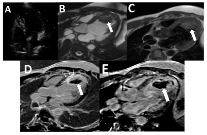
The 2022 AHA statement generally supports about 3 months after acute MI, with repeat imaging. In nonischemic cardiomyopathy with LV thrombus, treatment for at least 3–6 months is suggested, with discontinuation considered when thrombus resolves and EF improves to >35%, or when major bleeding intervenes. A DOAC is a reasonable alternative to warfarin in selected patients. Evidence remains less mature than for AF/VTE. These recommendations do not create an indefinite indication from apical akinesis or reduced EF alone.
For persistent thrombus, review adherence, anticoagulant exposure, morphology and imaging quality before declaring failure. A newly formed mural thrombus still warrants treatment consideration. A persistent organized/calcified mural lesion and a mobile protruding thrombus create different discussions. Use specialist review rather than a single automatic stop rule.
Other sources have different rules
| Finding | Management boundary |
|---|---|
| LA / LAA thrombus | Defer elective cardioversion. Institute or optimize therapeutic anticoagulation and repeat imaging after an appropriate treatment interval, at least 3–6 weeks before reconsidering cardioversion. After cardioversion, continue anticoagulation for at least 4 weeks and longer when stroke risk warrants it. |
| Mechanical valve | VKA remains the anticoagulant. DOACs are inappropriate. Suspected obstructive thrombosis needs urgent multimodality imaging and Heart Team assessment. Valve design, location and patient factors determine INR targets and intervention. |
| Bioprosthetic valve | Distinguish clinical thrombosis with obstruction/embolism from an incidental imaging finding. Do not prescribe lifelong anticoagulation merely because a bioprosthesis is present. |
| Vegetation / valvular mass | Consider infection and nonbacterial thrombotic endocarditis. Cancer, APS and systemic disease provide context, but microbiology and imaging must support the diagnosis before the treatment pathway is chosen. |
Continue in Ischemic Cardiomyopathy, Atrial Fibrillation and Structural & Valvular Cardiology.
The PE/VTE guide carries the full diagnostic, dosing and reperfusion pathways. This chapter connects the acute episode to recurrence prevention and explains why a new symptom on treatment requires investigation.
Make the duration decision twice
Primary treatment of most PE or proximal DVT lasts 3–6 months. The later decision is whether ongoing prevention is warranted. A major transient provoking factor that resolves supports stopping after primary treatment in many patients. Unprovoked, recurrent or persistent-risk VTE often favors extended therapy when bleeding risk is acceptable. Reassess the benefit, burden and preference over time rather than describing “indefinite” as unreviewable.
Do not use residual ultrasound abnormality, an isolated D-dimer or a thrombophilia result as a self-sufficient stop/continue instruction. Record the provoking factor, whether it resolved and what would trigger reassessment.
Work through apparent breakthrough thrombosis
- Confirm a new event Compare current and baseline imaging. Residual clot and post-thrombotic symptoms alone do not establish recurrence.
- Verify dose and phase Check indication, treatment phase, actual dose, weight and kidney function. AF dose-reduction criteria are not VTE criteria.
- Verify actual exposure Ask about missed doses, cost, vomiting, absorption, interacting drugs and procedural holds. Rivaroxaban 15/20 mg requires food.
- Find a new driver Consider active cancer, APS, HIT, venous compression and tumor extension rather than only bland clot.
The sequence organizes assessment, not elapsed clinical time. Urgent stabilization proceeds in parallel.
For confirmed recurrent PE despite adherent therapeutic-intensity treatment, the 2026 guideline considers a change to another drug class reasonable. Recurrence on a reduced-dose DOAC instead can justify full-dose treatment in the same class. In cancer-associated PE recurring on therapeutic LMWH, a 20–25% LMWH dose increase is reasonable with specialist assessment. These recommendations do not make an unconfirmed event or inadequate exposure “drug failure.” An IVC filter is not a substitute for this assessment.
Full treatment phases, doses, special populations and follow-up: Pulmonary Embolism & VTE. For long-term therapy, verify adherence, access and organ function at follow-up.
A positive result may identify susceptibility without establishing why this event happened. Before ordering a panel, state whether the result would alter the agent, treatment duration, future prophylaxis or family counseling.
- DecisionWhat action differs if the test is positive?
- ValidityWill the illness or anticoagulant distort the assay?
- ConsequenceWho will interpret and act on the result?
A focused approach to inherited testing
ASH advises against testing to determine duration after unprovoked VTE when the usual treatment strategy already favors ongoing anticoagulation, and against routine testing after surgically provoked VTE. Selected nonsurgical, pregnancy/hormone-associated and unusual-site scenarios have different conditional recommendations. Most recommendations rest on very low certainty evidence. Hematology input is useful when the result could change a planned stop decision.
Factor V Leiden, prothrombin G20210A and natural-anticoagulant deficiencies chiefly concern venous risk. For an arterial event, inherited screening usually does not change management. An atypical arterial presentation instead warrants a mechanism-based assessment, including acquired causes when supported by the clinical picture.
APS is an acquired disorder with practical consequences
| Question | Interpretation |
|---|---|
| Which assays? | Lupus anticoagulant, anticardiolipin IgG/IgM and anti-β2-glycoprotein I IgG/IgM. One positive antibody does not establish clinical APS. |
| Persistent or transient? | Relevant positivity needs confirmation at least 12 weeks apart and an appropriate clinical history. Anticoagulants can distort lupus-anticoagulant testing. |
| When should testing wait? | Acute illness, pregnancy and anticoagulants affect several functional assays. For protein C, protein S and antithrombin deficiency testing after acute thrombosis, BSH recommends waiting until at least 3 months of treatment. Coordinate the assay and drug-interference plan with the laboratory. Never stop necessary treatment simply to obtain a panel. |
| Does the result change therapy? | For APS with first unprovoked venous thrombosis, EULAR recommends long-term VKA, usually INR 2–3. Use VKA for arterial APS, with individualized intensity and bleeding-risk assessment. Avoid DOACs in definite APS with arterial events. Avoid rivaroxaban in triple-positive APS. |
Explain a result in ordinary language: which risk it identifies, which action changes today, and what remains uncertain. A negative inherited panel does not erase prior VTE, active cancer, a strong family history or a persistent provoking condition. Leave a plan for pending confirmatory testing rather than labeling a provisional laboratory finding as lifelong disease.
Thrombocytopenia does not identify the mechanism. Platelets may be destroyed, consumed or sequestered, and a patient can have serious bleeding and thrombosis simultaneously. Use the timeline, smear and organ pattern.
HIT: probability before antibody testing
Calculate the 4Ts: platelet fall, timing, thrombosis and alternative causes. A fall exceeding 50% can matter even when the absolute count remains normal. A reliable low score (0–3) generally needs neither testing nor empiric HIT treatment. Missing data warrant reassessment rather than false precision.
At intermediate/high probability, stop all heparin sources, including flushes, and obtain a PF4/heparin immunoassay while starting an appropriate nonheparin anticoagulant. Use a functional assay after a positive immunoassay when available and indicated. For intermediate probability without another therapeutic indication and with high bleeding risk, prophylactic intensity may be appropriate. High probability generally calls for therapeutic intensity. Defer starting warfarin until platelet recovery, usually ≥150 × 109/L. If acute HIT develops during VKA treatment, stop VKA and give IV vitamin K while initiating nonheparin anticoagulation. Avoid routine platelet transfusion unless bleeding or another compelling indication warrants it.
The other two urgent patterns
| Pattern | Next action |
|---|---|
| Possible TTP / TMA | Thrombocytopenia with microangiopathic hemolysis and neurologic, renal or cardiac injury needs urgent hematology review. Send ADAMTS13 activity/inhibitor testing before plasma treatment if feasible. With high suspicion for TTP, start plasma exchange and corticosteroids without waiting for results. Specialist-directed caplacizumab/rituximab decisions follow. |
| Possible DIC | Find the underlying illness and trend platelets, PT, fibrinogen and fibrin-related markers in context. Systemic coagulation activation, altered fibrinolysis and endothelial injury may evolve into hemorrhage or organ dysfunction. Use the current disease-appropriate DIC assessment rather than one isolated laboratory threshold. |
Do not wait for the classic five-feature TTP description. Likewise, a low count is not a guarantee against thrombosis, and a TMA label is not a guarantee against bleeding. In a supported circulation, mechanical hemolysis and critical illness complicate the interpretation. The syndrome requires clinical synthesis.
HIT history, current immunoassay and functional-assay status are distinct. Record them separately when planning procedures or future heparin exposure.
A history of hemostatic challenges often explains more than a screening panel. Ask what happened after dental extraction, surgery, childbirth and injuries, and whether there was an opportunity for abnormal bleeding to appear.
Describe the phenotype before naming the disorder
Mucocutaneous bleeding, epistaxis, easy bruising and heavy menstrual bleeding suggest a primary-hemostasis problem, including von Willebrand disease. Deep muscle bleeding, hemarthrosis or delayed postoperative bleeding raises concern for a coagulation-factor problem. These are useful patterns, not diagnostic partitions. Medications and acquired disease can blur them. A structured bleeding assessment tool helps record severity and prior challenges. A low score must not block specific testing when pretest probability is high, such as an affected first-degree relative.
| Result | What it can and cannot say |
|---|---|
| CBC / smear | Count the platelets, compare the trajectory and look for clumping or hemolysis. A normal count does not establish normal platelet function. |
| PT / INR | Useful for VKA monitoring. Prolongation can reflect drug effect, vitamin K deficiency, liver dysfunction or factor consumption. It does not identify the cause on its own. |
| aPTT | Consider heparin, intrinsic-pathway deficiency, inhibitors and assay interference. Warfarin can also prolong aPTT. A prolonged value is not synonymous with a bleeding disorder. |
| Mixing study | Correction can support a deficiency and failure to correct an inhibitor/drug effect, but incubation and assay interference matter. Discuss the pattern with the laboratory before assigning a diagnosis. |
| Fibrinogen | Functional activity and antigen answer different questions. Interpret trends and discrepancies in the clinical and assay context. |
Target testing when the history remains convincing
For possible vWD, obtain vWF antigen, platelet-dependent vWF activity and factor VIII activity. vWF rises with stress and acute illness. Testing should reflect baseline health when possible. Normal screening PT/aPTT does not reliably exclude vWD, and a low factor VIII result is not by itself proof of hemophilia A. Specific testing and subtype interpretation belong with hematology.
Preserve the patient's actual bleeding history, not just “no bleeding disorder.” A negative history in someone never challenged by surgery, childbirth or dental work carries a different meaning from an uneventful major procedure.
Major bleeding involves a critical site, hemodynamic instability, a hemoglobin fall ≥2 g/dL or transfusion of ≥2 units of red cells. Hold the anticoagulant and control the source. Reserve reversal for life-threatening bleeding or major bleeding that persists despite initial measures.
- StabilizeHemodynamics, access, blood products and critical-site assessment.
- IdentifyDrug, last actual dose, kidney function and relevant assays.
- ControlUrgent source control plus indicated reversal through the current pathway.
Match reversal to the exposure
| Anticoagulant | Major / life-threatening bleeding direction |
|---|---|
| Warfarin | 4-factor PCC plus IV vitamin K using an INR/weight-based protocol. If PCC is unavailable, use plasma. Kcentra contains heparin and is contraindicated in known HIT or DIC. |
| Dabigatran | Idarucizumab 5 g IV (two 2.5 g vials). Account for kidney failure, persistent exposure and possible rebound. |
| Factor Xa inhibitor | Use the current Xa-reversal pathway. 4-factor PCC is a nonspecific, off-label option in U.S. practice, with thrombotic risk. Verify the product and contraindications. |
| UFH / LMWH | Protamine reverses recent UFH exposure and only partly reverses LMWH. Timing and the administered dose determine the protocol. Specialist/pharmacy input is required. |
Plan resumption with an explicit owner
After source control, reassess the indication, interruption risk and proposed agent/dose. Remove unnecessary aspirin/NSAIDs and correct dosing or interaction problems. Review organ function, adherence and affordability. Use bleeding scores to target surveillance, not to independently deny indicated anticoagulation.
Intracranial bleeding and hemorrhagic transformation require imaging- and indication-specific review. Selected early DOAC evidence after AF-related ischemic stroke does not establish timing for mechanical valves, substantial parenchymal hemorrhage or endocarditis. Involve stroke and anticoagulation teams.
For elective procedures, account for drug clearance, procedural bleeding risk and confirmed hemostasis. Routine DOAC bridging is unnecessary. High-risk VKA scenarios require a separate plan agreed with the procedural team.
Full interruption and site-specific bleeding pathways: Antithrombotics Around Procedures & Bleeding. Clinical source check: 17 October 2026.