Ischemic cardiomyopathy describes persistent ventricular dysfunction attributable to coronary disease and ischemic myocardial injury. Establish the causal link; coronary atherosclerosis and a low ejection fraction can coexist without one fully explaining the other.
Three descriptions belong in the diagnosis
- Coronary diseasePrior infarction, anatomy, previous PCI or CABG, and current angina.
- Myocardial substrateLV/RV function, infarct scar, potentially reversible dysfunction and valve consequences.
- Clinical trajectoryCongestion, perfusion, exercise limitation, rhythm and response to therapy.
There is no single EF cutoff that defines the ischemic cause. Severe-LV-dysfunction trials commonly enrolled LVEF ≤35%; those eligibility thresholds must remain attached to their results. The 2026 universal HF definition emphasizes cause and trajectory and is a consensus classification document, not a replacement treatment guideline.
Separate the mechanisms
| Mechanism | Clinical interpretation |
|---|---|
| Infarction and scar | Myocyte loss can produce regional thinning, aneurysm, remodeling and a substrate for reentrant VT. An unrecognized infarction may be evident on CMR despite no recorded MI. |
| Stunning / hibernation | Viable but dysfunctional tissue may recover; temporary dysfunction after ischemia and chronically impaired contraction are not equivalent to irreversible scar. Recovery of a segment does not prove a survival benefit from intervention. |
| Mixed disease | Hypertension, valve disease, tachyarrhythmia, alcohol/toxins, genetic or inflammatory cardiomyopathy may add to an ischemic substrate. Reconsider attribution when the coronary territory and myocardial phenotype do not match. |
First identify acute ischemia, hypoperfusion, pulmonary edema or unstable rhythm. A chronic ischemic label must not normalize a new emergency.

Escalate the acute presentations
- New or persistent ischemic symptoms, dynamic ECG change or suspected ACS: obtain serial ECG/troponin assessment and use an ACS pathway. Troponin elevation alone describes myocardial injury; integrate its change with ischemic evidence.
- Hypotension with altered mentation, cool extremities or oliguria; severe hypoxemia; sustained VT: urgent monitored assessment and stabilization. A negative viability study never delays emergency care.
- A new murmur or abrupt deterioration after MI: urgent echocardiography for acute MR, septal rupture or pericardial complications.
Build a baseline that can be compared later
History and examination
Retrieve actual angiograms, bypass targets, infarct history and prior EF measurements. Record exertional angina versus dyspnea, orthopnea, edema, syncope, shocks, functional limits and hospitalization frequency.
Examine BP including orthostasis when relevant, heart rate/rhythm, JVP, perfusion, lungs, edema and murmurs. Reconcile medication use, affordability and adverse effects.
Initial tests
ECG and transthoracic echo; CBC, electrolytes, creatinine/eGFR, liver tests, glucose/HbA1c, lipid profile, TSH and iron studies as appropriate. Natriuretic peptides support HF diagnosis and risk assessment, interpreted with obesity, AF and kidney function.
Echo should report EF and volumes, regional wall motion, RV function, MR and other valves, pulmonary pressure estimate, and possible thrombus.
When to widen the differential
Global severe dysfunction with only limited CAD; noncoronary LGE; marked LV hypertrophy; early conduction disease; a family history of cardiomyopathy or sudden death; toxin exposure; or persistent tachycardia/PVC burden should prompt targeted evaluation. CMR and phenotype-directed investigation can identify a second process. A prior stent is evidence of treated CAD, not proof that every subsequent EF decline is ischemic.
Clarify whether the question is coronary anatomy, inducible ischemia, myocardial cause, or procedural planning. A sequence of tests is useful only when each resolves a remaining uncertainty.
| Test | Useful question and practical limits |
|---|---|
| Transthoracic echo | How impaired is the ventricle, and what complications accompany it? Regional dysfunction supports an ischemic pattern but is not specific; a normal resting regional pattern does not exclude CAD. |
| Coronary CT angiography | Can obstructive CAD be excluded or defined noninvasively in a suitable stable patient? Consider prior stents, heavy calcification, heart rate, contrast exposure and expected image quality. A nondiagnostic scan is not a negative study. |
| Stress imaging | Is there inducible ischemia in a territory that might be treated? Stress echo, PET/SPECT or stress CMR depend on patient characteristics and expertise. Attenuation, LBBB, limited exercise and balanced ischemia can complicate interpretation. |
| CMR | Does scar distribution support infarction or another myocardial process? Quantify LV/RV function and characterize LGE; add perfusion or thrombus sequences when indicated. Check device compatibility and contrast suitability. |
| Invasive angiography | What anatomy can be treated, especially with high clinical likelihood, refractory symptoms or a plausible revascularization indication? Balance diagnostic value against procedural and kidney risk; review grafts and distal targets. |
Current HF and chronic-coronary guidance support evaluation of an ischemic cause and a patient-specific choice between noninvasive and invasive approaches. Stress ischemia assessment may assist selection when revascularization is being considered; testing need not postpone appropriate HF treatment.
Write down the decision before ordering
- New low EF, uncertain CAD: establish whether relevant coronary disease exists and whether another cause is plausible.
- Known CAD, new symptoms: assess urgency, treatment adherence, ischemia and anatomy relevant to the proposed intervention.
- Known diffuse CAD, prohibitive procedural risk: identify a specific management consequence before adding stress or viability imaging.
Viability testing can describe tissue and the likelihood of contractile recovery. Randomized evidence has not established viability as a reliable selector of survival benefit from revascularization.

| Finding | What it supports |
|---|---|
| CMR LGE | Subendocardial or transmural enhancement in a coronary distribution supports infarction. Increasing transmural scar makes segmental recovery less likely; an intermediate segment contains both injured and potentially functional tissue. |
| Dobutamine echo | Contractile reserve supports viable myocardium. Image quality, loading and inducible ischemia affect interpretation. |
| PET / SPECT | Perfusion and metabolism or tracer retention characterize viable tissue. PET perfusion-metabolism mismatch can support hibernation; preparation and acquisition quality matter. |
Two randomized-program cautions
STICH viability follow-up
Among 601 tested participants, viability by SPECT and/or dobutamine echo did not significantly modify the long-term effect of CABG on mortality (interaction P=0.34). Viable myocardium was associated with EF improvement, but that improvement did not explain survival. Selection for testing was nonrandom; CABG versus medical therapy was randomized. Few participants lacked viability; CMR was not tested.
REVIVED viability analysis
In 610 core-lab participants, the amount of viable myocardium did not identify a PCI-responsive subgroup for death/HF hospitalization. Nonviable tissue and scar carried prognostic information. Enrollment already required viability, limiting inference for patients with little or no viable myocardium.
Use imaging for a concrete discussion
Report territory, scar distribution/transmurality, ventricular volumes and uncertainty. Explain whether the expected benefit is symptom relief, regional recovery or a longer-term clinical outcome. A numerical scar threshold can support a recovery estimate; it is not a validated universal survival threshold.
CABG plus medical therapy has randomized long-term survival evidence in selected patients with LVEF ≤35% and coronary disease amenable to surgery. The survival result emerged over extended follow-up.

| Trial | Design and result |
|---|---|
| STICH · 2011 | 1,212 participants, LVEF ≤35%, CABG-amenable CAD; CABG plus medical therapy versus medical therapy. At median 56 months, all-cause death was 36% versus 41%: HR 0.86 (95% CI 0.72–1.04), P=0.12. The primary mortality endpoint was not statistically significant; cardiovascular death and death/cardiovascular hospitalization favored CABG. |
| STICHES · 2016 | Extended follow-up of the same randomized cohort, median 9.8 years. All-cause death: 58.9% versus 66.1%, HR 0.84 (95% CI 0.73–0.97), P=0.02. Cardiovascular death: 40.5% versus 49.3%, HR 0.79 (95% CI 0.66–0.93). |
What the evidence supports
The 2021 US revascularization guideline recommends CABG to improve survival in suitable patients with multivessel CAD and severe LV systolic dysfunction (LVEF <35%; Class I, B-R). It considers CABG reasonable in selected patients with multivessel disease and LVEF 35–50%, a distinct evidence category.
What must accompany the recommendation
- Time matters: CABG carries early operative risk before its long-term advantage. Compare that risk with likely longevity, frailty and recovery burden.
- Eligibility matters: these were surgical candidates with suitable coronary anatomy, not an unselected population with every form of advanced HF.
- The comparator matters: STICH did not compare CABG with PCI. It also preceded routine ARNI and SGLT2-inhibitor therapy.
- Explain the number: the observed mortality difference was 7.2 percentage points across extended follow-up, not an annual reduction and not a personalized forecast.
In stable severe ischemic LV dysfunction, PCI should not be presented as a proven way to prevent death or HF hospitalization simply because viable myocardium is present.

REVIVED-BCIS2 · 2022
700 patients with LVEF ≤35%, extensive CAD amenable to PCI and demonstrable viability were randomized to PCI plus optimal medical/device therapy or optimal therapy alone. At median 41 months, death from any cause or HF hospitalization occurred in 37.2% versus 38.0%: HR 0.99 (95% CI 0.78–1.27), P=0.96. PCI did not improve EF at 6 or 12 months. Early quality-of-life differences diminished by 24 months. Most participants had little or no angina, limiting conclusions about symptom relief in refractory angina.
What remains a reason to consider PCI?
- Acute coronary syndromes: use the acute presentation, coronary anatomy and current ACS guidance.
- Persistent angina despite appropriate therapy: a treatable culprit territory may justify symptom-directed revascularization.
- When CABG is unsuitable: the Heart Team may consider PCI for an individualized objective. Surgical ineligibility does not create evidence of a PCI survival benefit.
Two extrapolations to avoid
| Tempting inference | Why it fails |
|---|---|
| “A positive viability test rescues the PCI indication.” | REVIVED required viability; its core-lab analysis did not identify a benefit from PCI according to the amount of viable myocardium. |
| “ISCHEMIA settles revascularization in low EF.” | ISCHEMIA excluded LVEF <35% and NYHA III–IV HF; its stable-coronary population cannot answer the severe ischemic-LV-dysfunction question. |
In the consent discussion, name the intended endpoint: relief of angina, treatment of ACS, or another specific clinical objective. Improved angiographic appearance is not itself proof of improved patient outcomes.
Decide on the clinical objective before choosing the procedure. Record why CABG, PCI or medical therapy best matches the anatomy, expected benefit, procedural risk and patient priorities.
| Review domain | Information that changes the recommendation |
|---|---|
| Coronary anatomy | Left main or multivessel disease, proximal territories, distal graft targets, diffuse disease, chronic occlusions, prior grafts/stents and feasibility of durable revascularization. |
| Ventricle and valves | EF and volumes, RV function, scar distribution, MR, pulmonary hypertension, aneurysm and thrombus. Link each imaging result to a proposed action. |
| Clinical objective | Long-term survival in a CABG-eligible phenotype; relief of limiting angina; an ACS indication; or clarification that advanced HF treatment is the more relevant next step. |
| Patient risk | Operative estimate plus frailty, kidney/lung/liver disease, neurologic function, bleeding risk, recovery capacity, life expectancy and patient goals. |
| Implementation | Ability to sustain GDMT and antithrombotic therapy, rehabilitation and follow-up; prior chest surgery; contrast strategy; realistic procedural expertise and support. |
Multidisciplinary review is particularly useful when the optimal strategy is unclear. The guideline preference for CABG in suitable severe-LV-dysfunction multivessel disease should prompt a surgical assessment, not automatic PCI because it appears less invasive.
When no procedure is the current plan
State whether the limitation is anatomy, operative risk, patient preference, absence of a symptom/prognostic indication, or competing advanced-HF needs. Continue HF and secondary-prevention treatment. Define what would reopen the discussion, such as new angina, an acute event or a meaningful change in candidacy.
For low output, recurrent admissions or progressive end-organ dysfunction, involve advanced HF concurrently. Repeated viability testing should not defer referral. For support dependence, see shock assessment and revascularization during supported recovery.
Start disease-modifying therapy promptly when the patient is clinically stable. A coronary workup or revascularization plan is not a substitute for HFrEF treatment.
| Foundation | Practical application |
|---|---|
| ARNI / ACEi / ARB | Use sacubitril/valsartan when appropriate; ACEi or ARB when ARNI is unsuitable. Monitor BP, kidney function and potassium. Never combine ARNI with an ACE inhibitor; allow the required 36-hour washout. Do not use ARNI or ACE inhibitors with a history of angioedema. Avoid ARNI, ACE inhibitors and ARBs during pregnancy. |
| Evidence-based beta blocker | Carvedilol, metoprolol succinate or bisoprolol. Start or increase once compensated and adequately perfused; do not initiate/up-titrate during shock or active hypoperfusion. Heart rate, conduction and congestion guide titration. |
| MRA | Spironolactone or eplerenone when eGFR >30 mL/min/1.73 m² and potassium <5.0 mmol/L, with close surveillance. Stop if potassium cannot be maintained <5.5 mmol/L. |
| SGLT2 inhibitor | Dapagliflozin or empagliflozin has HFrEF benefit regardless of diabetes. DAPA-HF tested dapagliflozin 10 mg daily on recommended background treatment. Assess volume and kidney status, and discuss genital infection, ketoacidosis and fasting/illness precautions. |
Implement a regimen, then titrate
Introduce the complementary classes early at tolerated doses rather than waiting to maximize one before adding the next. Individualize sequence to congestion, BP, heart rate, eGFR and potassium. Check symptoms and labs after relevant changes; document a reason and revisit any omitted class.
Relieve congestion
Use loop diuretics to achieve and maintain euvolemia. Adjust against weight, examination, symptoms, kidney function and electrolytes. Diuresis improves congestion but does not replace therapies that modify HF outcomes.
Add selectively
Persistent symptoms may warrant additional therapies, iron-deficiency treatment, CRT or advanced-HF assessment. Continue indicated HF therapy after EF improves; recovery does not erase the ischemic substrate.
The HF diagnosis adds to the secondary-prevention workload. Treat established atherosclerotic disease while accounting for bleeding, hypotension, kidney function and the burden of the complete regimen.
Lipid treatment: current 2026 goals
Use high-intensity or maximally tolerated statin therapy. In clinical ASCVD, the 2026 US dyslipidemia guideline recommends an LDL-C reduction of at least 50% with a goal <55 mg/dL for very-high-risk ASCVD and <70 mg/dL when not at very high risk. Assess the formal risk category; ischemic cardiomyopathy alone is not a shortcut to every category. Add evidence-based nonstatin treatment when needed and address intolerance systematically.
Give every antithrombotic an indication
| Context | Decision |
|---|---|
| Stable CAD, no OAC indication | Low-dose aspirin is a standard secondary-prevention option; P2Y12 monotherapy may be appropriate according to prior PCI, tolerance and current guidance. Low EF does not by itself require DAPT. |
| Recent ACS / PCI | Select agent and duration using ischemic and bleeding risk, stent/ACS history, planned surgery and adherence. Provide an explicit stop/review date; do not make DAPT indefinite by default. |
| AF, LV thrombus or another OAC indication | Coordinate anticoagulant and antiplatelet strategy. Minimize triple therapy; the 2026 anticoagulation statement emphasizes bleeding risk and a specific coronary indication for combined treatment. Chronic HFrEF in sinus rhythm alone is not an anticoagulation indication. |
Restore function and reduce exposure
Offer cardiac rehabilitation when eligible, smoking cessation treatment, a heart-healthy diet and progressive activity within symptoms. Address BP, diabetes, obesity, sleep and adherence. SGLT2 therapy is part of HFrEF care; a GLP-1 receptor agonist may have a separate ASCVD, diabetes or obesity indication and does not replace HFrEF foundations.
Related guide: Statins & Lipid Therapy.
Plan EF reassessment when the diagnosis is made. Primary-prevention ICD decisions depend on persistent dysfunction, time from MI/revascularization, medical treatment, symptoms and meaningful expected survival.
Primary-prevention framework
| Situation | Typical guideline pathway |
|---|---|
| Ischemic LVEF ≤35%, NYHA II–III | Consider ICD after an adequate course of GDMT when meaningful survival >1 year is expected. The decision includes competing pump-failure/noncardiac risk and patient preference. |
| Prior MI, LVEF ≤30%, NYHA I | A primary-prevention indication can remain despite few HF symptoms, after the required intervals. MADIT-II established a mortality benefit in a prior-MI, low-EF population. |
| Recently diagnosed / treated | Ordinarily reassess after ≥40 days from MI and ≥90 days from revascularization, with roughly ≥3 months of optimized/tolerated GDMT when relevant. Apply all applicable intervals; a predischarge EF is not necessarily the final primary-prevention EF. |
SCD-HeFT enrolled symptomatic NYHA II–III HF with LVEF ≤35%, including ischemic and nonischemic disease. ICD therapy reduced mortality; amiodarone did not provide the same survival benefit. An antiarrhythmic drug is not an equivalent substitute for an indicated defibrillator.
Ask separately about resynchronization
The strongest conventional CRT indication is persistent LVEF ≤35%, sinus rhythm, LBBB with QRS ≥150 ms and NYHA II–III or ambulatory IV symptoms despite GDMT. Other QRS/rhythm patterns and a substantial pacing requirement need individualized EP review; coronary scar can affect response but does not replace the electrical criteria.
If EF improves, document both the current EF and the residual infarct/rhythm history. At generator replacement, reassess benefit, shocks, comorbidity and goals rather than assuming an unchanged decision.
In a scarred ventricle, recurrent monomorphic VT may persist despite successful coronary treatment. Investigate reversible triggers and involve EP early for a durable rhythm strategy.
Evaluate the event, not only the device counter
- Review ECGs and device electrograms to distinguish VT/VF from AF, oversensing or other inappropriate therapy.
- Assess ischemic symptoms/ECG changes, congestion, electrolytes, drug effects and adherence. Acute ischemia can provoke arrhythmia; healed-scar reentry is a different mechanism.
- Syncope, sustained VT, recurrent shocks or electrical storm require urgent monitored care. Device programming, beta blockade, antiarrhythmic treatment and ablation decisions should be coordinated.
Ablation evidence in ischemic cardiomyopathy
| Trial | What it adds |
|---|---|
| VANISH · 2016 | In ischemic cardiomyopathy with an ICD and VT despite antiarrhythmic therapy, catheter ablation reduced the composite of death, VT storm or appropriate ICD shock compared with escalation of antiarrhythmic drugs. This composite result should not be recast as a proven mortality reduction alone. |
| VANISH2 · 2025 | 416 patients with prior MI, clinically significant VT and an ICD: initial ablation versus sotalol/amiodarone. At median 4.3 years, the composite of death, VT storm, appropriate ICD shock or treated sustained VT below ICD detection occurred in 50.7% versus 60.6%; HR 0.75 (95% CI 0.58–0.97). It supports discussing ablation earlier in this selected population. |
Do not promise that PCI eliminates VT
Investigate and treat acute ischemia when present, but chronic scar-related VT deserves its own EP pathway. REVIVED's arrhythmia analysis did not support PCI solely to reduce potentially fatal ventricular arrhythmias. Likewise, a low EF with unexplained syncope or NSVT can warrant risk evaluation even before a routine follow-up date.
Related guide: Tachyarrhythmia Management.
Identify which complication is driving risk or symptoms. Low EF, aneurysm, thrombus and secondary MR are related findings, but they do not imply the same treatment.
LV thrombus: image, treat, reimage
Use echo with an ultrasound-enhancing agent when needed; CMR is valuable when echo is inconclusive or embolic suspicion persists. For LV thrombus after an acute MI, oral anticoagulation is typically given for about 3 months with repeat imaging. For thrombus newly detected after a distant MI/chronic ischemic cardiomyopathy, the 2022 AHA statement discusses at least 3–6 months; whether to continue indefinitely is uncertain. Tailor to persistence, morphology, embolism and bleeding. A DOAC is a reasonable alternative to warfarin in selected patients, with a less mature evidence base. Coordinate any concurrent antiplatelets.
Secondary mitral regurgitation
Reassess MR after decongestion, GDMT and CRT when indicated. Persistent important MR with symptoms warrants a valve/Heart Team discussion, especially if CABG is planned. Transcatheter edge-to-edge repair benefits carefully selected patients; it is not a treatment for every dilated ventricle with MR. COAPT showed fewer HF hospitalizations and lower mortality with repair plus optimized therapy in its selected symptomatic secondary-MR population.
Aneurysm and surgical remodeling
Differentiate a true LV aneurysm from a pseudoaneurysm when suspected; a possible contained rupture requires urgent specialist assessment. Address symptoms, thrombus and VT individually. In STICH's reconstruction comparison, routinely adding surgical ventricular reconstruction to CABG reduced LV volume but did not improve death or cardiac hospitalization: HR 0.99 (95% CI 0.84–1.17). A smaller ventricle is not automatically a better clinical outcome.
Related guides: Structural & Valvular Cardiology and MI Mechanical Complications.
Recurrent deterioration despite tolerated therapy should trigger advanced-HF referral even while coronary options are being reviewed. Waiting for irreversible end-organ injury can narrow treatment choices.
Practical referral signals
| Signal | Why it changes the next step |
|---|---|
| Recurrent admissions / escalating diuretics | Persistent congestion or repeated rescue care despite outpatient treatment suggests a failing strategy. |
| Low BP / inability to tolerate GDMT | Loss of previously tolerated therapies may mark progression, particularly when accompanied by low output or organ dysfunction. |
| Kidney/liver deterioration, weight loss or frailty | Evaluate reversible contributors while determining whether HF is driving end-organ compromise and loss of reserve. |
| Severe functional limitation / inotropes / recurrent VT | Symptoms with minimal activity, dependence on intravenous support or recurrent ventricular arrhythmia require broader assessment than EF alone. |
What the referral evaluates
Advanced-HF assessment integrates CPET when feasible, hemodynamics when indicated, RV function, pulmonary vascular disease, end-organ function, frailty, comorbidity, psychosocial support and goals. Peak oxygen uptake or a risk score supports a decision; no single value establishes transplant eligibility. Consider transplant, durable LVAD, continued medical care and symptom-focused options through a multidisciplinary process.
Coronary disease still matters
Distal vascular disease, previous sternotomy, prior grafts and systemic atherosclerosis can affect procedures and candidacy. Share coronary images and the Heart Team rationale with the advanced-HF team.
Palliative care belongs throughout
Discuss symptom relief, acceptable treatment burden and future device preferences alongside disease-directed therapy. Palliative support can coexist with transplant or LVAD evaluation.
Each visit should answer whether congestion, function, ischemic symptoms and treatment tolerance are improving, and whether a previously deferred decision is now due.
| When / trigger | What to close |
|---|---|
| After discharge or major medication changes | Arrange early review; after HF hospitalization, generally within 7 days. Reassess volume, BP, heart rate, medication access and kidney/electrolyte results. Give drug-specific lab and illness instructions. |
| During titration | Use frequent contacts tailored to risk. Record actual tolerated doses, barriers, diuretic response, symptoms and the next laboratory check. Revisit omitted foundational therapies. |
| After a recovery opportunity | Reassess EF after the appropriate GDMT and MI/revascularization intervals to address ICD/CRT candidacy. Repeat earlier for an important clinical change; document the dates that determine eligibility. |
| Stable chronic follow-up | Review angina, exercise capacity, HF events, rhythm/device history, bleeding, lipid response and adherence. Routine periodic ischemia/anatomic testing without a clinical or functional change is not recommended. |
Two cases that test the central distinction
Case A · viable myocardium
A stable patient has EF 28%, extensive CAD and viability, without limiting angina. The viability result alone does not justify a PCI survival claim. Optimize therapy and assess CABG suitability with the Heart Team; attach trial population and time horizon to the discussion.
Case B · persistent deterioration
A patient has recurrent HF admissions after prior revascularization, low BP and worsening kidney function. Reassess triggers, medications and anatomy when actionable, while referring for advanced-HF evaluation. Another procedure is not the default answer. If support is required, use the shock and support-dependence pathway.
Evidence review: 5 September 2026; shock, support exit and viability interpretation updated 27 September 2026. Source identifiers appear with claims and in chapter references. Trial thresholds remain tied to their studied populations.
Adequate perfusion on inotropes or temporary mechanical circulatory support (tMCS) establishes supported stability. Native-heart recovery requires evidence that perfusion remains adequate as support is reduced.
Stable CAD with mildly reduced EF and adequate output does not by itself establish shock or require advanced-HF consultation. Heart Team decisions remain anatomy- and indication-based; refractory HF, low output or support dependence changes the referral and exit-planning question.
Acute culprit or decompensated chronic HF?
An acute coronary occlusion causing new pump failure differs from chronic, scarred iCM decompensating into low output. CAD and an elevated troponin alone do not establish an acute culprit. Assess the ECG, symptoms, coronary anatomy, LV/RV function and mechanical complications while treating hypoperfusion.
INTERMACS describes the advanced-HF trajectory
| Profile | Meaning for the next decision |
|---|---|
| 1 · Critical shock | Life-threatening hypoperfusion despite escalating support. Rescue circulation and assess neurologic and organ reversibility. |
| 2 · Progressive decline | Deterioration despite inotropes, including worsening congestion, kidney function or nutrition. Stabilization and definitive planning are urgent. |
| 3 · Stable but dependent | Clinical stability on inotropes or temporary support, with documented repeated failure to wean. Dependence remains despite an acceptable BP or lactate. |
| 4 · Symptoms at rest | Persistent resting or daily-activity symptoms and recurrent decompensation, generally without continuous inotropes. Advanced HF, not automatically shock. |
Record current shock severity and trajectory separately, using SCAI staging when appropriate. INTERMACS and SCAI describe different dimensions; they are not interchangeable scales.
Reassess what the native circulation can sustain
- Trend support doses/settings, clinical perfusion, lactate, congestion and kidney/liver function; document the support present when measurements were obtained.
- Combine echo and hemodynamics with a supervised reduction-of-support assessment when appropriate. Reassess RV function as LV or biventricular support falls.
- Normal lactate or improved EF on full support does not prove durable recovery. No single value determines successful weaning.
Next: coronary intervention during supported recovery and a durable exit plan. Device-specific assessment: MCS Weaning & Decannulation.
After initial rescue, ask whether coronary intervention can plausibly change the HF trajectory and whether the patient can tolerate it. Evaluate recovery and advanced-therapy candidacy in parallel.
Make the supported Heart Team review explicit
| Question | Information that changes the plan |
|---|---|
| What can be treated? | Acute culprit, residual ischemia, graftable distal targets and the territory supplied. Connect scar/recovery findings to feasible anatomy. |
| Is there an operative opportunity? | Direction of support requirements; organ and neurologic recovery; RV reserve; congestion, ventilation, bleeding and antiplatelet exposure. |
| What if separation fails? | Planned perioperative support and whether durable LVAD, transplantation or another goal-concordant pathway is feasible. |
This is a clinical synthesis, not a validated allocation score. Necessary revascularization may occur while supported. There is no fixed number of supported days, EF, lactate or cardiac-index cutoff that establishes CABG timing; weaning criteria are not prerequisites for acute salvage.
Keep the stable-trial boundaries attached
STICH / STICHES
The design excluded shock within 72 hours of randomization, defined by IABP or IV inotrope need, and recent MI judged an important cause of LV dysfunction. Its long-term CABG benefit is not direct randomized evidence for actively support-dependent shock. The 72-hour exclusion is not a clinical waiting rule.
REVIVED
Excluded acute decompensated HF or sustained ventricular arrhythmia within 72 hours, and MI within 4 weeks. Its neutral elective-PCI result does not settle treatment of a specific acute shock presentation, or compare CABG with durable LVAD.
CABG versus LVAD: observational evidence
A two-center study of CAD with LVEF ≤25% matched 168 pairs from 881 patients treated in 2011–2022. CABG versus LVAD survival was 77.4% versus 68.9% at 1 year (P=0.10) and 74.0% versus 54.4% at 2 years (P=0.001). Recent MI within 7 days, prior operations and concomitant procedures were excluded.
Temporary support needs a purpose and a reassessment plan. Repeated inability to reduce support should accelerate a durable-exit discussion without assuming that LVAD or transplant eligibility is established.
| Possible exit | What must become credible |
|---|---|
| Native-heart recovery | Sustained perfusion and acceptable filling pressures as support falls, with a corrected or improving cause. Some patients need transition from tMCS to inotropes before a longer wean. |
| Durable LVAD | Predominantly LV failure with adequate RV reserve, recoverable organ dysfunction and an acceptable operative/device-care burden. |
| Heart transplantation | Multidisciplinary candidacy assessment, including pulmonary vascular disease, end-organ function, comorbidity, rehabilitation potential and support. |
| Time-limited decision or comfort-focused care | Explicit milestones, acceptable burdens and an agreed response if recovery or candidacy does not emerge. Palliative care can accompany every pathway. |
Assess LVAD/transplant candidacy while reversible causes and coronary options are addressed. Severe persistent RV failure can make isolated LVAD inadequate. Neither a viability result nor a mandatory elective PCI attempt should postpone the broader evaluation.
Evidence that informs support, not allocation
ALTSHOCK-2 · 2025
101 HF-CS patients eligible for heart-replacement therapy, SCAI B–D, were randomized to early IABP or standard care. The trial stopped for futility. Survival or successful bridge at 60 days was 43/53 (81%) versus 36/48 (75%), P=0.45. Only 39 had ischemic disease. Routine early IABP had no demonstrated benefit; this does not test individualized rescue or CABG versus LVAD.
HeartMate 3 recipients · 2025
In 5,787 INTERMACS 1–3 recipients implanted in 2020–2024, 66.7% had tMCS at implantation. Unadjusted 1-year survival ranged from 70.6% to 85.7% across support groups. These are outcomes among patients who reached LVAD, excluding deaths and recoveries before implantation; selection and residual confounding preclude causal device rankings.
Detailed pathways: MCS Weaning & Decannulation, Durable LVAD and Heart Transplantation.
Ask what an imaging result could change before ordering it. Tissue survival, contractile recovery, symptoms and survival benefit are distinct endpoints; a test that informs one does not automatically establish another.
Interpret the physiology before using a label
| Common shortcut | More useful interpretation |
|---|---|
| “Viable, so revascularize” | First identify feasible coronary targets and an intended benefit. Match the dysfunctional territory to the vessel that can actually be treated; viability alone does not create a procedural indication. |
| “Fixed defect means scar” | A fixed perfusion defect is not synonymous with complete scar. Consider attenuation/artifact and viable tissue with severe flow reduction; integrate gated function, tracer protocol, metabolism or CMR when the distinction changes care. |
| “FDG uptake guarantees recovery” | FDG reflects glucose utilization; dobutamine tests contractile reserve. Perfusion–metabolism mismatch can support hibernation, but preparation, diabetes, loading and image quality matter. Neither signal guarantees global EF recovery or a survival benefit. |
| “A thin wall is dead” | Thinning raises the probability of substantial scar but is not a futility gate. In a CMR study, 18% of thinned regions had limited scar; selected revascularized regions recovered thickness and function. Integrate scar transmurality and the wider clinical picture. |
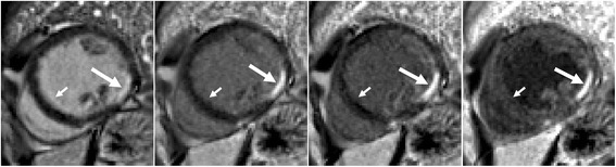
Acute infarction changes the interpretation
Early FDG uptake can reflect inflammation: a 15-patient post-MI study used myocyte-suppression preparation. Acute and chronic protocols are not interchangeable.
Acute imaging is not universally prohibited. A 28-patient PET/CMR study at 5–7 days after PCI assessed later segmental recovery, not imaging-guided treatment selection. Match timing and protocol to the question.
Read the randomized evidence precisely
STICH selected viability testing nonrandomly, but CABG versus medical therapy was randomized. Selection limits subgroup inference. Neither STICH nor REVIVED validated a binary viability rule for revascularization survival benefit.
See viability and scar, Nuclear Cardiology and Cardiac MRI for modality-specific interpretation. Do not delay indicated emergency reperfusion for viability imaging.