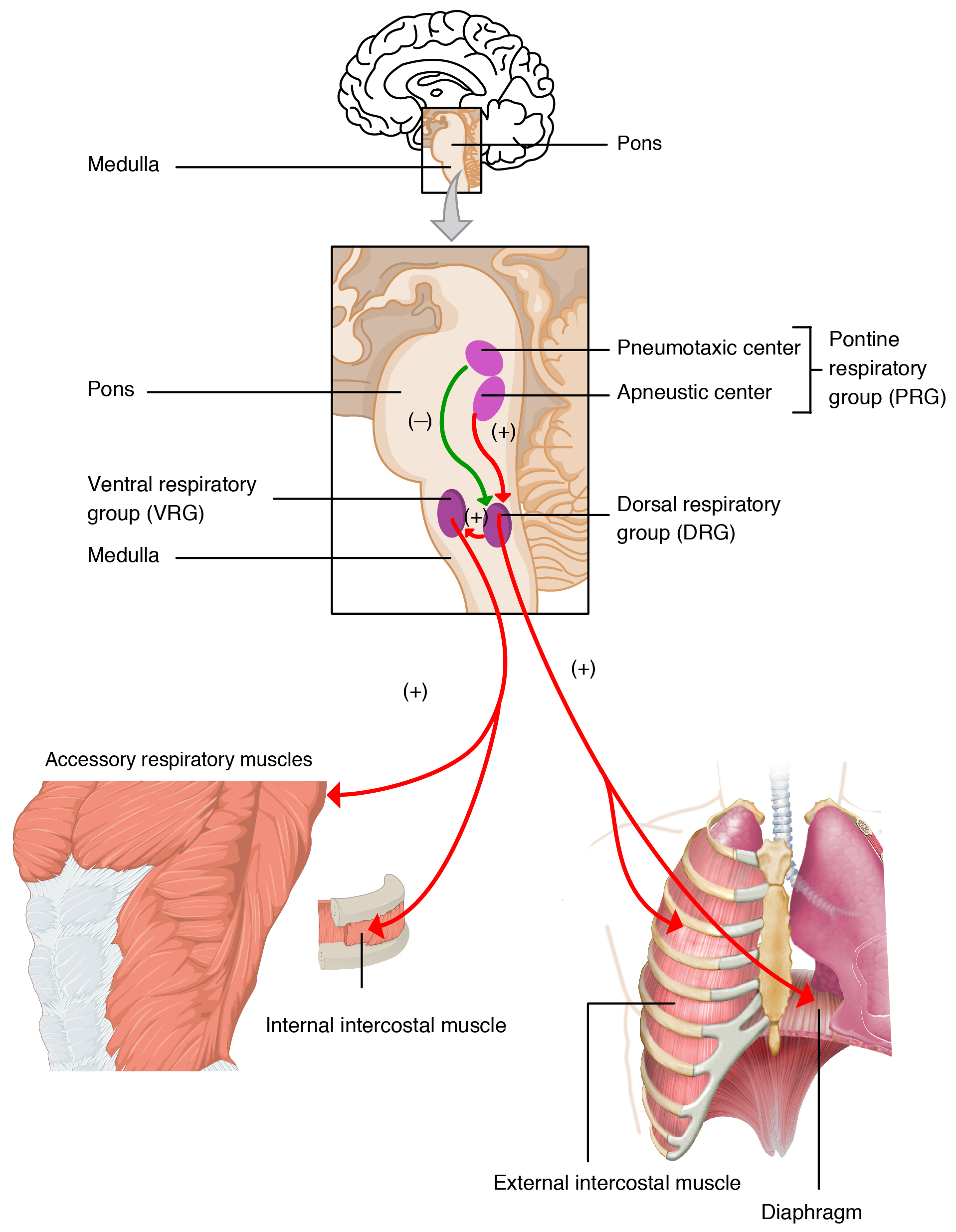
In some patients with chronic lung disease, giving oxygen raises PaCO₂. The long-taught explanation, that oxygen removes the hypoxic drive and the patient stops breathing, is not supported as the main mechanism, and the response to hypoxemia is still titrated oxygen.
1
At the bedside
Titrate · repeat gases · reassess
- Titrate oxygen to 88–92% in COPD or other risk of hypercapnic respiratory failure pending blood gases. Adjust to the patient’s gas results and prior history. Critical illness may initially require high-concentration oxygen while urgent assessment and ventilatory support are arranged.
- Repeat blood gases within 30–60 minutes after starting or increasing oxygen in patients at risk of hypercapnia, even if the initial PaCO₂ is normal. Reassess sooner if deteriorating. Rising PaCO₂ with acidosis requires urgent assessment for ventilatory support. Step oxygen down to the target rather than abruptly stopping it.
- This chapter concerns CO₂ retention. The hypothetical example in chapter 5 is hypocapnic and asks a different physiologic question.
2
What the physiology studies found
Mechanisms and their limitsAubier 1980: COPD in acute respiratory failure, air then 100% oxygen
Minute ventilation fell early (by 18 ± 2% of control, through both tidal volume and rate), then recovered to 93 ± 6% of the room-air value by 15 minutes. PaCO₂ nevertheless rose by 23 ± 5 mmHg, with no significant correlation between the change in ventilation and the change in PaCO₂. The authors attributed the rise mainly to increased VA/Q inhomogeneity.
Hanson 1996: computer model compared with a prior case series
Oxygen alters hypoxic pulmonary vasoconstriction and modulates the Haldane effect, changing physiologic dead space. The model found these changes sufficient to account for the hypercarbia.
Robinson 2000: 22 patients in an acute exacerbation
Twelve retained CO₂ (PaCO₂ rose 8.3 ± 5.6 mmHg) and ten did not. Ventilation fell in retainers (9.0 to 7.2 L/min, P = 0.007) but not in non-retainers, while V/Q dispersion widened in both groups. The authors concluded that an overall reduction in ventilation characterizes oxygen-induced hypercapnia. Alveolar dead space rose in retainers only.
Abdo and Heunks 2012: review
The review argues against loss of hypoxic drive as the principal explanation and recommends titrated oxygen. Reduced ventilation can still contribute.
The honest synthesis is multifactorial. V/Q redistribution with added dead space, the Haldane effect and, in some patients, a modest fall in ventilation all contribute, and the studies disagree about their relative weight. Aubier found ventilation back near baseline while PaCO₂ still climbed. Robinson found a ventilation fall that separated retainers from non-retainers. Loss of hypoxic drive alone does not adequately explain these findings.
3
What the outcome data show
Austin trial · Cochrane review
Austin and colleagues used cluster randomization of paramedics to give either 8–10 L/min oxygen by non-rebreather mask or oxygen titrated to a saturation of 88–92% in 405 patients with a presumed COPD exacerbation (214 with confirmed COPD). Mortality was 9% on the high-flow regimen and 4% on titrated oxygen (relative risk 0.42, 95% CI 0.20–0.89), and 9% versus 2% in confirmed COPD (relative risk 0.22, 0.05–0.91). Lower acidosis and hypercapnia were found in the small per-protocol blood-gas subgroup. These secondary outcomes did not differ significantly in the intention-to-treat analysis.
Grade the evidence honestly. The 2020 Cochrane review found this to be the only eligible trial and judged the evidence low-certainty, with unclear to high risk of bias.