A monitor alarm starts an investigation. Preserve the original tracing, establish whether the rhythm explains the symptoms, and separate what was recorded from what remains suspected.
- 1 · EventSymptoms, pulse, blood pressure, duration and circumstances.
- 2 · RecordingOriginal ECG, onset and termination, device or monitor strips.
- 3 · ContextBaseline ECG, heart disease, medication changes and family history.
- 4 · QuestionWhat is the rhythm, why did it occur, and what evidence is missing?
Obtain a usable rhythm record
- During tolerated tachycardia: obtain a standard 12-lead ECG. Save the complete recording at its original calibration, with the associated vital signs.
- After termination: obtain a sinus-rhythm ECG and compare prior tracings for baseline bundle-branch block, Q waves, pre-excitation, conduction disease and repolarization abnormalities.
- On a monitor or implanted device: review the actual strips or stored electrograms. A detection label alone does not adjudicate VT; examine signal quality, multiple channels and the relationship to symptoms.
Describe before you localize
The rhythm description
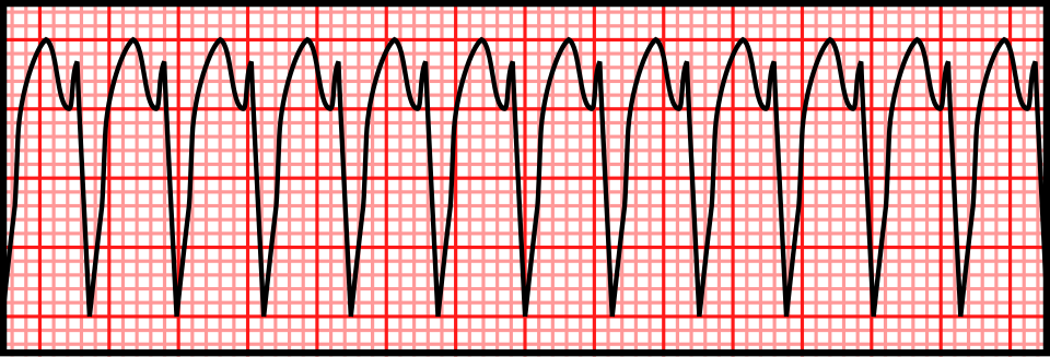
Rate, regularity, QRS width and morphology, atrial activity, onset/termination, and whether successive ventricular complexes look alike. State when a short or single-lead recording cannot answer these questions.
The recording check
Check the pulse and other recording channels for artifact. An intact underlying rhythm may expose noise despite an automated VT label.
For emergency treatment: Tachyarrhythmia & Temporary Pacing.
A regular wide-complex tachycardia may be VT, supraventricular tachycardia with aberrancy, or a pre-excited tachycardia. The complete tracing and clinical context determine how much weight to give an individual sign.
| Finding | What it contributes | What it cannot establish |
|---|---|---|
| A–V dissociation, convincing capture or fusion | Strong evidence for a ventricular mechanism when correctly identified. | Absent or hidden atrial activity does not exclude VT. |
| Precordial morphology | Absence of an RS complex across the precordial leads, prolonged RS timing, and selected bundle-pattern criteria can support VT. | One apparently typical bundle pattern does not resolve every case. |
| Initial R wave in aVR | A useful VT clue within the Vereckei approach. | A negative first step is not a rule-out test. Later steps require accurate timing and waveform measurement. |
| Algorithm result | Organizes ECG evidence and makes the reasoning explicit. | External evaluation still found substantial misclassification; diagnostic performance depends on the tracings and readers studied. |
Morphology suggests a region, not a complete mechanism
Axis, bundle pattern and precordial transition help an electrophysiologist plan further evaluation. In scar-related re-entry, the surface QRS often reflects the exit into working myocardium; the critical circuit can lie elsewhere. A surface localization is therefore a hypothesis, not a demonstrated ablation target.
Look for independent atrial timing
For the acute rhythm approach: SVT, AF/flutter, VT and pre-excitation.
Investigate the immediate trigger and the underlying heart in parallel. Identifying a correctable abnormality does not, by itself, prove that it fully explains the arrhythmia.
Reversible contributors
- Check potassium, magnesium, renal function and clinically relevant metabolic abnormalities.
- Assess hypoxaemia, fever, acute illness and suspected myocardial ischaemia.
- Review prescribed drugs, recent dose changes, drug interactions and non-prescribed exposures.
- Consider drug accumulation and QT prolongation in the appropriate rhythm context.
Underlying substrate
- Prior infarction, coronary disease or revascularization.
- Ventricular dysfunction, chamber enlargement, valve disease or congenital repair.
- Inflammatory, infiltrative or inherited cardiomyopathy.
- Syncope, earlier ventricular arrhythmias and premature sudden deaths in the family.
Make each test answer a question
| Test / review | Diagnostic purpose |
|---|---|
| Echocardiography | Assess LV and RV function, regional wall motion, dimensions and valve/congenital abnormalities. It is the initial structural assessment in suspected or documented VA when heart disease is a concern. |
| Coronary assessment | Choose urgency and modality from symptoms, ECG, clinical likelihood and the suspected mechanism. Acute ischaemia and chronic infarct scar are distinct questions; a history of CAD alone does not answer either. |
| Targeted history / tests | Use the phenotype to direct additional laboratory, imaging and family evaluation. Avoid treating every ventricular arrhythmia as an indication for the same broad test panel. |
Related guides: Chest Pain & ACS · Electrolytes & Acid–Base · HF diagnosis and workup.
A normal echocardiogram does not exclude an arrhythmic substrate. CMR can add ventricular characterization, scar distribution and evidence of inflammation when the presentation warrants further investigation.
| Clinical question | How imaging helps | Interpretive boundary |
|---|---|---|
| Infarct versus non-ischaemic disease | Integrate scar distribution, ventricular morphology and function with coronary and clinical findings. | An imaging pattern informs a diagnosis; it does not replace the rest of the evaluation. |
| Active inflammation? | New arrhythmia with inflammatory clues, conduction disease or ventricular dysfunction may require a myocarditis-focused evaluation with biomarkers and CMR. | Late gadolinium enhancement alone does not establish active inflammation or a specific cause. |
| Is tissue needed? | Discuss biopsy when high-risk presentation or a suspected treatable cause makes tissue information consequential. | Biopsy is indication driven; it is not a routine next step for every unexplained VT. |
| Does scar explain the VT? | CMR can inform the substrate assessment and an EP procedure plan. | Scar location is not, by itself, proof of the clinical circuit. |
Phenotype references: Myocarditis · Cardiac Sarcoidosis · Genetic Cardiomyopathies.
Match recording duration to symptom frequency. Interpret a negative recording in light of whether the suspected event actually occurred.
| Question | Useful approach | Read the limitation |
|---|---|---|
| Frequent palpitations or ectopy? | Quantify burden, morphology and symptom correlation on ambulatory ECG. | Review tracings; a single lead may not establish mechanism. |
| Rare episodes or unexplained syncope? | Consider longer external monitoring or a selected implantable recorder. | Do not postpone evaluation of a documented high-risk event. |
| Unexplained cardiac arrest? | After baseline investigation, supervised exercise testing can reveal an adrenergic phenotype or dynamic repolarization findings. | This recommendation concerns undiagnosed arrest survivors; it is not a mandate to provoke every incidental ventricular run. |
| Possible inherited electrical disease? | Combine phenotype, family history and specialist-selected testing. | Neither an isolated normal ECG nor one abnormal test settles every syndrome. |
Provocation belongs in an experienced setting
Exercise, sodium-channel-blocker challenge and other provocative tests answer different questions. In unexplained cardiac arrest, selection depends on the suspected phenotype and findings already available. False-positive diagnoses and induced arrhythmias are meaningful limitations; provocative drug testing is not an indiscriminate screening panel.
For exercise physiology beyond arrhythmia provocation: Cardiopulmonary Exercise Testing. For syncope evaluation: Syncope, Bradycardia & Pacing.
A complete workup connects the rhythm to a plausible cause, records the remaining uncertainty, and gives the next clinician the evidence needed to resolve it.
Genetics follows the phenotype
- Identify an inherited-disease question: a compatible cardiomyopathy or channelopathy phenotype, premature disease, suggestive extracardiac findings or a family history may justify cardiogenetics assessment.
- Before testing: obtain counselling, define the suspected disease and choose genes with established disease associations. An expansive panel is not automatically a more informative test.
- After testing: interpret pathogenic/likely pathogenic findings in the clinical setting. A variant of uncertain significance is not a confirmed diagnosis and should not drive predictive cascade testing as though it were disease causing.
- Keep clinical follow-up: a negative genetic result does not erase a convincing phenotype or an affected family. Offer appropriate clinical screening to first-degree relatives when inherited disease is suspected, even when no causative variant is found. Plan variant review with the specialist team.
State the electrophysiology question
What EP can clarify
Uncertain tachycardia mechanism, the relationship of a documented morphology to a substrate, or selected questions requiring intracardiac assessment. Surface ECG and imaging should accompany the referral.
What an EP study is not
A universal exclusion test for inherited electrical disease. In unexplained arrest, routine invasive EP testing is not the standard workup for a suspected channelopathy; testing must answer a specific mechanistic question.
Recorded event and symptoms → original tachycardia and baseline ECGs → device/monitor evidence → reversible contributors and their timing → LV/RV function and imaging → coronary assessment → family history → working diagnosis, alternatives and the unresolved question.
Keep diagnosis and risk decisions connected
Finishing the diagnostic record is not the same as completing long-term treatment or sudden-death risk assessment. The significance of the same ventricular rhythm depends on the disease, symptoms and substrate; document who will address those decisions.
Related guides: Genetic Cardiomyopathies · Arrhythmogenic Cardiomyopathy · Hypertrophic Cardiomyopathy.